


Idiomas
Páginas
Jurídico


Efecte de l’estrès oxidatiu en les taupaties
Anna Martínez Casals
ADVERTIMENT. La consulta d’aquesta tesi queda condicionada a l’acceptació de les següents condicions d'ús: La difusió d’aquesta tesi per mitjà del servei TDX (www.tesisenxarxa.net) ha estat autoritzada pels titulars dels drets de propietat intel·lectual únicament per a usos privats emmarcats en activitats d’investigació i docència. No s’autoritza la seva reproducció amb finalitats de lucre ni la seva difusió i posada a disposició des d’un lloc aliè al servei TDX. No s’autoritza lapresentació del seu contingut en una finestra o marc aliè a TDX (framing). Aquesta reserva de drets afecta tant al resum de presentació de la tesi com als seus continguts. En la utilització o cita de parts de la tesi és obligat indicar el nom de lapersona autora.
ADVERTENCIA. La consulta de esta tesis queda condicionada a la aceptación de las siguientes condiciones de uso: La difusión de esta tesis por medio del servicio TDR (www.tesisenred.net) ha sido autorizada por los titulares de los derechos de propiedad intelectual únicamente para usos privados enmarcados en actividades de investigación y docencia. No se autoriza su reproducción con finalidades de lucro ni su difusión y puesta a disposición desde un sitio ajeno al servicio TDR. No se autoriza la presentación de su contenido en una ventana o marco ajeno a TDR (framing). Esta reserva de derechos afecta tanto al resumen de presentación de la tesis como a sus contenidos. En la utilización o cita de partes de la tesis es obligado indicar el nombre de la persona autora.
WARNING. On having consulted this thesis you’re accepting the following use conditions: Spreading this thesis by the TDX (www.tesisenxarxa.net) service has been authorized by the titular of the intellectual property rights only for private uses placed in investigation and teaching activities. Reproduction with lucrative aims is not authorized neither its spreading and availability from a site foreign to the TDX service. Introducing its content in a window or frame foreign to the TDX service is not authorized (framing). This rights affect to the presentation summary of the thesis as well as to its contents. Inthe using or citation of parts of the thesis it’s obliged to indicate the name of the author.
Departament de Patologia i Terapèutica Experimental
Efecte de l’estrès oxidatiu en les taupaties
Anna Martínez Casals
Director de la tesi: Dr. Isidre Ferrer Abizanda
Gener 2010


Departament de Patologia i Terapèutica Experimental
Memòria presentada per Anna Martínez Casals, llicenciada en biologia, per optar al grau de doctora per la Universitat de Barcelona. La tesi ha estat realitzada sota la direcció del Dr. Isidre Ferrer Abizanda en el departament de Patologia i Terapèutica Experimental de la Universitat de Barcelona.
Director Dr. Isidre Ferrer Abizanda Anna Martínez Casals Professor titular UB

Als meus pares, a la Mari Pau i al meu germà
per la seva ajuda i recolzament incondicionals

AGRAÏMENTS
És ben curiós,…els agraïments són quasi sempre la primera part que una
persona es llegeix quan li cau una tesi a les mans. Suposo que deu ser perque tothom
és conscient que una tesi no només es fa gràcies al doctorant sinó que darrera hi ha
tot un engranatge que la fa possible.
Primer de tot agrair a l’Isidre l’oportunitat de fer la tesi en el seu grup, la seva energia
positiva i els seus enfoncs constructius en els experiments. Les pel·lícules i els llibres
recomanats sempre han estat un encert. Gràcies per les tres parts.
A la Marga i a la Rosi per donar-li caliu al lab, especialment a la Marga per l’ajuda
tècnica i també la moral!. A la Núria, al Jesús, al Salva i a la Loli amb qui he resolt
dubtes inesperats. A l’Esther Pérez, el Gabriel, la Sandra, el Gerard, l’Anna Gómez, la
Laia, l’Anna Janué i el Guido amb els qui s’ha format una família de becaris,
compartint moments de tota mena dins i fora del laboratori: gràcies gent!. A les
postdocts que em van rebre a l’arribar al grup, l’Esther Dalfó, la Berta i la Marta
Barrachina i a la carisima Beatrice amb qui vaig tenir sort de poder coincidir amb ella
abans del trasllat a l’altre lab,…gels bidimensionals, riures i no tants riures! A la
Judith per la paciència que ha tingut quan vaig entrar en el món murí i a la Susanna
que sempre ha trobat un moment per ajudar-me. A la nova generació de postdocts,
amb les que he coincidit poc però amb les que he viscut la recta final de la tesi com la
Marta Martínez, la Gema, l’Ester Aso i l’Anton.
No puc passar pas per alt els agraïments pels altres labs: la gent del 4145, la Mireia, el
Joan, l’Ezequiel i la Laura (gràcies per socòrrem en els meus moments d’hipoglucèmia
vespertins), el Jonathan, l’Adriana, l’Artur i l’Anna Priscila. Als “bioquímics”,
especialment a la Maria José i a la Roser que sempre han tingut un moment pels
meus dubtes.
Als amics de la universitat l’Esther, la Gemma, la Sandra, la Ruth, la Mayra, el Juan,
el Javi, la Maria José amb els qui fa anys que vaig fent camí i és tot un plaer
compartir la vida amb vosaltres. A la Irene i al Carlos que han viscut fil per randa
aquesta tesi i que m’han ajudat molt més del que es pensen. A les meves “xatis”, on
cadascuna m’ha recolzat a la seva manera. A la família Casorran-Kappert que ha
viscut algun que altre powerpoint dels meus. A l’Ivan per confiar sempre i tant en mi.
A la Blanca per ajudar-me en els meus inicis-inicis en el món del laboratori i per
continuar al meu costat. A l’Alba amb qui vaig començar ballant i ara caminem
plegades. A la Roser García per la seva visió sempre tan positiva de la vida.

AGRAÏMENTS
Al Frede amb qui he obert una porta nova, on a dins hi he trobat que el pragmatisme i
el realisme poden anar de la mà de la imaginació i dels somnis.
A la família per ser com sou i, especialment, al meu germà Sergi MCasals per ajudar-
me a posar una cara a aquesta tesi.

I. ÍNDEX
II. ABREVIATURES
III. INTRODUCCIÓ
1. Estrès oxidatiu
1.1 BREU HISTÒRIA DELS RADICALS LLIURES
1.2 ESTRÈS OXIDATIU I RADICALS LLIURES
1.2.1 Definició
1.2.2 Classes de radicals lliures
1.2.3 Orgànuls generadors de radicals lliures
1.2.4 Sistemes antioxidants
A. Sistemes enzimàtics
B. Sistemes no enzimàtics
1.3 CARACTERÍSTIQUES DEL CERVELL
1.4 OXIDACIÓ PROTEÏCA
1.4.1 Peroxidació lipídica
1.4.2 Adductes de lipoxidació (ALEs)
1.4.3 Glicoxidació
1.4.4 Adductes de glicoxidació (AGEs)
1.5 TEIXIT HUMÀ POST MORTEM
1.6 ENVELLIMENT
2. Malalties neurodegeneratives
2.1 TAUPATIES: Tau
2.1.1 Malaltia de Steele-Richardson-Olszewski o
Paràlisi Supranuclear Progressiva (PSP)
A. Clínica
B. Característiques neuropatològiques
C. Etiologia
D. Estrès oxidatiu i PSP
1
5
9
11
11
12
12
12
15
16
16
18
19
23
24
26
28
28
29
29
30
35
37
37
38
40
40
2.1.2 Degeneració frontotemporal lobar amb parkinsonisme
lligada al cromosoma 17 associada a mutacions de la tau
(DFTL-tau)
A. Clínica
B. Característiques neuropatològiques
C. Etiologia
D. Estrès oxidatiu i DFTL-tau
2.2 NO TAUPATIES
2.2.1 Degeneració frontotemporal lobar amb inclusions de tau
negatives i d’ubiqüitina positives (DFTL-U):
proteinopatia TDP-43
A. Clínica
B. Característiques neuropatològiques
C. Etiologia
2.2.1.1 Degeneració frontotemporal lobar associada a patologia
de motoneurona i esclerosi lateral amiotròfica (DFTL-ELA)
A. Clínica
B. Característiques neuropatològiques
C. Etiologia
D. Estrès oxidatiu i DFTL-U, DFTL-ELA
IV. OBJECTIUS
V. RESULTATS
1. Brain banks: benefits, limitations and cautions concerning the
use of post-mortem brain tissue for molecular studies
2. Glycolitic enzymes are targets of oxidation in aged human frontal
cortex and oxidative damage of these proteins is increased in
progressive supranuclear palsy
40
42
42
44
45
46
46
47
47
48
49
49
50
51
51
53
57
61
77

3. Type-dependent oxidative damage in frontotemporal lobar
degeneration: cortical astrocytes are targets of oxidative damage
4. Protein targets of oxidative damage in human neurodegenerative
diseases with abnormal protein aggregates
VI. DISCUSSIÓ
VII. CONCLUSIONS
VIII. BIBLIOGRAFIA
87
105
133
151
155


II. ABREVIATURES

II.ABREVIATURES
AA : àcid araquidònic
AGEs : adductes de glicoxidació
AL : àcid linoleic
ALEs : adductes de lipoxidació
ALN : �-àcid linolenic
Arg : arginina
BHE : barrera hematoencefàlica
CEL : carboxietil-lisina
CML : carboximetil-lisina
CN : cabdells neurofibril·lars, NFTs de neurofibrillary tangles
Cys : cisteïna
DHA : àcid docosahexanoic
DFTL : degeneració frontotemporal lobar
DFTL-tau : degeneració frontotemporal lobar amb parkinsonisme lligada al
cromosoma 17 associada a mutacions de la tau
DFTL-U : degeneració frontotemporal lobar amb inclusions de tau negatives i
d’ubiqüitina positives
DFTL-ELA : degeneració frontotemporal lobar associada a patologia de motoneurona i
esclerosi lateral amiotròfica
ELA : esclerosi lateral amiotròfica
FGC-1 : fosfoglicerat cinasa-1
GFAP : glial fibrillary acidic protein
GPx : glutatió peroxidasa
GSH : �-L-glutamil-L-cisteïnglicina
His : histidina
HNE : 4-hidroxi-trans-2-nonenal
Hsps : heat shock proteins
LCPUFA : long-chain polyunsaturated fatty acids
Lys : lisina
MA : malaltia d’Alzheimer
MAP : microtubule-associated phosphoprotein
MDA : malondialdehid
MDA-L : malondialdehid-lisina
MH : malaltia de Huntington
MP : malaltia de Parkinson
ON : òxid nítric
ONS : òxid nítric sintasa
Prx : peroxiredoxines
7

II.ABREVIATURES
Pro : prolina
PSP : paràlisi supranuclear progressiva
PUFA: polyunsaturated fatty acids
RCS : reactive carbonyl species
RNS : reactive nitrogen species
ROS : reactive oxygen species
SLIs : skein-like inclusions
SOD : superòxid dismutasa
TDP-43 : TAR DNA-binding protein-43
Trx : tioredoxina
8

III. INTRODUCCIÓ

III.INTRODUCCIÓ
1. Estrès oxidatiu
1.1 BREU HISTÒRIA DELS RADICALS LLIURES
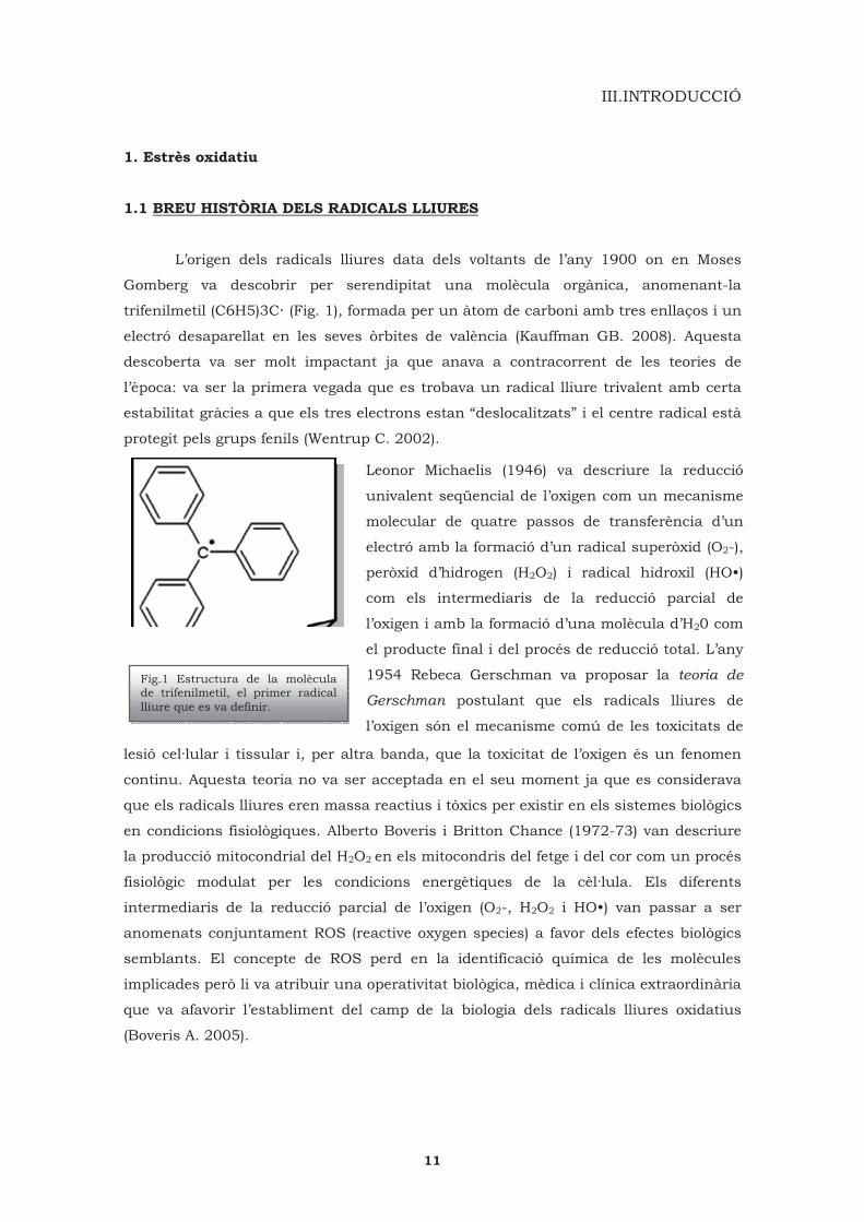
L’origen dels radicals lliures data dels voltants de l’any 1900 on en Moses
Gomberg va descobrir per serendipitat una molècula orgànica, anomenant-la
trifenilmetil (C6H5)3C· (Fig. 1), formada per un àtom de carboni amb tres enllaços i un
electró desaparellat en les seves òrbites de valència (Kauffman GB. 2008). Aquesta
descoberta va ser molt impactant ja que anava a contracorrent de les teories de
l’època: va ser la primera vegada que es trobava un radical lliure trivalent amb certa
estabilitat gràcies a que els tres electrons estan “deslocalitzats” i el centre radical està
protegit pels grups fenils (Wentrup C. 2002).
lesió cel·lular i tissular i, per altra banda, que la toxicitat de l’oxigen és un fenomen
continu. Aquesta teoria no va ser acceptada en el seu moment ja que es considerava
que els radicals lliures eren massa reactius i tòxics per existir en els sistemes biològics
en condicions fisiològiques. Alberto Boveris i Britton Chance (1972-73) van descriure
la producció mitocondrial del H2O2 en els mitocondris del fetge i del cor com un procés
fisiològic modulat per les condicions energètiques de la cèl·lula. Els diferents
intermediaris de la reducció parcial de l’oxigen (O2-, H2O2 i HO•) van passar a ser
anomenats conjuntament ROS (reactive oxygen species) a favor dels efectes biològics
semblants. El concepte de ROS perd en la identificació química de les molècules
implicades però li va atribuir una operativitat biològica, mèdica i clínica extraordinària
que va afavorir l’establiment del camp de la biologia dels radicals lliures oxidatius
(Boveris A. 2005).
Fig.1 Estructura de la molècula de trifenilmetil, el primer radical lliure que es va definir.
Leonor Michaelis (1946) va descriure la reducció
univalent seqüencial de l’oxigen com un mecanisme
molecular de quatre passos de transferència d’un
electró amb la formació d’un radical superòxid (O2-),
peròxid d’hidrogen (H2O2) i radical hidroxil (HO•)
com els intermediaris de la reducció parcial de
l’oxigen i amb la formació d’una molècula d’H20 com
el producte final i del procés de reducció total. L’any
1954 Rebeca Gerschman va proposar la teoria de
Gerschman postulant que els radicals lliures de
l’oxigen són el mecanisme comú de les toxicitats de
11
III.INTRODUCCIÓ
1.2 ESTRÈS OXIDATIU I RADICALS LLIURES
1.2.1 Definició
Finalment l’any 1985 Helmut Sies va definir el concepte d’estrès oxidatiu com
“un desequilibri en el que hi ha un augment d’oxidants o una disminució
d’antioxidants en comparació amb la situació definida com a normal”. A partir
d’aquesta definició es van crear diferents hipòtesis de l’origen de l’estrès oxidatiu: M.
Bergel, basant-se en experiments de rates exposades a determinades infeccions
(Micobacterium leprae i Micobacterium tubercuosis) a les que se’ls hi administraven
dietes prooxidants (alt contingut de lípids autoxidables i vaig contingut de vitamina E),
va concloure que l’estrès oxidatiu és un desequilibri que permet l’aparició de la
malaltia ja que les rates tractades es tornaven més susceptibles. Per altra banda, G.
Deucher considerava que les malalties porten a una “perturbació metabòlica” i
desencadenen en una situació d’estrès oxidatiu prenent com a referència diferents
casos d’hepatitis i de SIDA on els virus estan associats a nivells baixos d’antioxidants
en els diferents teixits i en el plasma (Boveris A. 2005).
La importància de l’estrès oxidatiu en les diferents patologies s’ha fet palesa en la gran
quantitat d’estudis dedicats a aquest camp. Els radicals lliures han estat directa o
indirectament implicats en la patogènesi de diverses malalties neurodegeneratives com
per exemple en la malaltia d’Alzheimer (MA) (Markesbery WR. 1999, Markesbery i col.
2007, Marlatt i col. 2008), la malaltia de Parkinson (MP) (Chinta i col. 2008, Danielson
i col. 2008), la malaltia de Huntington (MH) (Polidori i col. 1999), l’esclerosi lateral
amiotròfica (ELA) (Cleveland DW. 1999) i d’altres patologies neurodegeneratives.
1.2.2 Classes de radicals lliures
En la majoria de cèl·lules aeròbiques la cadena mitocondrial respiratòria és
una de les principals fonts generadores de ROS havent-hi, però, d’altres orgànuls
productors de molècules reactives (veure apartat 1.2.3). Els nivells moderats de ROS
juguen un paper essencial en la modulació de diferents funcions cel·lulars (expressió
de gens, transducció de senyals, defensa contra patògens,...) (Dalle-Donne i col. 2005).
Per tant, en condicions fisiològiques de la cèl·lula es generen metabòlits potencialment
tòxics de l’oxigen en baixes concentracions on les vies productores es poden alterar i
augmentar dramàticament els nivells de ROS.
12

III.INTRODUCCIÓ
Les principals classes de ROS (Fig.2) inclouen (Dalle-Donne i col. 2005) :
1. anió superòxid (O-2), és un producte de vida mitja curta, intermediari en la
reducció de l’oxigen a H2O2, formant-se de manera ràpida i espontània en una reacció
depenent de pH. La seva inestabilitat en medis aquosos ve donada per la seva ràpida
dismutació a H2O2. En condicions normals s’estima que aproximadament sobre l’1%
del flux d’electrons mitocondrials comporta la formació d’O-2, tot i que si hi han
interferències en la cadena de transport d’electrons poden fer augmentar encara més
la seva producció (Dalle-Donne I., Scaloni A., Butterfield DA. 2006).
2. peròxid d’hidrogen (H2O2), té una vida mitja llarga sent un dels productes
més abundants amb gran capacitat de travessar fàcilment les membranes cel·lulars.
Dins de la cèl·lula l’O-2 es converteix ràpidament a H2O2 gràcies a l’enzim superòxid
dismutasa (Dalle-Donne I., Scaloni A., Butterfield DA. 2006).
3. radical hidroxil (HO•), la vida mitja del HO· és curta actuant normalment
en el lloc on s’ha produït. És un dels ROS més reactius per les macromolècules
biològiques on aquesta gran reactivitat implica una menor selectivitat, distància de
difusió i incapacitat per poder actuar com a missatger cel·lular (Mikkelsen i col. 2003).
El grup HO· funciona també com a un mutagen produït per l’exposició de radiacions
ionitzants. Pot formar-se a partir de l’O-2 i del H2O2 (reacció d’Haber-Weiss) o per mitjà
d’un ió metàl·lic (Fe2+, Fe3+) i del H2O2 (reacció de Fenton) (Dalle-Donne I., Scaloni A.,
Butterfield DA. 2006; Alberts B, Bray D, Lewis J. 1996). El HOCl també pot reaccionar
ràpidament amb O-2 i formar grups HO· (Candeias i col. 1993).
4. àcid hipoclorós (HOCl), el H2O2 oxida el Cl-, en una reacció catalitzada per
la mieloperoxidasa i l’eosinòfil peroxidasa, per obtenir HOCl (Thomas i col. 1995).
5. àcid hipobromós (HOBr), gràcies a l’acció dels mateixos enzims que
participen en la formació de l’HOCl, aquest últim pot oxidar el Br- (Thomas i col. 1995)
o bé pot oxidar-lo el H2O2 obtenint HOBr (Henderson i col. 2001).
S’han descrit d’altres tipus de ROS com el radical peroxi (RO•2), el radical alcoxi (RO•),
el radical hidroperoxil (HO•2), l’oxigen (O2), l’ozó (O3).
Per altra banda, les RNS (reactive nitrogen species) són també molècules necessàries
fisiològicament per la cèl·lula però a la vegada són potencialment destructives
(Mikkelsen i col. 2003). La producció biològica de l’òxid nítric (ON) actua com a un
missatger, present en tots els vertebrats, participant en la modulació del flux sanguini,
en processos de trombosi i també en l’activitat neural. Paradoxalment la producció
d’aquesta mateixa molècula pot arribar a ser altament nociva per les neurones en pocs
13

III.INTRODUCCIÓ
minuts en determinades patologies, com per exemple en la isquèmia cerebral.
L’element clau en la balança entre un estat fisiològic i un altre de patològic radica en
la reacció de NO amb O-2 formant molècules oxidants molt potents (peroxinitrit).
Les molècules majoritàries de RNS (Fig.2) són (Dalle-Donne i col. 2005):
1. òxid nítric (ON), s’han descrit tres isoformes diferents d’òxid nítric sintasa
(ONS) catalitzadores de la síntesi d’ON: ONS neuronal (ONSn, ONS-1), ONS induïble
(ONSi, ONS-2) i ONS endotelial (ONSe, ONS-3) (Stuehr DJ. 1999). Les isoformes ONSn
i ONSe produeixen quantitats relativament baixes d’ON si es compara amb la tercera
ONS, ONSi, que és la majoritària (Mikkelsen i col. 2003). Com a conseqüència del
metabolisme de l’ON les cisteïnes de les proteïnes poden patir modificacions nitroses:
l’ON pot formar adductes amb els grups –SH produint grups S-nitrosotiols (Hogg N.
2002). ON reacciona amb les ROS i les converteix en derivats redox molt reactius que
poden atacar a les proteïnes, als lípids i al DNA (Dalle-Donne i col. 2005).
2. peroxinitrit (ONOO-), en condicions fisiològiques existeix en un equilibri
ràpid i dinàmic amb el seu àcid conjugat peroxinitrós (ONOOH). Si es produeix un
augment en la concentració d’ON en el teixit, s’inicia una competència entre aquesta
molècula i l’enzim antioxidant SOD per tal d’eliminar l’O-2 i formar ONOO-. La reacció
entre O-2 i l’ON es produeix tres cops més ràpidament que amb la superòxid dismutasa
(Dalle-Donne i col. 2005).
3. diòxid de nitrogen (.NO2), aquest radical lliure és molt més reactiu que
l’ON. A pH neutre el ONOO- es troba, en part, protonat i aquesta forma protonada
(ONOOH) es descomposa, majoritàriament, de manera ràpida a NO-3 (molècula
inofensiva) i també forma intermediaris altament reactius com el HO· i el .NO2
(Mikkelsen i col. 2003). Per altra banda, el ONOO- reacciona amb el CO2 per formar
radicals carbonats (CO3-·) i .NO2 (Dalle-Donne I., Scaloni A., Butterfield DA. 2006).
També s’agrupen com a RNS l’alquil peroxinitrit (ROONO), el triòxid de dinitrogen
(N2O3), el tetraòxid de dinitrogen (N2O4), l’àcid nitrós (HNO2), el catió nitrosil (NO+),
l’anió nitrosil (NO-), el catió nitroni (NO+2).
14

III.INTRODUCCIÓ
1.2.3 Orgànuls generadors de radicals lliures
1.2.3 Orgànuls generadors de radicals lliures
Els radicals lliures poden originar-se a partir de diferents fonts: tan poden
formar-se a partir de factors medi ambientals, dietes, estils de vida com poden ser
produïts per l’exposició a radiacions ionitzants o raigs ultraviolats, xocs tèrmics,
metalls, pesticides, compostos orgànics tòxics persistents, partícules de l’aire,...
(Limon-Pacheco i col. 2009) o poden crear-se in vivo en diferents orgànuls cel·lulars.
Pel que fa a aquest últim grup una d’elles, potser la font més important productora de
ROS cel·lular, és el mitocondri durant la fosforilació oxidativa. Aquest procés es basa
en que l’energia d’oxidoreducció de la cadena de transport electrònic del mitocondri,
utilitzant el sistema enzimàtic NADH deshidrogenasa, es converteix en un enllaç
altament energètic d’ATP. El flux d’electrons dels diferents substrats, a través de
proteïnes transferidores d’electrons o per sistemes enzimàtics, resulta en la reducció
de quatre electrons de l’oxigen molecular a l’H2O catalitzada per la citocrom c oxidasa.
En aquest procés es generen ROS com a subproductes de les reaccions de
transferència d’electrons tals com l’O-2, HO·, RO·2, RO·, HO·2, HOCl, HOBr, O2 (Dalle-
Donne I., Scaloni A., Butterfield DA. 2006).
El peroxisoma conté un gran nombre d’oxidases que redueixen l’O2 a H2O2,
representant una important quantitat del H2O2 total de la cèl·lula (Wanders i col.
2006). La catalasa utilitza el H2O2 produït per diferents oxidases per oxidar diferents
substrats com per exemple el fenol, l’àcid fòrmic, el formaldehid, l’alcohol,...
Un compartiment intracel·lular que juga un important paper en la biosíntesi
cel·lular (proteïnes de transmembrana, lípids,...) és el reticle endoplasmàtic sent una
font generadora de H2O2. El reticle endoplasmàtic llis conté diferents enzims
necessaris per catalitzar drogues liposolubles i d’altres productes metabòlics
Fig. 2 Esquema representatiu de les diferents classes de ROS, RNS i la relació entre elles. Modificació dels diagrames de (Dalle-Donne i col. 2005, Mikkelsen i col. 2003).
15

III. INTRODUCCIÓ
perjudicials. Els enzims de les famílies citocrom P450 i b5 estan implicats en la
generació d’O-2 i H2O2 evitant l’excés de metabòlits i de tòxics (Dalle-Donne I., Scaloni
A., Butterfield DA. 2006).
El nucli està separat del citoplasma per una membrana doble que conté
citocrom oxidases i un sistema de transport d’electrons i pel que fa al citoplasma, es
manté sota condicions reduïdes gràcies a la capacitat redox dels tiols intracel·lulars.
Conté enzims solubles com la xantina oxidasa i la flavoproteïna deshidrogenasa que
poden generar ROS per diferents reaccions, a més a més existeixen petites molècules
com la dopamina, l’epinefrina,... que tenen la capacitat d’autoxidar-se passant a ser
una important font de ROS intracel·lular.
Físicament la membrana plasmàtica separa el compartiment intracel·lular del
medi extracel·lular, generant un potencial de transmembrana a través del qual es
forma un pas de protons cap a fora de la membrana i d’electrons que es dirigeixen cap
a ella. Aquesta barrera conté NADH oxidoreductases i NADPH oxidases que permeten
controlar les bombes de protons, canals iònics, regular el pH,... i també presenta un
complex redox (semblant al del mitocondri i al de l’aparell de Golgi) com és el coenzim
Q (Skulachev VP. 1996).
1.2.4 Sistemes antioxidants
Halliwell va redefinir recentment el terme d’antioxidant com a “qualsevol
substància que retardi, previngui o elimini el dany oxidatiu d’una molècula diana”
(Halliwell B. 2007).
La gran diferència en la vida mitja dels oxidants, des dels nanosegons del HO· fins als
segons del RO·2, ROONO o bé de l’ON, ajuda a fer una idea de la bateria tan diversa
que existeix d’antioxidants (Sies H. 1993). Les cèl·lules del nostre organisme han
desenvolupat diferents sistemes i mecanismes per combatre directament les ROS,
reparar les lesions oxidatives i restaurar l’homeòstasi de la cèl·lula.
A. Sistemes enzimàtics
S’han descrit diferents sistemes enzimàtics antioxidants tals com la superòxid
dismutasa, la catalasa, la glutatió peroxidasa, la tioredoxina i algunes xaperones
moleculars que ajuden a prevenir la lesió oxidativa produïda per les ROS.
Les superòxid dismutases (SODs) formen una família d’enzims que catalitzen
molt eficientment la reacció de dismutació de l’O-2 obtenint oxigen i H2O2 com a
resultat del procés (McCord i col. 1969, McCord i col. 2005), d’aquesta manera s’evita
16

III.INTRODUCCIÓ
la formació d’altres molècules oxidants molt potents com poden ser els peroxinitrits o
els HO·. L’activitat d’aquests enzims necessita un metall de transició en el seu lloc
actiu per tal de dur a terme el trencament catalític de l’anió superòxid (Culotta i col.
2006). Aquesta família està formada per tres tipus de SODs: la SOD-1/[Cu-Zn]
(mutacions de la SOD-1 en un petit percentatge donen lloc a casos familiars d’ELA), la
SOD-2/[Mn] i la SOD-3 (Fattman i col. 2001, Gao i col. 2008, Marklund SL. 1984).
Un altre sistema enzimàtic antioxidant és la catalasa, un enzim bifuncional on,
per una banda, catalitza la degradació de dues molècules de H2O2 obtenint dues
molècules d’H2O i O2 sense la producció de radicals lliures i, per altra banda,
metabolitza una gran varietat de substrats tals com l’etanol, el metanol, fenols o
nitrits (Oshino i col.1973). Aquest enzim es troba localitzat principalment en la matriu
dels peroxisomes però també en el citosol i el nucli. Alguns grups han demostrat la
seva presència en el mitocondri de certs òrgans tals com en el cor (Radi i col. 1991) o
bé en el fetge (Salvi i col. 2007) protegint contra lesions produïdes per H2O2.
La glutatió peroxidasa (GPx) comprèn una família de múltiples enzims que
catalitzen la reducció de H2O2 o hidroperòxids orgànics a H2O o als corresponents
alcohols, utilitzant el glutatió reduït com a donador d’electrons (Dringen R. 2000a,
Dringen i col. 2000b).
Una altra família d’enzims amb acció antioxidant correspon a la de la
tioredoxina (Trx) que participa en diverses reaccions redox a través de l’oxidació
reversible del centre actiu ditiol a disulfit de la Trx. Les proteïnes d’aquest grup
comparteixen una seqüència semblant en el lloc actiu -Cys-Xxx-Yyy-Cys- formant-n’he
part les Trxs però també la Trx reductasa i les peroxiredoxines (Prx) depenents de Trx.
(Mustacich i col. 2000).
Les Hsps (heat shock proteins) són proteïnes altament conservades entre
espècies que estan constitutivament expressades (representen del 5-10% de les
proteïnes totals) en condicions normals. Són necessàries pel creixement i pel
manteniment normal de la cèl·lula i protegeixen en situacions de lesions metabòliques
i oxidants (Calabrese i col. 2002). Aquestes proteïnes s’activen en situacions de xoc
tèrmic (d’aquí el nom de Heat shock proteins) però també en front d’alteracions de
l’estat redox intracel·lular, exposició a metalls pesats, anàlegs d’aminoàcids, drogues
citotòxiques, falta de glucosa, infecció per patògens i teixits lesionats on les xaperones
es sobreexpressen. Una de les seves funcions és la de protegir les dianes de les
proteïnes cobrint els seus llocs sensibles, en algunes situacions aquest mecanisme no
és suficient i persisteix el dany on les xaperones segresten les proteïnes
desnaturalitzades fins que són replegades o bé degradades (Papp i col. 2003). Aquests
mecanismes estan connectats amb d’altres a través de diferents vies.
17
III. INTRODUCCIÓ
B. Sistemes no enzimàtics
Per altra banda hi ha d’altres sistemes no-enzimàtics, diferents proteïnes que
poden actuar com antioxidants tals com la vitamina E, la vitamina C, la melatonina, la
ceruloplasmina, la ferritina, la transferrina, els carotenoides, la bilirubina, l’àcid úric,
els flavonoides.
La vitamina E és un terme genèric que fa referència a tots els tocoferols, i als
seus derivats, que presenten una activitat biològica RRR-�-tocoferol. A la natura,
s’han trobat 8 compostos amb aquesta activitat on el que té un efecte antioxidant més
eficient és l’�-tocoferol (s’utilitza sovint el terme vitamina E com a sinònim) (Bruun-
Jensen i col. 1994). Es pensa que la seva principal funció és protegir als lípids,
especialment als PUFA (polyunsaturated fatty acids), en situacions d’estrès oxidatiu
prevenint la propagació del dany dels radicals lliures en les membranes biològiques
(Traber i col. 1996).
La vitamina C (L-àcid ascòrbic) és un dels antioxidants més importants
solubles en aigua. La capacitat antioxidant de la vitamina C està relacionada amb la
seva estructura única: en condicions fisiològiques de pH existeix com un anió
monovalent (ascorbat) podent donar un electró per formar un radical ascorbil (Niki E.
1991, Rice ME. 2000). L’àcid ascòrbic protegeix també de la peroxidació que afecta a
les membranes, potenciant l’activitat de l’�-tocoferol (Sies i col. 1995).
La melatonina és una hormona pineal que actua com a un potent antioxidant
podent travessar fàcilment la barrera que separa el circuit sanguini del cervell i també
els diferents compartiments cel·lulars. És capaç d’estimular els enzims antioxidants
endògens cel·lulars (actuant com a un antioxidant indirecte) i funciona sinèrgicament
amb d’altres (Pappolla i col. 2000).
La ceruloplasmina és una proteïna de la fracció �2-globulina del sèrum
sanguini humà que transporta el 95% del coure a la sang. Actua com una ferroxidasa
i protegeix als PUFA de les membranes dels glòbuls vermells de les formes actives dels
ROS (Goldstein i col. 1979, Vassiliev i col. 2005). Per altra banda la ceruloplasmina
pot controlar els nivells de ferro ferròs (Fe2+) oxidant-lo per produir O2 i H2O evitant
reaccions perilloses i reactives.
La ferritina és una proteïna que emmagatzema una gran quantitat d’àtoms de
ferro (4000-4500 àtoms de Fe3+) (Arosio i col. 2002). Al funcionar les ferritines com un
magatzem de ferro, detoxifiquen jugant un paper central en l’administració biològica
del ferro degut a la insolubilitat del ió fèrric (Fe3+) lliure i a la seva alta toxicitat ja que
participa en diverses reaccions ajudant a la formació de ROS molt reactius a través de
la via de Fenton (Arosio i col. 2002, Orino i col. 2001, Riederer i col. 1989).
18

III.INTRODUCCIÓ
La transferrina, la seva principal funció és la del transport de ferro a les cèl·lules en
proliferació i, amb els factors de creixement, ajuda també a reduir la concentració de
ions Fe2+ lliure.
Els carotenoides són compostos naturals amb propietats lipofíliques, se n’han
identificat més de 500 on el més important és el �-carotè. La majoria dels carotenoides
tenen un extens sistema de dobles enllaços conjugats que proporciona l’activitat
antioxidant amb capacitat d’inhibir diferents reaccions dels radicals lliures (Hsu i col.
2002, Sies i col. 1995).
La bilirubina contribueix en la prevenció de la lesió cel·lular provocada per
ROS així com també per RNS. Presenta un grup format per diferents ions de metall
que estan quelants en un anell de porfirina. Aquests ions de metalls quelants
presenten una gran capacitat de canviar reversiblement l’estat d’oxidació (Fe2+ a Fe3+ o
viceversa) atribuint al compost hemo un efecte molt eficient com a catalitzador biològic
(Maines MD. 1988).
En els humans l’àcid úric s’excreta com a un producte final del metabolisme
de la purina (Schlesinger i col. 2008), pot actuar com a un important antioxidant
fisiològic oxidant-se i participant en la inhibició de la peroxidació lipídica (Ames i col.
1981, Davies i col. 1986).
Els flavonoides presenten una gran habilitat per dur a terme reaccions de
reducció. Les propietats antioxidants dels flavonoides són importants en les dietes
animals com a respostes inhibidores de diverses oxidases (per exemple, inhibint 12-
lipoxigenasa que és un enzim responsable de l’oxidació dels àcids grassos) (Andersen i
col. 2005, Bohm B. 1998).
1.3 CARACTERÍSTIQUES DEL CERVELL
El cervell representa el 2% del pes del cos, consumint el 20% de l’oxigen total
corporal la qual cosa el fa un òrgan sensible a l’oxigen i particularment vulnerable a
l’estrès oxidatiu (Acker i col. 2004, Adibhatla i col. 2009).
Les membranes biològiques són estructures dinàmiques que generalment estan
formades per bicapes de molècules amfipàtiques (fosfolípids) que es mantenen juntes a
través d’enllaços no-covalents. En les cèl·lules eucariotes, els lípids de membrana més
abundants són els fosfolípids que es distribueixen asimètricament a través de la
bicapa formant fines capes semipermeables que limiten totes les cèl·lules. L’estructura
del fosfolípid es basa en una molècula de glicerol a la que se li uneixen dues cues
d’àcids grassos (apolar o hidrofòbica) i un grup fosfat (polar o hidrofílica) (Fig. 3).
19

III.INTRODUCCIÓ
Grup nitrogenatÀcid fosfòric
Àcid gras
Àcid gras
que generen curvatura suau a la cadena i l’altra normalment no té dobles enllaços (és
a dir, saturada) (Alberts B, Bray D, Lewis J. 1996). Així doncs les cadenes
hidrocarbonades poden ser saturades, monosaturades o poliinsaturades variant de
14-22 en el nombre de carbonis. En les cèl·lules eucariotes, la mitjana de la longitud
de la cadena de les membranes biològiques es manté sobre 18 àtoms de carboni i la
distribució relativa entre àcids grassos saturats i insaturats segueix un ratio de 40:60,
respectivament (Pamplona R. 2008). Les diferències de longitud i de grau de saturació
entre les cues hidrocarbonades són importants perquè influeixen en la capacitat de les
molècules de fosfolípids a l’hora d’empaquetar-se una amb l’altra i, per tant, alterant
la fluïdesa de la membrana ja que els glicerolfosfolípids proporcionen estabilitat,
fluïdesa i permeabilitat a les membranes neuronals (Alberts B, Bray D, Lewis J. 1996).
Per altra banda la molècula de fosfat dels fosfolípids, s’uneix a través d’un enllaç
fosfodièster a un grup d’àtoms que normalment contenen nitrogen.
S’han trobat quatre classes de glicerolfosfolípids en les membranes neurals: els tres
primers tipus (1,2-diacil glicerolfosfolípid, 1-alk-1’-enil-2-acil glicerolfosfolípid o
plasmalogen i 1-alquil-2-acil glicerolfosfolípid) tenen una estructura de glicerol amb
un àcid gras, normalment insaturat, en el C2 i una base de fosfat en el C3 del glicerol
unida a una molècula nitrogenada (colina, etanolamina, serina o inositol). El quart
grup, esfingomielina, conté ceramida unida a una molècula de fosfocolina a través del
Fig. 3 Esquema d’un fosfolípid: un dels extrems de la molècula de glicerol està esterificat amb l’àcid fosfòric i l’altra part terminal està unida a àcids grassos saturats o monoinsaturats, on en la posició C2 està majoritàriament esterificat a un àcid gras poliinsaturat (Glomset JM. 2006).
Pel que fa a les molècules d’àcids
grassos, presenten dues regions
característiques: un grup d’àcid
carboxílic, ionitzat en solució
(COO-), extremadament hidrofílic
i que reacciona fàcilment amb un
grup hidroxil o un grup amino
d’una altra molècula formant
èsters i amides i, per altra
banda, una llarga cadena
hidrocarbonada hidrofòbica
químicament no reactiva. Se
n’uneixen dues, de cadenes
hidrocarbonades, on una de les
cues té un o més dobles enllaços
cis (és a dir, insaturada) que
20

III.INTRODUCCIÓ
seu grup hidroxil primari (Farooqui i col. 2000, Van Meer i col. 2008). La gran varietat
de caps i cadenes alifàtiques permeten l’existència de més de 100 espècies diferents de
fosfolípids en les cèl·lules eucariotes: la fosfatidilcolina, fosfatidiletanolamina,
fosfatidilserina, fosfatidilinositol i cardiolipina així com l’esfingomielina i els
glicoesfingolípids són els fosfolípids més importants en els teixits de mamífers
(Pamplona R. 2008). En les membranes neurals la distribució de fosfolípids és
normalment asimètrica on el glicerolfosfolípid etanolamina i la fosfatidilserina es
troben concentrats en la cara interna de la bicapa lipídica i el glicerolfosfolípid colina i
l’esfingomielina estan en la cara externa de la bicapa (Farooqui i col. 2000).
Després del teixit adipós, el cervell humà és el segon òrgan que conté més
àcids grassos (Bourre JM. 2006). Els lípids constitueixen d’un 50-60% del pes sec
d’un cervell adult, on un 35% aproximadament el formen els PUFA de cadena llarga
LCPUFA (long-chain polyunsaturated fatty acids) com per exemple l’àcid araquidònic
(20:4n-6; AA) i l’àcid docosahexanoic (22:6n-3; DHA) sent àcids grassos essencials
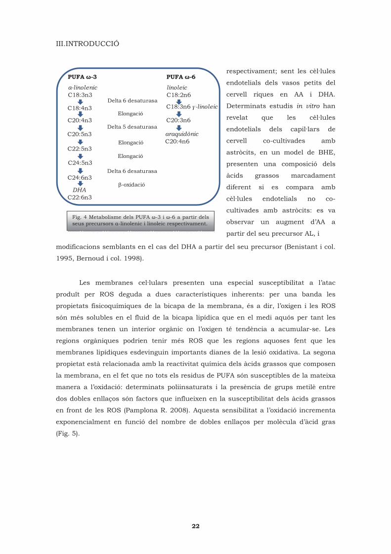
(Floyd RA. 1999, Wallis i col. 2002). Aquests LCPUFA són derivats de la biosíntesi dels
seus àcids grassos precursors com l’àcid linoleic (18:2n-6; AL) i �-àcid linolenic (18 :
3n-3; ALN) respectivament (Fig. 4), però també es poden obtenir directament de la
dieta com per exemple dels ous, el peix o la carn (Brenna i col. 2007, Wainwright PE.
2002). Aquests àcids grassos no s’emmagatzemen ni produeixen energia sinó que
participen en l’arquitectura de les membranes cel·lulars. Els PUFA, i els seus derivats,
són també coneguts com a àcids grassos omega-6 (�-6) i omega-3 (�-3) (Youdim i col.
2000). L’abreviatura de �-6 i �-3 dels àcids grassos insaturats indica la posició del
primer doble enllaç quan es conta des del carboni metil més distal del final de la
cadena d’àcids grassos. Una deficiència simultània d’AL i d’ALN és incompatible amb
la vida (Bourre JM. 2006).
Les lipoproteïnes del plasma, formades pel fetge, transporten el DHA �-3 pel torrent
sanguini on algunes molècules travessen la barrera hematoencefàlica (BHE) i
s’esterifiquen (Glomset JM. 2006). Aquesta barrera està constituïda per cèl·lules
endotelials especialitzades que recobreixen el sistema vascular cerebral i tenen una
gran importància en el manteniment de l’homeòstasi de les neurones, de les cèl·lules
glials i en el bloqueig de l’accés de substàncies tòxiques endògenes o exògenes.
Aquestes cèl·lules endotelials cerebrals conformen una barrera cel·lular entre la sang i
l’espai intersticial que permet mantenir estable la composició del líquid intersticial
indispensable pel bon funcionament neuronal. La BHE juga un important paper en el
control de pas dels PUFA i també en la conversió dels AL i ALN a AA i DHA,
21
III.INTRODUCCIÓ
�-linolenicC18:3n3
C18:4n3
C20:4n3
C20:5n3
C22:5n3
C24:5n3
C24:6n3
DHAC22:6n3
PUFA �-3
linoleicC18:2n6
C18:3n6 �-linoleic
C20:3n6
araquidònicC20:4n6
PUFA �-6
Delta 6 desaturasa
Elongació
Delta 5 desaturasa
Elongació
Elongació
Delta 6 desaturasa
�-oxidació
modificacions semblants en el cas del DHA a partir del seu precursor (Benistant i col.
1995, Bernoud i col. 1998).
Les membranes cel·lulars presenten una especial susceptibilitat a l’atac
produït per ROS deguda a dues característiques inherents: per una banda les
propietats físicoquímiques de la bicapa de la membrana, és a dir, l’oxigen i les ROS
són més solubles en el fluid de la bicapa lipídica que en el medi aquós per tant les
membranes tenen un interior orgànic on l’oxigen té tendència a acumular-se. Les
regions orgàniques podrien tenir més ROS que les regions aquoses fent que les
membranes lipídiques esdevinguin importants dianes de la lesió oxidativa. La segona
propietat està relacionada amb la reactivitat química dels àcids grassos que composen
la membrana, en el fet que no tots els residus de PUFA són susceptibles de la mateixa
manera a l’oxidació: determinats poliinsaturats i la presència de grups metilè entre
dos dobles enllaços són factors que influeixen en la susceptibilitat dels àcids grassos
en front de les ROS (Pamplona R. 2008). Aquesta sensibilitat a l’oxidació incrementa
exponencialment en funció del nombre de dobles enllaços per molècula d’àcid gras
(Fig. 5).
Fig. 4 Metabolisme dels PUFA �-3 i �-6 a partir dels seus precursors �-linolenic i linoleic respectivament.
respectivament; sent les cèl·lules
endotelials dels vasos petits del
cervell riques en AA i DHA.
Determinats estudis in vitro han
revelat que les cèl·lules
endotelials dels capil·lars de
cervell co-cultivades amb
astròcits, en un model de BHE,
presenten una composició dels
àcids grassos marcadament
diferent si es compara amb
cèl·lules endotelials no co-
cultivades amb astròcits: es va
observar un augment d’AA a
partir del seu precursor AL, i
22

III.INTRODUCCIÓ
Les espècies moleculars de fosfoglicèrids que contenen DHA �-3 esterificat es troben
acumulades en la substància gris dels cervells, per altra banda el plasmalogen
representa del 80-90% de la classe de glicerofosfolípid etanolamina en la substància
blanca i en la mielina i forma part del 50% dels diferents tipus de lípids de tot el
cervell (Farooqui i col. 2000).
1.4 OXIDACIÓ PROTEÏCA
Diverses evidències demostren que els ROS i els RNS poden produir
modificacions a tota classe de macromolècules tals com a les proteïnes, als lípids, als
glúcids i també als àcids nucleics (DNA i RNA)(Stadtman i col. 2003).
Les proteïnes són les dianes més importants dels radicals lliures ja que són els
components majoritaris dels sistemes biològics amb una gran rellevància funcional.
S’ha estimat que d’un 50-75% dels radicals reactius reaccionen amb les proteïnes i
que algunes tenen una vida mitja llarga suggerint que els residus modificats de les
proteïnes poden ser considerats com un dels marcadors majoritaris, sensibles de lesió
oxidativa en les cèl·lules de mamífers (Dean i col. 1997).
Les espècies reactives poden reaccionar directament amb les proteïnes o bé poden
reaccionar amb d’altres molècules com els carbohidrats i els lípids generant productes
anomenats RCS (reactive carbonyl species) que poden unir-se a les proteïnes. La
carbonilació de proteïnes és una modificació irreversible i irreparable que consisteix en
la introducció de derivats carbonílics (aldehids i cetones) en residus nucleofílics dels
diferents aminoàcids (Aldini i col. 2007).
Fig. 5 Susceptibilitat relativa dels PUFA a la peroxidació lipídica en funció del nombre de dobles enllaços. Modificat de (Pamplona R. 2008) però l’original és d’Holman RT (1954).
23
III.INTRODUCCIÓ
Les proteïnes poden oxidar-se per diferents vies:
• la lesió oxidativa de les proteïnes pot provenir de l’oxidació directa dels aminoàcids
per ROS, RNS i clorurs catalitzada per metalls, generant productes com els
semialdehids �-glutàmic (GSA, producte d’oxidació de l’arginina -Arg- i de la prolina -
Pro-) i �-aminoadípic (AASA, producte d’oxidació de la Cys) (Requena i col. 2001).
• a través del trencament d’unions peptídiques per vies d’�-amidació (Berlett i col.
1997, Dean i col. 1997).
• les espècies reactives carboníliques produïdes durant la peroxidació lipídica
(principalment aldehids �-� insaturats) poden afegir-se a les proteïnes en els residus
de Cys, histidina -His- i lisina -Lys- a través de reaccions secundàries. D’aquesta
manera es formen els ALEs (advanced lipoxidation end products) (Adibhatla i col.
2009).
• els grups carbonils, generats com a conseqüència de la reacció de residus de sucre
en processos de glicació, s’introdueixen en les proteïnes a través de reaccions
secundàries d’oxidació amb els residus nucleofílics de determinats aminoàcids
generant-se els AGEs (advanced glycation end products) (Thorpe i col. 2003).
Els adductes que provenen de la lipoxidació (ALEs) o bé de la glicoxidació (AGEs) es
poden detectar amb anticossos específics que permeten la identificació de les proteïnes
modificades. També es pot utilizar l’anticòs de la 2,4-dinitrofenilhidrazina (DNP) que
reconeix els grups carbonils associats als adehids i a les cetones.
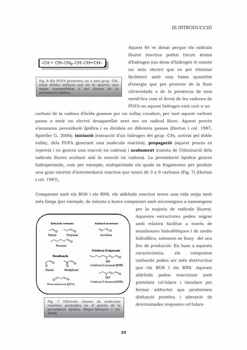
1.4.1 Peroxidació lipídica
Una de les conseqüències més importants de la lesió produïda en un teixit pels
radicals lliures és la peroxidació lipídica: els radicals lliures, gràcies a la seva
estructura i reactivitat (veure apartat 1.1 i 1.2), poden treure un electró d’una
molècula d’àcid gras insaturat generant una cadena de reaccions que permeten la
formació d’espècies reactives (Forman i col. 2008, Pamplona R. 2008, Spiteller G.
2006).
Les cadenes de PUFA (2 o més dobles enllaços) són molt més fàcils d’atacar pels
radicals que les cadenes saturades (sense dobles enllaços) o monosaturades (un doble
enllaç) (Fig. 6). Per exemple el DHA, un PUFA �-3 amb 6 dobles enllaços, és
extremadament susceptible a una lesió oxidativa i té 8 cops més tendència a ser
atacat que l’AL que només té 2 dobles enllaços.
24
III.INTRODUCCIÓ
carboni de la cadena d’àcids grassos per un enllaç covalent, per tant aquest carboni
passa a tenir un electró desaparellat sent ara un radical lliure. Aquest procés
s’anomena peroxidació lipídica i es divideix en diferents passos (Horton i col. 1987,
Spiteller G. 2006): iniciació (extracció d’un hidrogen del grup -CH2, activat pel doble
enllaç, dels PUFA generant una molècula reactiva), propagació (aquest procés es
repeteix i es genera una reacció en cadena) i acabament (consta de l’eliminació dels
radicals lliures acabant així la reacció en cadena). La peroxidació lipídica genera
hidroperòxids, com per exemple, endoperòxids els quals es fragmenten per produir
una gran varietat d’intermediaris reactius que tenen de 3 a 9 carbonis (Fig. 7) (Horton
i col. 1987).
Comparant amb els ROS i els RNS, els aldehids reactius tenen una vida mitja molt
més llarga (per exemple, de minuts a hores comparant amb microsegons a nanosegons
Fig. 6 Els PUFA presenten un o més grup -CH2 entre dobles enllaços que els hi aporten una major susceptiblitat a ser dianes de la peroxidació lipídica.
Aquest fet ve donat perque els radicals
lliures reactius poden treure àtoms
d’hidrogen (un àtom d’hidrogen té només
un únic electró que es pot eliminar
fàcilment amb una baixa quantitat
d’energia que pot provenir de la llum
ultraviolada o de la presència de ions
metàl·lics com el ferro) de les cadenes de
PUFA on aquest hidrogen està unit a un
Fig. 7 Diferents classes de molècules reactives produïdes en el procés de la peroxidació lipídica (Negre-Salvayre i col. 2008).
per la majoria de radicals lliures).
Aquestes estructures poden migrar
amb relativa facilitat a través de
membranes hidrofòbiques i de medis
hidrofílics, extenent-se lluny del seu
lloc de producció. En base a aquesta
característica, els compostos
carbonils poden ser més destructius
que els ROS i els RNS. Aquests
aldehids poden reaccionar amb
proteïnes cel·lulars i tissulars per
formar adductes que produeixen
disfunció proteïca i alteració de
determinades respostes cel·lulars
25

III.INTRODUCCIÓ
(Petersen i col. 2004, Zarkovic K. 2003). L’augment de la peroxidació lipídica modifica
la permeabilitat de la membrana així com l’oxidació de proteïnes estructuralment
importants, portant aquesta situació a una alteració transmembranal dels ions i dels
processos cel·lulars metabòlics (Youdim i col. 2000).
Així doncs les reaccions de lipoxidació comporten canvis estructurals i funcionals en
les proteïnes tals com (Pamplona R. 2008):
� alteracions de les propietats físicoquímiques (canvis de conformació,
hidrofobicitat, elasticitat, solubilitat, mobilitat electroforètica,…)
� formació d’unions intra i intermoleculars i també d’agregats
� disminució o inhibició de l’activitat enzimàtica
� alteracions en la degradació proteïca
� alteracions en el tràfic de proteïnes
� modificació de les propietats de la matriu extracel·lular
1.4.2 Adductes de lipoxidació (ALEs)
En base a les seves característiques estructurals els aldehids de cadena curta més
reactius, que s’han generat en la peroxidació lipídica, es poden classificar en diferents
famílies (Fig. 7) on les més importants són: el grup dels dicarbonils (on principalment
s’inclou el malondialdehid -MDA- i el carboximetil-lisina -CML-) i els 4-hidroxi-2-
alquenals (l’ALE més representatiu és el 4-hidroxi-trans-2-nonenal -HNE-) (Negre-
Salvayre i col. 2008).
Dicarbonils
Pel que fa al MDA, és un dels aldehids més abundant que es forma a partir de
la peroxidació lipídica de l’àcid araquidònic, eicosapentanoic o del DHA (Aldini i col.
2007). És molt abundant i es pot unir covalentment a les proteïnes, formant adductes
amb els residus de Lys o bé amb les amines que es troben en els caps polars dels
fosfolípids, així com la fosfatidilserina o la fosfatidiletanolamina (Uchida K. 2003b). La
Lys i l’Arg són els únics aminoàcids que reaccionen amb el MDA sent la Lys (MDA-L) el
marcador més sensible pel que fa als canvis en els PUFA. En diferents malalties
neurodegeneratives s’han trobat alteracions dels nivells de MDA en les mostres
patològiques comparant amb les controls. En el cas de la MA s’han publicat estudis
post mortem que han desmostrat nivells augmentats de MDA en diferents regions com
26
III.INTRODUCCIÓ
per exemple en l’hipocamp i l’amígdala (Lovell i col. 1995), el lòbul temporal, el frontal
i l’occipital (Dib i col. 2002).
El CML és un producte de la glicoxidació però més tard també es va descriure per ser
un derivat de les reaccions de peroxidació lipídica dels PUFA (Fu i col. 1996) trobant-
se augmentat en l’envelliment, en mostres humanes de MA (Pamplona i col. 2005), en
l’arterosclerosi i en la diabetes (Ahmed i col. 2009, Basta i col. 2009, Fu i col. 1996).
4-hidroxi-2-alquenals
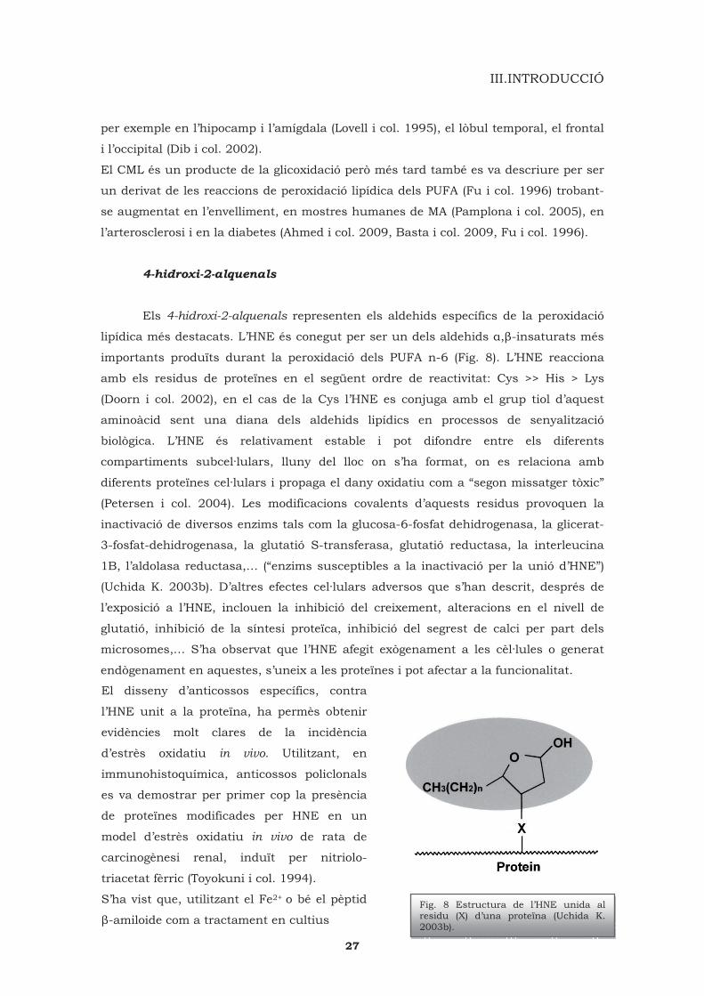
Els 4-hidroxi-2-alquenals representen els aldehids específics de la peroxidació
lipídica més destacats. L’HNE és conegut per ser un dels aldehids �,�-insaturats més
importants produïts durant la peroxidació dels PUFA n-6 (Fig. 8). L’HNE reacciona
amb els residus de proteïnes en el següent ordre de reactivitat: Cys >> His > Lys
(Doorn i col. 2002), en el cas de la Cys l’HNE es conjuga amb el grup tiol d’aquest
aminoàcid sent una diana dels aldehids lipídics en processos de senyalització
biològica. L’HNE és relativament estable i pot difondre entre els diferents
compartiments subcel·lulars, lluny del lloc on s’ha format, on es relaciona amb
diferents proteïnes cel·lulars i propaga el dany oxidatiu com a “segon missatger tòxic”
(Petersen i col. 2004). Les modificacions covalents d’aquests residus provoquen la
inactivació de diversos enzims tals com la glucosa-6-fosfat dehidrogenasa, la glicerat-
3-fosfat-dehidrogenasa, la glutatió S-transferasa, glutatió reductasa, la interleucina
1B, l’aldolasa reductasa,… (“enzims susceptibles a la inactivació per la unió d’HNE”)
(Uchida K. 2003b). D’altres efectes cel·lulars adversos que s’han descrit, després de
l’exposició a l’HNE, inclouen la inhibició del creixement, alteracions en el nivell de
glutatió, inhibició de la síntesi proteïca, inhibició del segrest de calci per part dels
microsomes,… S’ha observat que l’HNE afegit exògenament a les cèl·lules o generat
endògenament en aquestes, s’uneix a les proteïnes i pot afectar a la funcionalitat.
Fig. 8 Estructura de l’HNE unida al residu (X) d’una proteïna (Uchida K. 2003b).
El disseny d’anticossos específics, contra
l’HNE unit a la proteïna, ha permès obtenir
evidències molt clares de la incidència
d’estrès oxidatiu in vivo. Utilitzant, en
immunohistoquímica, anticossos policlonals
es va demostrar per primer cop la presència
de proteïnes modificades per HNE en un
model d’estrès oxidatiu in vivo de rata de
carcinogènesi renal, induït per nitriolo-
triacetat fèrric (Toyokuni i col. 1994).
S’ha vist que, utilitzant el Fe2+ o bé el pèptid
�-amiloide com a tractament en cultius
27
III.INTRODUCCIÓ
cel·lulars de neurones i sinaptosomes, hi ha un gran increment en l’HNE lliure i en
l’unit a proteïnes. En cèl·lules humanes de neuroblastoma SH-SY5Y es va trobar un
augment en la producció intracel·lular de ROS, induït per les prostaglandines classe
J2, acompanyat per l’acumulació de productes de peroxidació lipídica com l’HNE i
l’acroleïna (Kondo i col. 2001). En cultius cel·lulars d’hepatòcits de rates RL34 es va
trobar que l’HNE pot induir una forta producció intracel·lular de ROS, trobant-se
aquest mateix efecte en d’altres tipus cel·lulars tals com les abans comentades SH-
SY5Y (Uchida i col. 1999).
1.4.3 Glicoxidació
Les reaccions no-enzimàtiques dels carbohidrats amb les proteïnes han estat
un interessant tema durant diverses dècades pel que fa a la bioquímica del menjar i
de la nutrició. La dieta és una de les principals fonts externes d’AGEs, sobretot en
menjar cuinat amb elevats nivells de carbohidrats, proteïnes i àcids grassos. La
formació dels AGEs està potenciada per l’exposició a la calor, incrementant el seu
contingut amb la temperatura de cocció i la seva durada (Miyata i col. 1999, Vlassara i
col. 2003). El concepte del producte de glicoxidació va ser originàriament introduït per
caracteritzar el resultat de les reaccions seqüencials de glicació i d’oxidació (d’aquí el
terme glicoxidació): aquesta formació està considerada com el resultat de reaccions
químiques de segon ordre amb un índex depenent de la concentració de carbohidrats
precursors ([CHO], estrès glicatiu “glycative stress”) i de ROS ([O2], estrès oxidatiu)
(Miyata i col. 1999, Thorpe i col. 2003).
Hi han múltiples vies per formar AGEs a partir de carbohidrats però, breument, els
passos generals de la reacció de Maillard, descrita in vivo, es poden resumir: en la
primera etapa es forma una base de Schiff i adductes d’Amadori entre els residus dels
carbohidrats i els grups amino lliures de la proteïna. L’adducte d’Amadori pateix
reorganitzacions no-oxidatives i reaccions d’hidròlisi on en el pas següent, en
presència d’ions de metalls de transició, es formen els productes reactius
intermediaris. En la segona etapa de la reacció s’uneix el compost carbonil al residu
de les proteïna (Lys, Arg,…) formant un ampli ventall d’AGEs (Thorpe i col. 2003).
1.4.4 Adductes de glicoxidació (AGEs)
El grup dels AGEs està format per una important sèrie de molècules reactives
(Monnier VM. 2003, Thorpe i col. 2003) . El CML n’és un d’ells, tot i que s’ha comentat
que també pot provenir de la peroxidació lipídica, formant-se a partir de diferents
classes de carbohidrats, el CEL (carboxietil-lisina) és un altre AGE que prové de la
triosa fosfat,…
28
III.INTRODUCCIÓ
1.5 TEIXIT HUMÀ POST MORTEM
Els estudis de les malalties neurodegeneratives prenen encara més importància
si es poden realitzar en mostres de cervell humà post mortem que en models animals
o en experiments in vitro. El fet de treballar amb teixit cerebral humà té molts
advantatges: s’analitza directament el substrat real de les diferents malalties del
sistema nerviós, però també s’han de tenir en compte certs factors que poden alterar
el teixit i, conseqüentment, els resultats i les conclusions que se n’obtinguin.
Així doncs hi ha diferents elements que poden interferir (degradant o modificant) en la
preservació del DNA, el RNA, les proteïnes i els lípids. Es poden dividir en dos grups
en funció de si es donen abans o després de la mort de l’individu:
• pre mortem, durada de l’estat agònic, hipòxia o acidosis (podent alterar el pH del
teixit), febre, drogues, substàncies tòxiques.
• post mortem, el temps que transcorre entre la mort, el processament de la mostra i la
seva congelació, un altre factor a tenir en compte és la temperatura del cos, les
característiques de les solucions de fixació i del material de congelació.
S’han fet estudis de degradació post mortem per tal de caracteritzar determinades
proteïnes (Siewi col. 2004, Wu i col. 2002), observant que són sensibles però de
diferent manera i que la temperatura és un factor important a tenir en compte (Ferrer
i col. 2007a). Pel que fa al DNA i al RNA, s’han publicat diversos estudis relacionats
amb la seva preservació (Ervin i col. 2007, Ferrer i col. 2007b). Ara bé, degut a
l’increment cada cop més evident d’estudis relacionats amb l’estrès oxidatiu (utilitzant
marcadors de lipoxidació, glicoxidació,...) i, per altra banda, de factors epigènetics
(metilació i acetilació de cues d’histones, metilació de gens promotors,…) es fa patent
una caracterització d’aquests aspectes en mostres post mortem humanes.
1.6 ENVELLIMENT
L’envelliment és un procés natural que comporta una progressiva davallada de
les funcions biològiques, després que l’organisme hagi arribat al seu màxim en la
competència reproductiva. Es considera un deteriorament progressiu, funcional i
depenent del temps, que condueix cap a la mortalitat (Farooqui i col. 2009). S’han
formulat diverses teories per explicar l’envelliment però el mecanismes biològics
encara no estan ben establerts.
Com s’ha comentat anteriorment, l’estrès oxidatiu fa referència a conseqüències
citotòxiques produïdes per diferents espècies reactives. La lesió oxidativa implica a
29

III.INTRODUCCIÓ
una sèrie de malalties, tenint un impacte important en el procés d’envelliment del cos.
Les cèl·lules del sistema nerviós estan afectades per l’envelliment (Beckman i
col. 1998, Rikans i col. 1997), com les cèl·lules d’altres òrgans, i pateixen un augment
en la quantitat de lesions oxidatives de les proteïnes, els lípids i els àcids nuclèics
(Stadtman ER. 2006) tant com alteracions en l’homeòstasi energètica. En aquest
procés de deteriorament, els astròcits generen una gran quantitat d’ON que resulta ser
molt perjudicial per les neurones veïnes i pels oligodendròcits. El mecanisme
molecular exacte que involucra la lesió neuronal per l’ON encara no es coneix. En
models murins d’envelliment (SAMP8) s’observen modificacions carboníliques
associades amb l’edat (Nabeshi i col. 2006) i també disminució de l’activitat de la SOD
(Alvarez-Garcia i col. 2006). Aquests canvis, que es donen durant l’envelliment normal,
es troben accentuats en poblacions de neurones vulnerables en diferents malalties
neurodegeneratives (Farooqui i col. 2009, Lowe J. 2003, Mattson i col. 2006).
2. Malalties neurodegeneratives
En les últimes dècades ha augmentat l’atenció de la comunitat científica en el
camp de les malalties neurodegeneratives, en general, ja que aquestes tenen un
important impacte tant a nivell individual (per exemple, dels propis pacients i de les
persones que les cuiden) com de la societat (Lowe J. 2003). Degut al gran increment
d’individus als que se’ls hi diagnostica demència, la societat exigeix una major atenció
a nivell social i de salut. La prevenció podria ser una oportunitat per disminuir la
càrrega d’una sèrie de malalties tan devastadores com costoses (Fratiglioni i col.
2009).
Les malalties neurodegeneratives es caracteritzen per una disfunció
progressiva i una pèrdua selectiva de poblacions de neurones i sinapsis en àrees
concretes del sistema nerviós (cervell i medul·la espinal), determinant d’aquesta
manera la seva clínica (alteracions cognitives, desordres motors o ambdues
característiques). Un individu adult presenta una limitada capacitat neurogènica del
sistema nerviós, per tant la mort cel·lular neuronal marca una irreversible i
catastròfica fase en el procés neurodegeneratiu.
La neurodegeneració està considerada multifactorial, provocada per diferents
possibles elements: causes genètiques, medi ambientals, factors endògens relacionats
amb l’envelliment, defectes en la degradació proteïca per errors en el sistema
ubiqüitina-proteasoma-autofàgia, formació de radicals lliures donant lloc a situacions
d’estrès oxidatiu, alteracions bioenergètiques per la disfuncionalitat del mitocondri,
30

III.INTRODUCCIÓ
transtorns en l’aparell de Golgi neuronal i en el seu transport, mutacions de les
xaperones moleculars, disfunció de les neurotrofines, processos neuroinflamatoris
(Forman i col. 2004, Jellinger KA. 2009, Skovronsky i col. 2006). Tots aquests
mecanismes estan interrelacionats entre ells formant un cercle viciós que pot
comportar la disfunció i la mort cel·lulars però queden pendents d’aclarir les diferents
vies moleculars i els seus papers patogènics (Jellinger KA. 2009).
Les diverses classes de malalties neurodegeneratives comparteixen la característica de
presentar agregats i dipòsits (extra o intracel·lulars) de proteïnes anormals i/o mal
plegades que respresenten els marcadors de les diferents patologies
neurodegeneratives, anomenades proteinopaties (Fig. 9) (Jellinger KA.
2009).
Fig.9 Relació de malalties neurodegeneratives en funció de les inclusions. *CN: cabdell neurofibril·lar.
31
III.INTRODUCCIÓ
El plegament de les proteïnes és una part del procés normal de la síntesi
proteïca, indispensable per convertir-les en molècules fisiològicament funcionals i per
regular la seva activitat biològica gràcies a l’acció de les xaperones moleculars
(Muchowski i col. 2005). La manera que una cadena d’aminoàcids sintetitzada di novo
es transforma en una proteïna perfectament plegada depèn, en primer lloc, de les
propietats intrínsiques de la seqüència d’aminoàcids (patró de residus hidrofòbics i
polars) i de la influència de l’entorn cel·lular (Dobson CM. 2003). Així doncs, en
situacions fisiològiques les cèl·lules mantenen un equilibri entre el plegament, el
replegament i la degradació de les proteïnes però un trencament d’aquest equilibri, per
un excés de producció proteïca o una disminució de la degradació de les proteïnes
anormals, dóna lloc a una situació d’estrès proteolític que comporta l’acumulació i
l’agregació de proteïnes anormals. Les cèl·lules neuronals són particularment
susceptibles als efectes tòxics de les proteïnes mutants o mal plegades (Taylor i col.
2002). Les condicions metabòliques extremes, per un elevat ús d’oxigen i també per un
increment en la producció de neurotransmissors, les fa especialment vulnerables a les
alteracions relacionades amb l’agregació (Dohm i col. 2008).
La progressiva acumulació de proteïnes intracel·lulars pot donar lloc a: a) síntesi
anormal i mal plegament proteïc, b) interacció anormal amb d’altres proteïnes, c)
sobreproducció de proteïnes constitutives, d) alteracions en els processos de
degradació i de recanvi, e) afectacions en les modificacions post-translacionals de
proteïnes sintetitzades di novo, f) expressió incorrecta o alteració en l’splicing (procés
de tall i empalmament), g) activitat insuficient de les xaperones moleculars, h)
alteracions en el transport de proteïnes podent arribar a la mort cel·lular de les
cèl·lules afectades (Jellinger KA. 2009).
La inherent tendència de les proteïnes a agregar-se ha implicat un desenvolupament
forçat dels sistemes de defensa cel·lulars contra les proteïnes anormals (Ross i col.
2005). Així doncs hi ha diversos sistemes per tal de corregir i detectar les proteïnes
mal plegades:
• el reticle endoplasmàtic respon davant de proteïnes mal plegades a través de la
inducció de xaperones moleculars, podent replegar les proteïnes anormals i
transformar-les en no-tòxiques (Jellinger KA. 2009).
• el proteasoma és un complex molecular que pot desplegar proteïnes i processar-les
en petits fragments gràcies a la presència d’enzims proteolítics en el seu interior.
• l’autofàgia presenta diferents variants com la macroautofàgia, la microautofàgia i
l’autofàgia via xaperones. Les proteïnes citoplasmàtiques solubles, especialment
aquelles que tenen un recanvi lent, poden ser degradades per aquestes vies
32

III.INTRODUCCIÓ
lisosomals. Aquesta via pot ser activada per estrès oxidatiu o per estrès de nutrients.
• en el moment que les proteïnes anormals i agregades no poden ser replegades o
degradades, es presenta una altra alternativa: les cèl·lules poden segrestar els
agregats a través del transport dels microtúbuls reunint-los a prop del centríol en el
citoplasma. Aquest procés, genera un gran cos d’inclusió visible al microscopi,
anomenat agresoma. Després de que les proteïnes mal plegades estan acumulades en
un agresoma, aleshores estan a punt per la macroautofàgia: la cèl·lula pot raptar una
gran porció del seu citoplasma i empaquetar-lo en una estructura unida a la
membrana (autofagosoma) que més tard pot ser processada per la cèl·lula (Ross i col.
2004). La macroautofàgia pot eliminar agresomes en cultiu cel·lular.
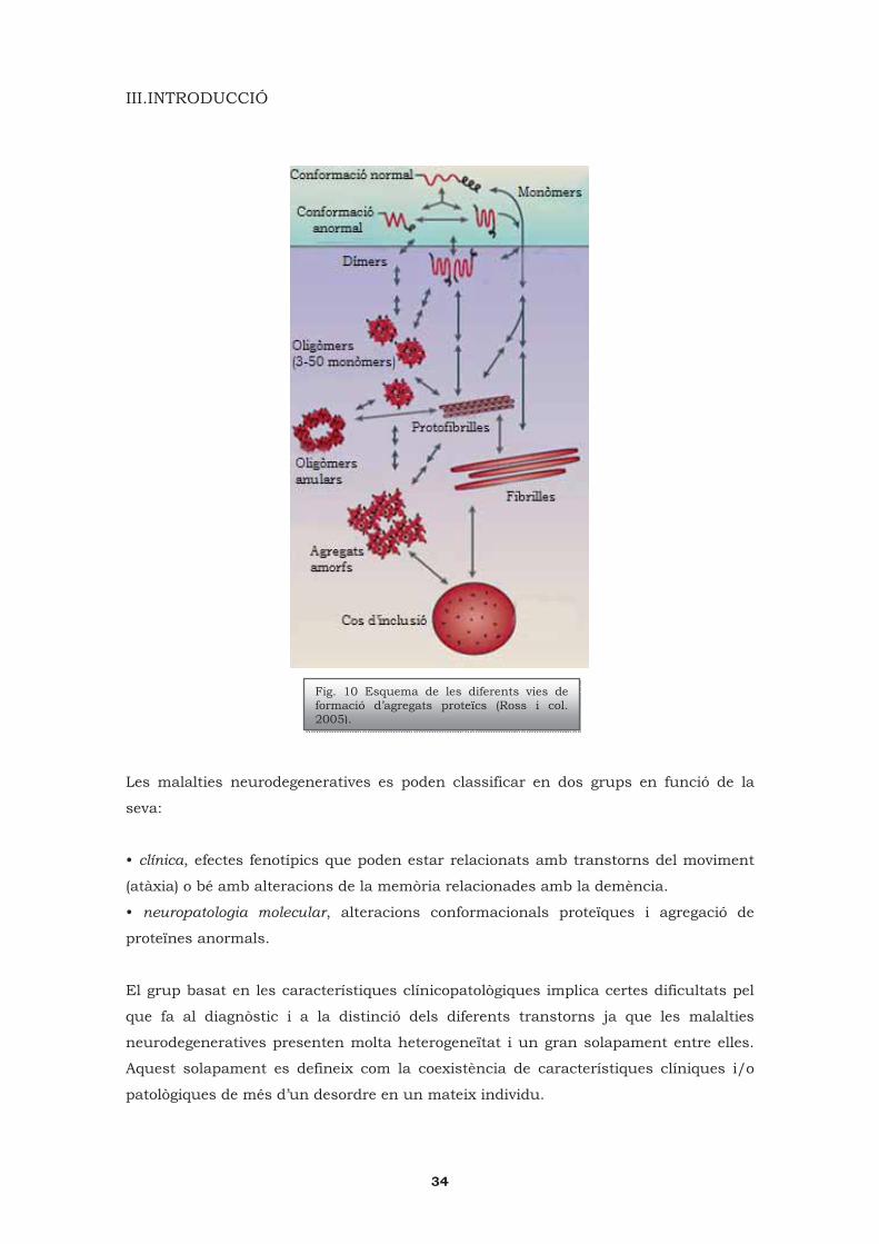
En general, l’agregació es considera un procés de molts passos que comença
quan una proteïna (monòmer) adopta una conformació anormal i hi ha una associació
de dues o més proteïnes anormals, o part d’elles, formant dímers i intermediaris
oligomèrics. Aquests poden donar lloc a estructures de �-làmina plegada inestables
que contenen oligòmers amb una morfologia semblant a les fibrilles. Aquestes
estructures fibrillars donen lloc als cossos d’inclusió que tenen tendència a ser
altament resitents a la proteòlisi (Fig. 10) (Dohm i col. 2008, Ross i col. 2005). La
majoria de les espècies intermèdies són inestables i han estat caracteritzades in vitro
en funció de les seves unions a determinats colorants i a les seves propietats
biofísiques.
Encara que els cossos d’inclusió proteïcs estan normalment associats a
característiques patològiques en les malalties neurodegeneratives, hi ha una gran
controvèrsia sobre el paper de l’agregació durant la progressió de la malaltia
(Bretteville i col. 2008). Diverses evidències associen l’agregació amb la toxicitat però
per altra banda hi han altres estudis que indiquen que les proteïnes agregades poden
exercir un paper neuroprotector com a una resposta fisiològica a un excés de
proteïnes mal plegades (Ross i col. 2005, Taylor i col. 2002). Actualment s’ha proposat
que les espècies primerenques en el procés d’agregació són més tòxiques que els
cossos d’inclusió o els agregats grans (Ross i col. 2005). Seguint aquesta línia, la mort
cel·lular neuronal podria estar principalment produïda per la presència d’intermediaris
tòxics oligomèrics mentres que els agregats insolubles visibles, típicament observats
en els autòpsies, apareixerien per ser el resultat de processos de detoxificació cel·lular
on el sistema UPS ha fallat i les proteïnes són segrestades i compartimentades en
inclusions, com els agresomes, amb la finalitat de neuroprotegir (Dohm i col. 2008).
33
III.INTRODUCCIÓ
Les malalties neurodegeneratives es poden classificar en dos grups en funció de la
seva:
• clínica, efectes fenotípics que poden estar relacionats amb transtorns del moviment
(atàxia) o bé amb alteracions de la memòria relacionades amb la demència.
• neuropatologia molecular, alteracions conformacionals proteïques i agregació de
proteïnes anormals.
El grup basat en les característiques clínicopatològiques implica certes dificultats pel
que fa al diagnòstic i a la distinció dels diferents transtorns ja que les malalties
neurodegeneratives presenten molta heterogeneïtat i un gran solapament entre elles.
Aquest solapament es defineix com la coexistència de característiques clíniques i/o
patològiques de més d’un desordre en un mateix individu.
Fig. 10 Esquema de les diferents vies de formació d’agregats proteïcs (Ross i col. 2005).
34
III.INTRODUCCIÓ
Com a conseqüència, s’ha d’intentar reconsiderar l’actual nosologia i tenir en compte
una nova divisió de les malalties neurodegeneratives (Armstrong i col. 2005). Pel que
fa al segon grup, cada cop s’estan realitzant més avanços en el camp de la
neuropatologia molecular que permeten fer una classificació en funció de la proteïna
anormal que s’acumula, representant un clar marcador bioquímic i histomorfològic de
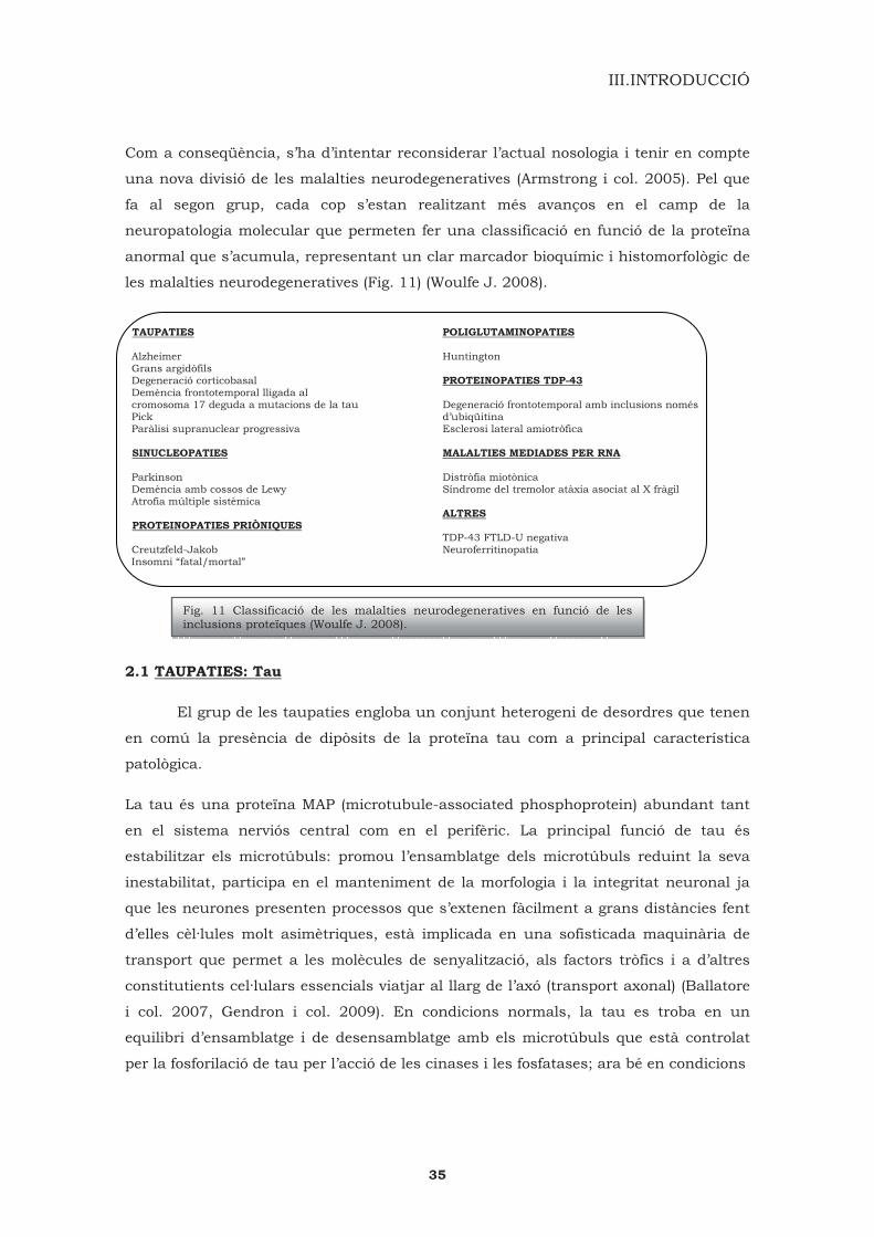
les malalties neurodegeneratives (Fig. 11) (Woulfe J. 2008).
TAUPATIES Alzheimer Grans argidòfils Degeneració corticobasal Demència frontotemporal lligada al cromosoma 17 deguda a mutacions de la tau Pick Paràlisi supranuclear progressiva SINUCLEOPATIES Parkinson Demència amb cossos de Lewy Atrofia múltiple sistèmica PROTEINOPATIES PRIÒNIQUES Creutzfeld-Jakob Insomni “fatal/mortal”
POLIGLUTAMINOPATIES Huntington PROTEINOPATIES TDP-43 Degeneració frontotemporal amb inclusions només d’ubiqüitina Esclerosi lateral amiotròfica MALALTIES MEDIADES PER RNA Distròfia miotònica Síndrome del tremolor atàxia asociat al X fràgil ALTRES TDP-43 FTLD-U negativa Neuroferritinopatia
2.1 TAUPATIES: Tau
El grup de les taupaties engloba un conjunt heterogeni de desordres que tenen
en comú la presència de dipòsits de la proteïna tau com a principal característica
patològica.
La tau és una proteïna MAP (microtubule-associated phosphoprotein) abundant tant
en el sistema nerviós central com en el perifèric. La principal funció de tau és
estabilitzar els microtúbuls: promou l’ensamblatge dels microtúbuls reduint la seva
inestabilitat, participa en el manteniment de la morfologia i la integritat neuronal ja
que les neurones presenten processos que s’extenen fàcilment a grans distàncies fent
d’elles cèl·lules molt asimètriques, està implicada en una sofisticada maquinària de
transport que permet a les molècules de senyalització, als factors tròfics i a d’altres
constitutients cel·lulars essencials viatjar al llarg de l’axó (transport axonal) (Ballatore
i col. 2007, Gendron i col. 2009). En condicions normals, la tau es troba en un
equilibri d’ensamblatge i de desensamblatge amb els microtúbuls que està controlat
per la fosforilació de tau per l’acció de les cinases i les fosfatases; ara bé en condicions
Fig. 11 Classificació de les malalties neurodegeneratives en funció de les inclusions proteïques (Woulfe J. 2008).
35

III.INTRODUCCIÓ
patològiques, aquest equilibri es trenca i la proteïna tau s’hiperfosforila provocant un
augment anormal dels nivells de tau lliure (no unida) on aquest increment de tau
citosòlica comporta canvis conformacionals patogènics que afavoreixen l’agregació i la
fibrillació de tau (Ballatore i col. 2007, Drewes i col. 1995, Mazanetz i col. 2007).
El gen de la tau es troba en el cromosma 17q21 amb 16 exons, dels quals 11
codifiquen per les isoformes més importants de la proteïna tau. A través de l’splicing
alternatiu dels exons 2,3 i 10 del mRNA es formen un grup de 6 isoformes en el cervell
humà d’un adult (Billingsley i col. 1997, Tolnay i col. 2003). Tau presenta dominis
d’unió als microtúbuls que estan formats per regions d’unió a la tubulina, altament
conservades i situades en la meitat C-terminal de la proteïna, havent-hi a continuació
una regió rica en prolina i després una regió N-terminal. Les 6 isoformes es diferencien
unes de les altres en el número de repeticions d’unió a la tubulina (3R o 4R) i també
per la presència o absència d’inserts en la regió N-terminal de la proteïna (Fig. 12)
(Ballatore i col. 2007, Gendron i col. 2009). En el cervell sa d’un adult, la proporció de
les isoformes de tau 3R i de 4R és d’aproximadament del 50% per cadascuna. Les
isoformes tau 4R interaccionen més eficientment amb els microtúbuls que les
isoformes tau 3R (Goedert i col. 1990). La proteïna tau anormalment hiperfosforilada
es dissocia dels microtúbuls empaquetant-se en fibres de manera molt densa donant
lloc a unes estructures anomenades FHE (filaments helicoïdals emparellats, PHF de
Paired Helical FilamentS) que per processament proteolític formen oligòmers de tau i
agregats insolubles, els CN (cabdells neurofibril·lars, NFTs de neurofibrillary tangles)
(Bamburg i col. 2009)
Fig. 12. Les sis isoformes de tau que s’expressen en el cervell d’un adult (Ballatore i col. 2007).
S’han descrit una important
llista de cinases implicades
en la fosforilació de tau, tant
in vitro com en models
animals i en mostres
humanes post mortem, tals
com la GSK3� (glycogen
synthase kinase 3�), CDK5
(cyclin-dependent kinase 5),
ERK 1/2 (extracellular
signal-regulated kinase 1/2),
MARK (microtubule affinity-
regulating kinase), PKA
(cAMP-dependent protein
kinase), on la MAP p38
(mitogen activated protein) i
36
III.INTRODUCCIÓ
la SAPK/JNK (stress-activated protein kinase) estan considerades cinases d’estrès ja
que s’activen davant d’estímuls estressants (Hartzler i col. 2002, Shelton i col. 2004,
Spittaels i col. 2000). Les proteïnes fosfatases són necessàries per contrarestar els
efectes de les proteïnes cinases: diversos estudis han compromès diverses fosfatases
en la regulació de la fosforilació de tau com per exemple la PP1, PP2A, PP2B i la PP2C
(protein phosphatase) (Liu i col. 2005).
Totes elles defosforilen tau in vitro però s’ha suggerit que la PP2A i la PP2B són
essencials per la regulació in vivo (Billingsley i col. 1997).
2.1.1 Malaltia de Steele-Richardson-Olszewski o Paràlisi Supranuclear Progressiva (PSP)
L’any 1964 tres investigadors de Toronto, John C. Steele, Clifford Richardson i
Jerzy Olszewski, van descriure la malaltia de PSP com una “degeneració heterogènia
on estan implicats el tronc de l’encèfal, els ganglis basals i el cerebel, amb presència
de paràlisi pseudobulbar, mirada vertical, distonia i demència”. És una malaltia amb
una prevalència de 5-6 casos per cada 100.000 individus on l’edat d’inici de la
malaltia està entre els 50-70 anys. L’afectació per sexes és semblant però hi ha un
major nombre de defuncions en els homes que en les dones (Santacruz i col. 1998,
Schrag, i col. 1999). Té una progressió ràpida i s’estimen de 5-10 anys de
supervivència des de l’inici dels símptomes (Albers i col. 2001).
A. Clínica
La PSP es manifesta clínicament per una anormalitat en el moviment dels ulls
(concretament al moure la mirada cap avall però quan avança la malaltia afecta a
totes les direccions), distonia cervical, síndrome parkinsonià amb presència de
rigidesa corporal, pèrdua de l’equilibri i un pas poc ferm, bradicinèsia (lentitud dels
moviments voluntaris), caigudes, desinhibició social, disfòria, ansietat i a vegades
agitació, irritabilitat, canvis d’humor i de la personalitat, apràxia (incapacitat
d'executar determinats gests o moviments, anteriorment apresos, de manera
voluntària i d'una manera apropiada) (Litvan i col. 1996, Morris i col. 2002, Rampello i
col. 2005). Aquests últims símptomes, d’alteracions importants del comportament i
cognitius, són deguts a la disfunció del lòbul frontal així com l’apatia, la depressió i la
bradifrenia (activitat mental lenta) estan associades amb un marcat hipometabolisme
de la glucosa d’aquest lòbul (Albers i col. 2001). Els pacients diagnosticats de PSP
també poden presentar demència, sent un tipus de demència subcortical diferent de la
37

III.INTRODUCCIÓ
cortical: es caracteritza per una alteració de la memòria, amb oblits freqüents però es
segueix mantenint la capacitat de gravar nous esdeveniments.
B. Característiques neuropatològiques
Macroscòpicament
A nivell patològic aquest desordre es pot diferenciar de les altres taupaties per
l’afectació i l’atrofia de les estructures corticals (lòbul frontal) però també de les
subcorticals concretament el tàlam, els nuclis subtalàmics, els ganglis basals, el
globus pallidus, la despigmentació de la substància negra, el locus ceruleus, l’estriat,
els nuclis basals de Meynert (Albers i col. 2001, Dickson DW. 1999, Hauw JJ. 2003),
trobant-se també afectat el complex oculomotor.
Microscòpicament
És una taupatia 4R, que resulta de l’splicing de l’exó 10, (Chambers i col.
1999) (Fig. 13) on la tau hiperfosforilada es troba acumulada en neurones, astròcits i
oligodendròcits (Lee i col. 2001).
En el cas de PSP, els agregats de tau positiu formen CNs i pre-cabdells en les
neurones distribuint-se, com els filaments del neuròpil, per l’estriat, en el globus
pallidus, en els nuclis subtalàmics, en els nuclis basals de Meynert podent-se trobar
també per l’hipocamp, para-hipocamp i isocòrtex. Aquests CNs tenen un aspecte
arrodonit o globós (Fig. 14a) formant inclusions intracitoplasmàtiques arrodonides, a
vegades extenent-se per tot el diàmetre de la neurona (Hauw JJ. 2003).
A part dels CNs, s’han descrit dos
marcadors bioquímics característics
de PSP relacionats amb la glia, els
cabdells tau positius de la glia:
• un dels més importants fa
referència als anomenats astròcits
tufted (Fig.14b): fibres empaquetades
densament de diferents diàmetres que
es marquen positivament amb tau en
Fig. 13 Gràcies a l’anàlisi per Western blot de fraccions insolubles FHE de pacients amb PSP, utilitzant anticossos contra tau, s’observa la presència de dues bandes molt intenses de 68 i 64 kDa corresponents a isoformes de tau 4R (Sergeant i col. 2005).
38

III.INTRODUCCIÓ
els astròcits. Aquest tipus cel·lular pren forma estrellada i és casi exclusiu d’aquesta
malaltia localitzant-se especialment en l’estriat, tàlam, nuclis subtalàmics i a l’escorça
frontal (Dickson DW. 1999, Togo i col. 2002). Un altre tipus d’astròcits fibrillars
anomenats thorn-shaped, amb les mateixes propietats histoquímiques que els
astròcits tufted però amb processos cel·lulars curts, es poden trobar a PSP però no són
específics d’aquesta detectant-se també en d’altres malalties com per exemple en la
degeneració corticobasal (DCB) (Hauw JJ. 2003).
• l’altra implica als oligodendròcits, els cossos d’inclusió (Fig. 14c): són inclusions
intracitoplasmàtiques oligodendroglials tau positives que rodegen els seus nuclis. Es
troben normalment a l’escorça i a prop de la matèria blanca, nuclis subtalàmics,
tàlam, substància negra.
a)
b)
Fig. 14 a) Imatge dels CNs globosos a les neurones; b) s’observen els astròcits tufted; c) es marquen els cossos en espiral a la malaltia de PSP.
39
III.INTRODUCCIÓ
C. Etiologia
L’etiologia de PSP no és coneguda però s’hipotetitzen diferents factors que
poden contribuir al seu desenvolupament: genètics, medi ambientals, dany produït
per l’estrès oxidatiu, inflamació. Encara que en la majoria de casos de PSP s’ha vist
que apareixen esporàdicament, s’han descrit algunes formes genètiques on poden
existir patrons dominants autosòmics o bé recessius. La regió cromosòmica de la
MAPT està formada per dos haplotips, H1 i H2 on en el cas de PSP s’ha trobat una
relació amb l’haplotip de tau H1 que està sobrerepresentat en mostres de pacients
comparant amb els controls (Baker i col. 1999, Dickson DW. 1999).
D. Estrès oxidatiu i PSP
L’estrès oxidatiu ha estat descrit com un factor que juga un paper en la
patogènesi de PSP, trobant-se marcadors de peroxidació lipídica (MDAL, 4-HNE)
augmentats en el nucli subtalàmic, en l’escorça frontal superior i en el mesencèfal
(Albers i col. 1999, Albers i col. 2000, Odetti i col. 2000) de mostres post mortem
humanes. Els sistemes de defensa antioxidant augmenten la seva expressió com a
resposta davant de la producció elevada de radicals lliures, observant-se en casos de
PSP un increment dels nivells i de l’activitat de Cu/Zn-SOD i del glutatió total
(Aoyama i col. 2006, Cantuti-Castelvetri i col. 2002). S’ha trobat un increment de la
concentració de Fe2+ en els ganglis basals i també la presència de ferritina en dipòsits
de tau hiperfosforilat en mostres de pacients de PSP, suggerint ambdues evidències
una possible relació entre l’estrès oxidatiu, la presència de metalls i els agregats de
tau (Albers i col. 2001).
2.1.2 Degeneració frontotemporal lobar amb parkinsonisme lligada al
cromosoma 17 associada a mutacions de la tau (DFTL-tau)
El conjunt de malalties agrupades com a degeneració frontotemporal lobar
(DFTL) representa la tercera causa més comuna de demència, després de la malaltia
d’Alzheimer i de la malaltia de cossos de Lewy (Neary i col. 1998). La DFTL agrupa un
grup de patologies que tenen en comú l’atrofia bilateral del lòbul frontal i la part
anterior del temporal. Les manifestacions clíniques més típiques són una demència
semàntica, una afàsia primària progressiva i una demència frontotemporal on aquesta
última és la més comú mostrant canvis progressius en la personalitat i en el
comportament (Brun i col. 1997, Neary i col. 1998, Neary i col. 2005). Alteracions en el
llenguatge i dificultats cognitives també solen detectar-se. Per tal de facilitar el
diagnòstic d’un grup tan complex com el de les DFTL el grup de McKhann i col. 2001
40

III.INTRODUCCIÓ
va simplificar els criteris englobant l’afàsia progressiva i la demència semàntica dins
del terme de demència frontotemporal i proposant 6 característiques clíniques: 1.
canvis primerencs i progressius en la personalitat i en el llenguatge; 2. problemes
socials; 3. progrés gradual i progressiu; 4. exclusió d’altres causes; 5. els dèficits no
passen només durant el delirium (estat de confusió agut); 6. exclusió de causes
psiquiàtriques, com per exemple la depressió.
L’examen macroscòpic del cervell de pacients amb DFTL mostra una atrofia del lòbul
frontal, del temporal o dels dos. En un elevat tant per cent dels casos s’observa
l’atrofia dels ganglis basals i també pèrdua de la pigmentació de la substància negra
(Cairns i col. 2007a). Pel que fa a nivell microscòpic, la majoria de grups de DFTL
presenten una microvacuolització i una pèrdua neuronal de l’escorça cerebral
observable gràcies a a tincions d’hematoxilina i eosina. També pot haver-hi pèrdua de
mielina de la substància blanca i gliosi astrocitària (Cairns i col, 2007a).
Així doncs aquest conjunt de patologies de DFTL es poden clasificar en tres
grups, on el primer d’ells presenta inclusions de tau hiperfosforilat com la DFTL amb
parkinsonisme lligada al cromosoma 17 associada a mutacions en el gen de la tau -
MAPT 17q21-q22- (DFTL-tau). El segon està format per la presència d’inclusions
d’ubiqüitina que són negatives per tau i per l’�-sinucleina (DFTL-U) i l’últim enlgoba
un grup de casos de DFTL sense característiques neuropatològiques particulars i amb
poca incidència.
DFTL-tau va ser definida l’any 1996 en el congrés International Consensus
Conference a Ann Arbor (Míchigan) on es van presentar 13 famílies amb síndromes
lligats al cromosoma 17q21-22. Es va acordar que era una malaltia neurodegenerativa
autosòmica dominant que presentava tres característiques fonamentals: canvis en el
comportament i la personalitat, discapacitats cognitives i símptomes motors. Està
causada per mutacions en el gen de la tau sense saber la seva prevalència ni la
incidència però havent-se identificat, aproximadament, sobre unes 100 famílies per tot
el món amb 38 mutacions diferents de tau (Lowe J. 2003, Wszolek i col. 2006). No hi
ha diferència de distribució entre gèneres, trobant-se l’edat d’inici de la malaltia entre
els 30 i els 50 anys. El pronòstic i l’índex de la progressió de la patologia varia molt
entre diferents pacients, el rang d’esperança de vida pot anar des d’alguns mesos a
diferents anys i, en casos excepcionals, es pot allargar fins a 2 dècades.
41

III.INTRODUCCIÓ
A. Clínica
En els pacients de DFTL-tau s’observen, com a símptomes clínics, canvis en la
personalitat i en el comportament (desinhibició, apatia, comportament compulsiu,
deixar de banda la higiene personal, agressió verbal o física). A nivell cognitiu, en
estadis primerencs, s’observen dificultats en la parla i problemes en les funcions
executives mentre que la memòria o l’orientació estan preservats; en estadiatges més
avançats de la patologia els pacients poden desenvolupar deteriorament progressiu de
la memòria, de l’orientació, ecolalia (repeticions de les frases o paraules pronunciades
per l’interlocutor) i finalment pot aparèixer demència i un mutisme del pacient. Pel que
fa als símptomes motors, el parkinsonisme a DFTL-tau es caracteritza per
bradicinèsia, inestibilitat en la postura, rigidesa i normalment absència de tremolors
(Kumar-Singh i col. 2007, Lowe J. 2003, Tsuboi Y. 2006, Wszolek i col. 2006). El
fenotip pot variar tant entre famílies que presenten diferents mutacions com dins de
famílies que porten la mateixa: a vegades el diagnòstic definitu necesita una
combinació de característiques clíniques, de trets patològics i anàlisis genètics
moleculars.
B. Característiques neuropatològiques
Macroscòpicament
S’observa una atrofia dels lòbuls frontal i temporal, podent ser asimètrica, on
en etapes avançades de la malaltia l’atrofia està present en diferents graus. Els lòbuls
temporal, parietal i occipital estan normalment menys lesionats. Hi ha afectació del
nucli caudat, el putamen, el globus pallidus, l’amígdala, l’hipocamp, els nuclis basals,
una important pèrdua de pigmentació de la substància negra i del locus ceruleus.
L’escorça cerebelar està normalment preservada encara que a vegades es pot veure
una lleugera atrofia (Ghetti B, Hutton ML, Wszolek ZK. 2003, Tsuboi Y. 2006).
Microscòpicament
Microscòpicament es detecta una pèrdua neuronal, una gliosi astrocitària i una
espongiosi en les capes superficials corticals. Els fenotips neuropatològics varien no
només en les característiques morfològiques sinó també en la intensitat i la
distribució, on aquesta variabilitat correlaciona amb el tipus de mutació (Spillantini i
col. 2000, Tsuboi Y. 2006):
• les mutacions en els exons 1 i 10, com també en l’intró que segueix l’exó 10, estan
42

III.INTRODUCCIÓ
associades amb dipòsits de tau a les neurones (predominantment en els axons) i a la
glia.
• les que afecten als exons 9, 11, 12 i 13 provoquen dipòsits de tau principalment a les
neurones.
S’ha demostrat que aquestes mutacions afecten a la capacitat de tau per unir-se als
microtúbuls i a la proporció de les diferents isoformes de tau (Fig. 15). Les mutacions
de tau que afecten a l’exó 10, i a l’intró que segueix a l’exó 10, incrementen la
proporció de la isoforma 4R (excepte la delK280 que elimina “virtualment” l’exó 10 i
augmenta la forma 3R).
Les mutacions més comunes són la P301L i la N279K (localitzades en l’exó 10 donant
un patró 4R) representant el 60% dels casos coneguts. En el cas de la P301L (canvi de
l’aminoàcid prolina per la leucina en la posició 301), a part dels dipòsits de tau a les
neurones, s’han observat neurones balonades a l’escorça i en alguns nuclis
subcorticals. A més a més, els astròcits i l’oligodendroglia es troben afectats: els
astròcits es troben en el neocòrtex i l’oligodendroglia apareix en forma de cossos en
espiral (Ghetti B, Hutton ML, Wszolek ZK. 2003).
Per tant, el marcador neuropatològic de les DFTL-tau és la presència de dipòsits de la
proteïna tau en les neurones i també en les cèl·lules glials (Fig.16).
Fig. 15 Diferents patrons electroforètics de la proteïna tau patològica (Sergeant i col. 2005).
43
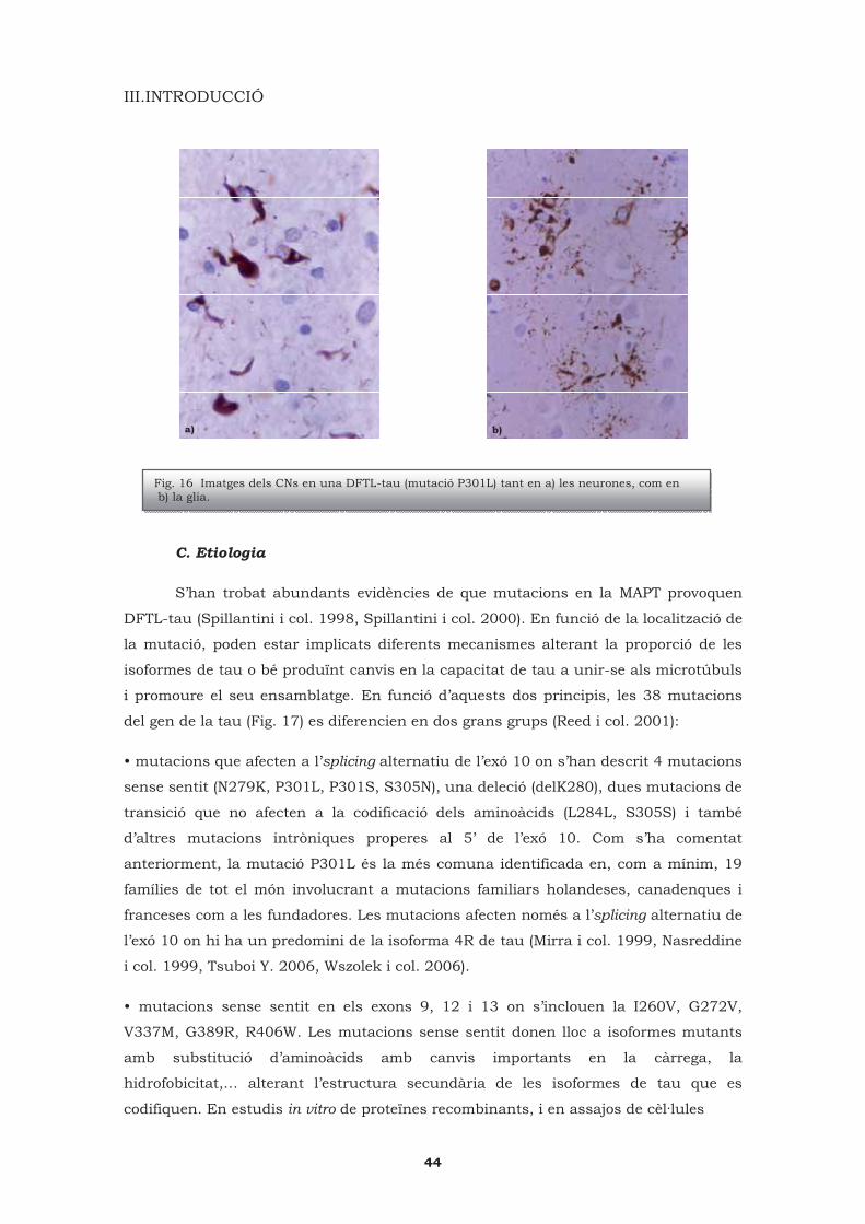
III.INTRODUCCIÓ
a) b)
C. Etiologia
S’han trobat abundants evidències de que mutacions en la MAPT provoquen
DFTL-tau (Spillantini i col. 1998, Spillantini i col. 2000). En funció de la localització de
la mutació, poden estar implicats diferents mecanismes alterant la proporció de les
isoformes de tau o bé produïnt canvis en la capacitat de tau a unir-se als microtúbuls
i promoure el seu ensamblatge. En funció d’aquests dos principis, les 38 mutacions
del gen de la tau (Fig. 17) es diferencien en dos grans grups (Reed i col. 2001):
• mutacions que afecten a l’splicing alternatiu de l’exó 10 on s’han descrit 4 mutacions
sense sentit (N279K, P301L, P301S, S305N), una deleció (delK280), dues mutacions de
transició que no afecten a la codificació dels aminoàcids (L284L, S305S) i també
d’altres mutacions intròniques properes al 5’ de l’exó 10. Com s’ha comentat
anteriorment, la mutació P301L és la més comuna identificada en, com a mínim, 19
famílies de tot el món involucrant a mutacions familiars holandeses, canadenques i
franceses com a les fundadores. Les mutacions afecten només a l’splicing alternatiu de
l’exó 10 on hi ha un predomini de la isoforma 4R de tau (Mirra i col. 1999, Nasreddine
i col. 1999, Tsuboi Y. 2006, Wszolek i col. 2006).
• mutacions sense sentit en els exons 9, 12 i 13 on s’inclouen la I260V, G272V,
V337M, G389R, R406W. Les mutacions sense sentit donen lloc a isoformes mutants
amb substitució d’aminoàcids amb canvis importants en la càrrega, la
hidrofobicitat,… alterant l’estructura secundària de les isoformes de tau que es
codifiquen. En estudis in vitro de proteïnes recombinants, i en assajos de cèl·lules
Fig. 16 Imatges dels CNs en una DFTL-tau (mutació P301L) tant en a) les neurones, com en b) la glia.
44

III.INTRODUCCIÓ
transfectades, es va trobar que aquestes mutacions poden provocar una disminució
en la interacció i estabilització dels microtúbuls amb la tau, reduïnt la capacitat
d’ensamblatge dels microtúbuls (Barghorn i col. 2000, Dayanandan i col. 1999),
incrementant la proporció de tau que no està unida als microtúbuls i augmentant
directa o indirectament la tendència de tau no unida per formar filaments. Aquestes
mutacions impliquen a exons que s’expressen constitutivament, per tant totes les sis
isoformes es troben afectades, bioquímicament s’ha confirmat que presenten tant tau
3R com tau 4R.
D. Estrès oxidatiu i DFTL-tau
Hi ha pocs estudis que s’hagin centrat en el paper que té l’estrès oxidatiu en la
DFTL-tau. S’han trobat evidències, en mostres de demències frontotemporals, on el
ferro es va trobar augmentat (sent important per l’estat redox de la cèl·lula) (Gerst i
col. 1999) i, per altra banda, en mostres d’escorça frontal de DFTL-tau hi havia una
diferent expressió de proteïnes relacionades amb l’estrès oxidatiu (SOD-2,
peroxiredoxin, Hsp-70). Per altra banda s’han generat animals transgènics, com els
ratolins que sobreexpressen la mutació de la tau humana P301L, on els ratolins
mutats presentaven alteració en l’expressió de proteïnes involucrades en l’estrès
oxidatiu (SOD, peroxiredoxin) i també amb proteïnes del metabolisme implicades amb
Fig. 17 Representació esquemàtica dels exons i dels introns del gen de la tau, localitzat en el cromosoma 17, on es mostren les diferents mutacions identificades (Tsuboi Y. 2006).
45

III.INTRODUCCIÓ
la cadena de respiració mitocondrial trobant-se també augmentats els nivells de H202
i d’O-2 (David i col. 2005).
2.2 NO TAUPATIES
2.2.1 Degeneració frontotemporal lobar amb inclusions de tau negatives i d’ubiqüitina positives (DFTL-U): proteinopatia TDP-43
Fa aproximadament tres anys es va identificar la proteïna TDP-43 (TAR DNA-
binding Protein) (Neumann i col. 2006) com un dels components majoritaris de les
inclusions d’ubiqüitina, tant en els casos esporàdics de DFTL-U com en els familiars i
també en els esporàdics d’ELA. La TDP-43 és una proteïna de 414 aminoàcids
codificada pel gen TARDBP en el cromosoma 1. Està altament conservada amb
expressió ubiqüa, localitzada principalment en el nucli en condicions normals i on
canvis en la seva distribució afecten a les neurones. La TDP-43 està implicada en la
regulació de la transcripció i de l’splicing però, estudis recents, l’han relacionat amb
d’altres processos cel·lulars com en la biogènesi dels mRNA, l’estabilització del mRNA,
regulació de la plasticitat neuronal actuant com a un factor de resposta de l’activitat
neuronal (Neumann M. 2009, Wang i col. 2008). Els mecanismes que porten a
l’acumulació de la TDP-43 patològica en les inclusions citoplasmàtiques, neurítiques i
nuclears no estan gens clars. En aquests desordres la TDP-43 està anormalment
fosforilada, ubiqüitinada i truncada generant fragments en el C-terminal (Cairns i col.
2007a).
Es va proposar el criteri de proteinopatia TDP-43 per ser reconeguda com la troballa
histològica més freqüent en les DFTL-U i amb la idea d’intentar resoldre les vies
moleculars que estan relacionades amb la naturalesa de les malalties ubiqüitinades
(Cairns i col. 2007a).
En aquest segon grup, que forma part del conjunt de DFTL, no s’han trobat
dades fiables pel que fa ni a la incidència ni a la prevalencia de la DFTL-U però el que
es pot afirmar és que representa aproximadament el 50% de les DFTL totals
comparant amb les taupaties (sobre un 40%) i amb el tercer grup de DFTL, que està
format per casos que no presenten inclusions, sense trets histològics particulars i sent
considerats excepcionals (Fig.18).
46
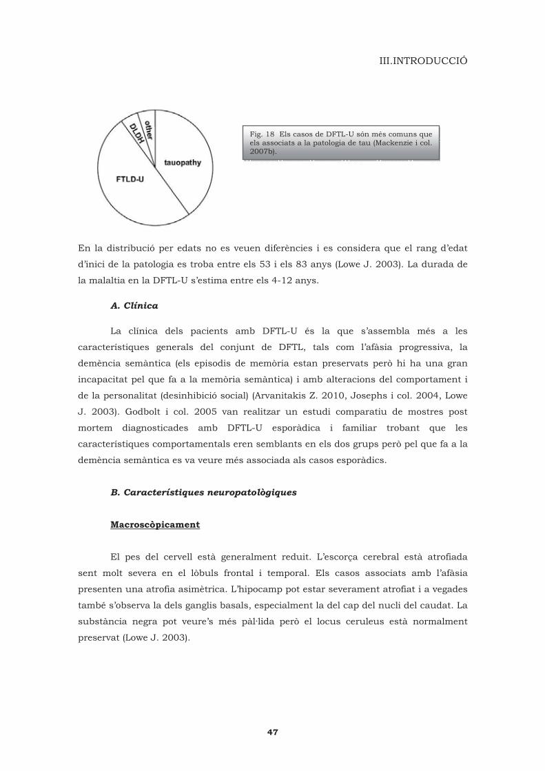
III.INTRODUCCIÓ
En la distribució per edats no es veuen diferències i es considera que el rang d’edat
d’inici de la patologia es troba entre els 53 i els 83 anys (Lowe J. 2003). La durada de
la malaltia en la DFTL-U s’estima entre els 4-12 anys.
A. Clínica
La clínica dels pacients amb DFTL-U és la que s’assembla més a les
característiques generals del conjunt de DFTL, tals com l’afàsia progressiva, la
demència semàntica (els episodis de memòria estan preservats però hi ha una gran
incapacitat pel que fa a la memòria semàntica) i amb alteracions del comportament i
de la personalitat (desinhibició social) (Arvanitakis Z. 2010, Josephs i col. 2004, Lowe
J. 2003). Godbolt i col. 2005 van realitzar un estudi comparatiu de mostres post
mortem diagnosticades amb DFTL-U esporàdica i familiar trobant que les
característiques comportamentals eren semblants en els dos grups però pel que fa a la
demència semàntica es va veure més associada als casos esporàdics.
B. Característiques neuropatològiques
Macroscòpicament
El pes del cervell està generalment reduit. L’escorça cerebral està atrofiada
sent molt severa en el lòbuls frontal i temporal. Els casos associats amb l’afàsia
presenten una atrofia asimètrica. L’hipocamp pot estar severament atrofiat i a vegades
també s’observa la dels ganglis basals, especialment la del cap del nucli del caudat. La
substància negra pot veure’s més pàl·lida però el locus ceruleus està normalment
preservat (Lowe J. 2003).
Fig. 18 Els casos de DFTL-U són més comuns que els associats a la patologia de tau (Mackenzie i col. 2007b).
47
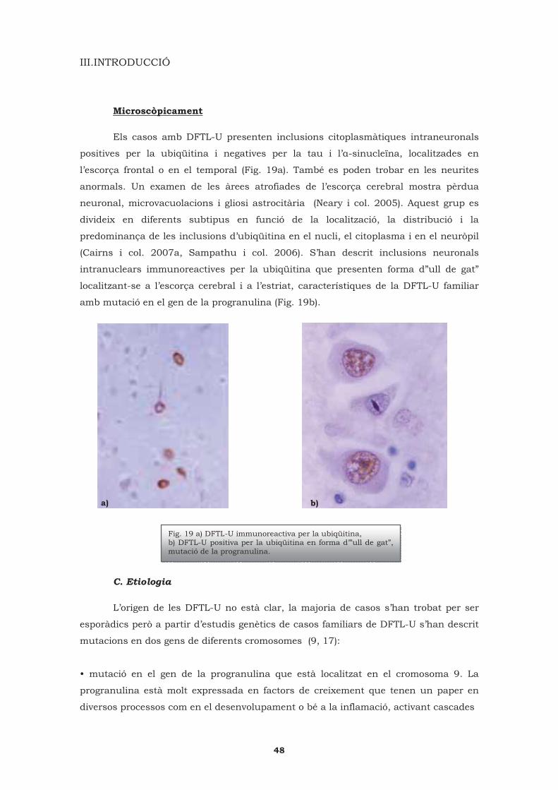
III.INTRODUCCIÓ
Microscòpicament
Els casos amb DFTL-U presenten inclusions citoplasmàtiques intraneuronals
positives per la ubiqüitina i negatives per la tau i l’�-sinucleïna, localitzades en
l’escorça frontal o en el temporal (Fig. 19a). També es poden trobar en les neurites
anormals. Un examen de les àrees atrofiades de l’escorça cerebral mostra pèrdua
neuronal, microvacuolacions i gliosi astrocitària (Neary i col. 2005). Aquest grup es
divideix en diferents subtipus en funció de la localització, la distribució i la
predominança de les inclusions d’ubiqüitina en el nucli, el citoplasma i en el neuròpil
(Cairns i col. 2007a, Sampathu i col. 2006). S’han descrit inclusions neuronals
intranuclears immunoreactives per la ubiqüitina que presenten forma d”ull de gat”
localitzant-se a l’escorça cerebral i a l’estriat, característiques de la DFTL-U familiar
amb mutació en el gen de la progranulina (Fig. 19b).
C. Etiologia
L’origen de les DFTL-U no està clar, la majoria de casos s’han trobat per ser
esporàdics però a partir d’estudis genètics de casos familiars de DFTL-U s’han descrit
mutacions en dos gens de diferents cromosomes (9, 17):
• mutació en el gen de la progranulina que està localitzat en el cromosoma 9. La
progranulina està molt expressada en factors de creixement que tenen un paper en
diversos processos com en el desenvolupament o bé a la inflamació, activant cascades
Fig. 19 a) DFTL-U immunoreactiva per la ubiqüitina, b) DFTL-U positiva per la ubiqüitina en forma d’”ull de gat”, mutació de la progranulina.
a) b)
48
III.INTRODUCCIÓ
que controlen la progressió del cicle cel·lular i la motilitat de les cèl·lules. S’expressa
en les neurones de l’escorça cerebral, de l’hipocamp i del cerebel produïnt efectes
neurotròfics però encara no s’ha determinat la seva funció normal en les neurones
(Gass i col. 2006). La DFTL-U està provocada per mutacions en aquest gen donant lloc
a al·lels nulls (haploinsuficiència): la majoria de mutacions identificades en el gen de
la progranulina són sense sentit o frameshift que donen lloc a codons prematurs
d’acabament en la seva seqüència de RNAm (Baker i col. 2006, Skoglund i col. 2009).
La identificació de la progranulina va ser particularment interessant ja que es
demostrava la coincidència de dos importants gens relacionats amb el mateix fenotip
de la malaltia en el cromosoma 17q21-22 17 (encara que aquesta mutació es troba en
la mateixa regió cromosomal que el gen de la tau, no s’han trobat mutacions de la tau)
(Baker i col. 2006, Kumar-Singh i col. 2007).
• mutació en el gen de la VCP (valosin-containing protein), sent una mutació sense
sentit en el cromososma 9, és un membre del tipus AAA ATPasa (ATPasa associada
amb diferents activitats) que actua com una xaperona associada a la degradació de
proteïnes en el reticle endoplasmàtic, resposta a l’estrès, mort cel·lular programada,
interaccions amb el sistema d’ubiqüitina-proteasoma,… (Cairns i col. 2007a).
2.2.1.1 Degeneració frontotemporal lobar associada a patologia de motoneurona i esclerosi lateral amiotròfica (DFTL-ELA)
La prevalència d’aquesta malaltia és de 5 per cada 100.000 individus i el risc
de patir ELA augmenta en un ordre de magnitud passats els 60 anys d’edat. El rang
d’edat d’inici de la patologia està entre els 39 i els 77 anys. La velocitat de la
progressió de la malaltia varia entre individus, podent estar influenciada pel lloc
d’inici, però sent normalment ràpida amb una supervivència de només 2-3 anys des
del començament dels símptomes (Barber i col. 2009, Lowe J. 2003). Aquesta durada
de la malaltia és molt menor que en el cas de les DFTL-U, pot ser per la ràpida
progressió de les complicacions mortals que presenta la patologia com la insuficiència
respiratòria que apareix com a un símptoma secundari.
A. Clínica
L’ELA té un patró de neurodegeneració amb un síndrome clínic ben definit que
normalment es diferencia bé dels altres desordres neurològics. Els casos esporàdics i
els familiars són clínicament indistingibles, la identificació dels genètics no és fàcil i es
necessita un historial familiar ben documentat.
49
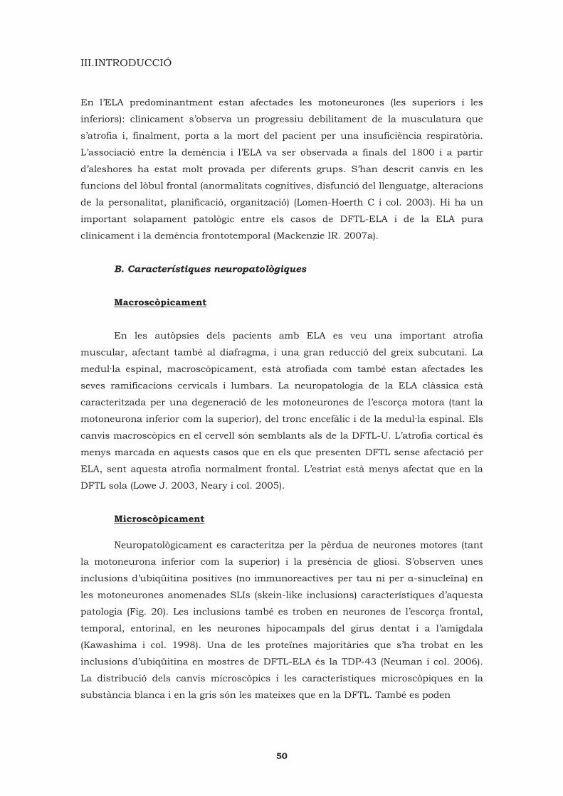
III.INTRODUCCIÓ
En l’ELA predominantment estan afectades les motoneurones (les superiors i les
inferiors): clínicament s’observa un progressiu debilitament de la musculatura que
s’atrofia i, finalment, porta a la mort del pacient per una insuficiència respiratòria.
L’associació entre la demència i l’ELA va ser observada a finals del 1800 i a partir
d’aleshores ha estat molt provada per diferents grups. S’han descrit canvis en les
funcions del lòbul frontal (anormalitats cognitives, disfunció del llenguatge, alteracions
de la personalitat, planificació, organització) (Lomen-Hoerth C i col. 2003). Hi ha un
important solapament patològic entre els casos de DFTL-ELA i de la ELA pura
clínicament i la demència frontotemporal (Mackenzie IR. 2007a).
B. Característiques neuropatològiques
Macroscòpicament
En les autòpsies dels pacients amb ELA es veu una important atrofia
muscular, afectant també al diafragma, i una gran reducció del greix subcutani. La
medul·la espinal, macroscòpicament, està atrofiada com també estan afectades les
seves ramificacions cervicals i lumbars. La neuropatologia de la ELA clàssica està
caracteritzada per una degeneració de les motoneurones de l’escorça motora (tant la
motoneurona inferior com la superior), del tronc encefàlic i de la medul·la espinal. Els
canvis macroscòpics en el cervell són semblants als de la DFTL-U. L’atrofia cortical és
menys marcada en aquests casos que en els que presenten DFTL sense afectació per
ELA, sent aquesta atrofia normalment frontal. L’estriat està menys afectat que en la
DFTL sola (Lowe J. 2003, Neary i col. 2005).
Microscòpicament
Neuropatològicament es caracteritza per la pèrdua de neurones motores (tant
la motoneurona inferior com la superior) i la presència de gliosi. S’observen unes
inclusions d’ubiqüitina positives (no immunoreactives per tau ni per �-sinucleïna) en
les motoneurones anomenades SLIs (skein-like inclusions) característiques d’aquesta
patologia (Fig. 20). Les inclusions també es troben en neurones de l’escorça frontal,
temporal, entorinal, en les neurones hipocampals del girus dentat i a l’amígdala
(Kawashima i col. 1998). Una de les proteïnes majoritàries que s’ha trobat en les
inclusions d’ubiqüitina en mostres de DFTL-ELA és la TDP-43 (Neuman i col. 2006).
La distribució dels canvis microscòpics i les característiques microscòpiques en la
substància blanca i en la gris són les mateixes que en la DFTL. També es poden
50
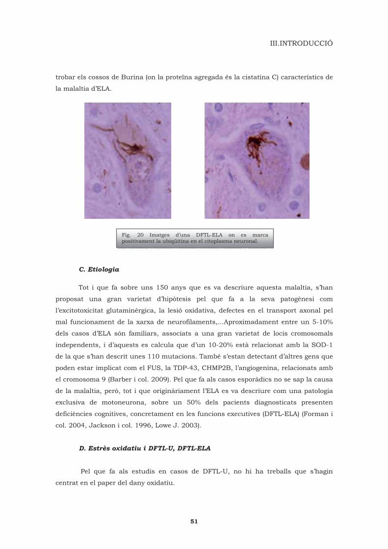
III.INTRODUCCIÓ
trobar els cossos de Burina (on la proteïna agregada és la cistatina C) característics de
la malaltia d’ELA.
C. Etiologia
Tot i que fa sobre uns 150 anys que es va descriure aquesta malaltia, s’han
proposat una gran varietat d’hipòtesis pel que fa a la seva patogènesi com
l’excitotoxicitat glutaminèrgica, la lesió oxidativa, defectes en el transport axonal pel
mal funcionament de la xarxa de neurofilaments,...Aproximadament entre un 5-10%
dels casos d’ELA són familiars, associats a una gran varietat de locis cromosomals
independents, i d’aquests es calcula que d’un 10-20% està relacionat amb la SOD-1
de la que s’han descrit unes 110 mutacions. També s’estan detectant d’altres gens que
poden estar implicat com el FUS, la TDP-43, CHMP2B, l’angiogenina, relacionats amb
el cromosoma 9 (Barber i col. 2009). Pel que fa als casos esporàdics no se sap la causa
de la malaltia, però, tot i que originàriament l’ELA es va descriure com una patologia
exclusiva de motoneurona, sobre un 50% dels pacients diagnosticats presenten
deficiències cognitives, concretament en les funcions executives (DFTL-ELA) (Forman i
col. 2004, Jackson i col. 1996, Lowe J. 2003).
D. Estrès oxidatiu i DFTL-U, DFTL-ELA
Pel que fa als estudis en casos de DFTL-U, no hi ha treballs que s’hagin
centrat en el paper del dany oxidatiu.
Fig. 20 Imatges d’una DFTL-ELA on es marca positivament la ubiqüitina en el citoplasma neuronal.
51

III.INTRODUCCIÓ
Diversos estudis patològics han demostrat evidències de l’augment de l’estrès oxidatiu
en l’ELA, centrant-se però en les ELAs familiars causades per la mutació de SOD-1
(tant en mostres humanes com desenvolupant models de ratolins transgènics per la
SOD-1 que permeten mimetitzar la patologia humana).
En el cas de l’ELA esporàdica els treballs per determinar l’efecte de l’estrès oxidatiu no
són tant abundants. Els nivells de carbonils proteics es van detectar augmentats, així
com els de 8-OHdG, en la medul·la espinal i en l’escorça motora de casos d’ELA
esporàdics (Ferrante i col. 1997). També es va trobar un increment dels nivells de 3-
nitrotirosina a la medul·la espinal (Beal i col. 1997). Gràcies a estudis
immunohistoquímics de les motoneurones de la medul·la espinal i de les cèl·lules
glials, es va observar un increment de la immunoreactivitat dels marcadors de
peroxidació lipídica i de glicoxidació proteïca en els patològics comparant amb els
controls (Shibata i col. 2001). D’altres estudis també van corroborar aquest increment
en el líquid cefaloraquidi de mostres patològiques utilitzant el 8-OHdG i l’HNE (Smith i
col. 1998). Ilieva i col. 2007 van utilitzar tècniques d’espectometria de masses per tal
de determinar la lesió oxidativa proteïca: es va detectar un augment d’aquesta en la
medul·la espinal i també en l’escorça frontal d’ELA esporàdics. També van demostrar
la presència d’estrès de reticle endoplasmàtic (augment de xaperones de reticle),
alteracions en la funcionalitat mitocondrial (complex I i III) i canvis en la composició
dels àcids grassos (Ilieva i col. 2007). Encara que els presents estudis han analitzat
l’escorça frontal per veure quin és l’efecte de l’estrès oxidatiu, no s’han realitzat
estudis per determinar quin és impacte en les proteïnes en mostres de pacients
diagnosticats amb DFTL-ELA.
52

IV. OBJECTIUS

IV.OBJECTIUS
1. Estudiar les modificaciones en el RNA i en les proteïnes que poden patir les
mostres de teixit de cervell humà per causa de factors post mortem.
2. Investigar la presència d’estrès oxidatiu en les proteïnes a la malaltia de
paràlisi supranuclear progressiva (PSP) comparant amb mostres de controls. Per altra
banda, identificar les proteïnes que resultin ser les possibles dianes d’oxidació.
3. Caracteritzar l’escorça frontal de mostres de les diferents classes de
degeneració frontotemporal lobar (DFTL), concretament la DFTL amb parkinsonisme
lligada al cromosoma 17 associada a mutacions de la tau -DFTL-tau-, la DFTL amb
inclusions positives per la ubiqüitina -DLFT-U- i la DLFT associada a patologia de
motoneurona i esclerosi lateral amiotròfica -DFTL-ELA-, en el camp de l’estrès oxidatiu
i identificar les proteïnes que siguin la diana dels diferents adductes d’oxidació.
4. Fer una revisió de les proteïnes modificades (oxidació, nitració) en les
diferents malalties neurodegeneratives, tant en mostres humanes com en models
animals i cultius cel·lulars, i de la metodologia de proteòmica redox utilitzada.
55


V. RESULTATS

V. RESULTATS
ARTICLES PUBLICATS
1. Brain banks: benefits, limitations and cautions concerning the use of post-mortem brain tissue for molecular studies
Isidre Ferrer, Anna Martinez, Susana Boluda, Piero Parchi, Marta Barrachina.
Cell Tissue Bank. 2008 Sep;9(3):181-94. Epub 2008 Jun 10. Review.
2. Glycolitic enzymes are targets of oxidation in aged human frontal cortex and oxidative damage of these proteins is increased in progressive supranuclear
palsy
A. Martínez, E. Dalfó, G. Muntané, I. Ferrer.
J Neural Transm. 2008;115(1):59-66. Epub 2007 Aug 21.
3. Type-Dependent Oxidative Damage in Frontotemporal Lobar Degeneration: Cortical Astrocytes Are Targets of Oxidative Damage
Anna Martínez, Margarita Carmona, Manuel Portero-Otin, Alba Naudí,
Reinald Pamplona, Isidre Ferrer.
J Neuropathol Exp Neurol. 2008 Dec;67(12):1122-36. Erratum in: J Neuropathol Exp
Neurol. 2009 Jan;68(1):112.
4. Protein Targets of Oxidative Damage in Human Neurodegenerative Diseases with Abnormal Protein Aggregates
Anna Martínez, Manuel Portero-Otin, Reinald Pamplona, Isidre Ferrer.
Brain Pathol. 2009 Aug 6. [Epub ahead of print].
59


V.RESULTATS
1. Brain banks: benefits, limitations and cautions concerning the use of post-mortem brain tissue for molecular studies
Isidre Ferrer, Anna Martinez, Susana Boluda, Piero Parchi, Marta Barrachina.
Cell Tissue Bank. 2008 Sep;9(3):181-94. Epub 2008 Jun 10. Review.
Els bancs de teixit, en aquest cas del sistema nerviós, representen el centre
d’unió entre el generós donatiu del teixit i els laboratoris d’investigació per tal de poder
augmentar el coneixement de les malalties neurodegeneratives, intentar detectar
noves dianes de diagnòstic i poder dissenyar noves estratègies terapèutiques.
Per tal de poder treballar amb la certesa de que els resultats obtinguts no estan
relacionats amb alteracions del teixit no vinculades amb la patologia, és necessari
saber les limitacions i les possibles millores en la metodologia utilitzada. Hi han
diferents factors que poden interferir en la preservació del DNA, el RNA, les proteïnes i
els lípids: pre mortem (estat agònic, pH) i post mortem (temps de retardament post
mortem, temperatura d’emmagatzemar la mostra, material d’extracció del teixit).
Per tal de saber la qualitat del DNA i del RNA l’ús del bioanalyzer (valors del
RIN) és molt més sensible que no els gels d’agarosa. A nivell de RNA, a través d’estudis
del temps de retardament post mortem i de la temperatura, es va veure que hi havia
diferència en el valor del RIN en diferents regions del cervell d’un mateix individu. Pel
que fa a les proteïnes, es van analitzar les modificacions d’oxidació i de nitració per
western blot en diferents temps de retardament post mortem de teixit humà. Amb els
diferents anticossos utilitzats, en general, es va observar que les diferents bandes es
mantenien estables fins a les 20h i que a partir d’aquest moment hi havia variacions
en el marcatge.
Així doncs és important caracteritzar els diferents factors (pre i post mortem)
que poden modificar les diverses molècules (DNA, RNA, proteïnes) per tal de poder
reduir aquestes variables i saber fins a quin punt poden o no influir en els
experiments que es volen realitzar i en els respectius resultats.
61

Brain banks: benefits, limitations and cautions concerningthe use of post-mortem brain tissue for molecular studies
Isidre Ferrer Æ Anna Martinez Æ Susana Boluda ÆPiero Parchi Æ Marta Barrachina
Received: 10 February 2008 / Accepted: 18 May 2008 / Published online: 10 June 2008
� Springer Science+Business Media B.V. 2008
Abstract Brain banks are facilities providing an
interface between generous donation of nervous
tissues and research laboratories devoted to increase
our understanding of the diseases of the nervous
system, discover new diagnostic targets, and develop
new strategies. Considering this crucial role, it is
important to learn about the suitabilities, limitations
and proper handling of individual brain samples for
particular studies. Several factors may interfere with
preservation of DNA, RNA, proteins and lipids, and,
therefore, special care must be taken first to detect sub-
optimally preserved tissues and second to provide
adequate material for each specific purpose. Basic
aspects related with DNA, RNA and protein preser-
vation include agonal state, post-mortem delay,
temperature of storage and procedures of tissue
preservation. Examination of DNA and RNA preser-
vation is best done by using bioanalyzer technologies
instead of less sensitive methods such as agarose gels.
Adequate RNA preservation is mandatory in RNA
microarray studies and adequate controls are necessary
for proper PCR validation. Like for RNA, the preser-
vation of proteins is not homogeneous since some
molecules are more vulnerable than others. This aspect
is crucial in the study of proteins including expression
levels and possible post-translational modifications.
Similarly, the reliability of functional and enzymatic
studies in human post-mortem brain largely depends
on protein preservation. Much less is known about
other aspects, such as the effects of putative deleteri-
ous factors on epigenetic events such as methylation of
CpGs in gene promoters, nucleosome preservation,
histone modifications, and conservation of microRNA
species. Most brains are appropriate for morphological
approaches but not all brains are useful for certain
biochemical and molecular studies.
Keywords DNA � RNA � Protein �Post-mortem delay � Brain bank � Oxidation �Epigenetics
Introduction
Brain banks have been created to obtain, classify,
preserve and distribute nervous system tissue speci-
mens and biological samples for research of diseases
of the nervous system under very precise ethical
settings. Brain banks are located worldwide and they
have been proved to be excellent platforms and
I. Ferrer (&) � A. Martinez � S. Boluda � M. Barrachina
Institut de Neuropatologia, Servei Anatomia Patologica,
IDIBELL-Hospital Universitari de Bellvitge, Universitat
de Barcelona, carrer Feixa LLarga sn, 08907 Hospitalet de
LLobregat, Spain
e-mail: [email protected]
I. Ferrer � A. Martinez � S. Boluda � M. Barrachina
CIBERNED, Barcelona, Spain
P. Parchi
Dipartamento di Scienze Neurologiche, Universita di
Bologna, Bologna, Italy
123
Cell Tissue Banking (2008) 9:181–194
DOI 10.1007/s10561-008-9077-0
63
facilities contributing significantly to improve our
understanding of neurological disorders. Moreover,
methodological protocols are geared to provide
optimal tissue for research under established ethical
and legal regulations (Tourtellotte et al. 1993; Bird
and Vonsattel 1993; Daniel and Lees 1993; New-
combe and Cuzner 1993; Esiri 1993; Hulette et al.
1997; Morgello et al. 2001; Sarris et al. 2002; Niwa
2002; Haroutunian and Davis 2002; Hulette 2003;
Murphy and Ravina 2003; Webster 2006; Nagatsu
and Sawada 2007; Schmidt et al. 2007; Ravid 2008).
Yet human brain banks are mainly based on
nervous tissue obtained after death and, therefore,
several factors may interfere with tissue and molec-
ular preservation of these samples. Some factors are
related with pre-mortem events such as prolonged
agonal state, hypoxia, acidosis, fever and seizures.
Others are related with long post-mortem delay
between death and sample processing for storage
and fixation, temperature of the corpse, characteris-
tics of the fixative solutions and processing of frozen
material. Finally, a third group of factors seems
unpredictable at the moment, and concerns unex-
pected variations from case to case or from region to
region from the same brain, despite similar pre-
mortem and post-mortem conditions. All these fac-
tors are of major concern in brain banks. Yet it is still
difficult to predict adequate or inappropriate condi-
tions for DNA, RNA and protein preservation solely
on the basis of these parameters.
Several specific focused studies have dealt with the
vulnerability of DNA, RNA and proteins in post-
mortem samples (Wu et al. 2002; Ma et al. 2003;
Hilbig et al. 2004; Kovacs et al. 2005; Bazinet et al.
2005). However, more comprehensive analyses are
often necessary to offer a wider perspective on the
impact of deleterious factors on tissue degradation.
Similarly, informations are needed on methods to
detect degradation and on practical procedures to
minimize pitfalls occurring when working with human
post-mortem brain tissues. In this line, several studies
have been undertaken, many of them within the scope
of the European Brain Bank Network (BrainNet II), to
identify possibilities and limitations of human brain
tissues for DNA, RNA and protein studies, as well as to
define methods that may improve tissue preservation
and processing. The present review includes an update
of general information and the result of personal
experience in the management of samples for
molecular studies that may be useful for future
research based on human post-mortem tissue obtained
from brain banks and brain collections.
DNA preservation
DNA is relatively resistant to post-mortem degrada-
tion under appropriate conditions. This property is
valuable in forensic pathology and paleontology,
allowing limited genetic studies in tissues maintained
for thousand of years (Marotta and Rollo 2002;
Konomi et al. 2002; Paabo et al. 2004; Calacal et al.
2005; Luciani et al. 2006). However, DNA is
vulnerable to degradation in liquid solutions, partic-
ularly fixatives used for tissue preservation in current
pathological practice.
It is the general belief that frozen material, even
when stored for years, is suitable for DNA studies
(Krajick 2002; Rollo et al. 2006). Sub-optimal DNA
preservation or even massive degradation, however,
can be expected in tissues stored in formalin at room
temperature for months or years (Koppelstaetter et al.
2005; Kunkle et al. 2006; Miething et al. 2006). This
is mainly due to the progressive acidification of
formalin into formic acid. The use of buffered formalin
stabilizes the solution for a longer time than non-
buffered formalin. Periodical renewal of the buffered
fixative solution in samples stored for months or years
reduces the deleterious effects of old formalin. Several
studies have recently appeared with methods aimed to
obtain the fixation of tissues with less damage to
nucleic acids and to optimize DNA extraction and
evaluation (Panaro et al. 2000; Coura et al. 2005;
Kline et al. 2005; Rivero et al. 2006; Stanta et al.
2006). DNA preservation in paraffin blocks is variable
but probably dependent on the type of fixative solution
and on the length of time of tissue storage in the
fixative before paraffin embedding.
A study by the European Brain Bank Network was
recently carried out in order to: (1) compare DNA
preservation in frozen, formalin-fixed and paraffin-
embedded tissues stored for different periods; (2)
study point mutations and triplet expansions in frozen,
formalin-fixed and paraffin-embedded material stored
for variable periods, and using different fixative
solutions; (3) compare different methods to optimize
DNA extraction and DNA amplification from sub-
optimally preserved brain tissue (Ferrer et al. 2007a).
182 Cell Tissue Banking (2008) 9:181–194
123
64

The study confirmed that DNA preservation is
good and suitable for genetic studies in samples
stored at -80�C for several years. Fresh-frozen tissue
is the optimal method to preserve banked brain tissue
for DNA recovery. The Alzheimer Disease Centres in
the United States, as well as other brain banks in
North America, Europe, Japan and Australia, already
employ a system of storing both fresh-frozen and
fixed tissue. The use of this model, in the context of a
multi-institutional program for the banking of brain
tissue, is further emphasized.
Acceptable results were also obtained in paraffin
blocks provided that these blocks were produced after
short fixation times in 4% buffered formalin. Sub-
optimal or bad results occur in buffered formalin-
fixed tissues stored for long periods. The shorter the
time of buffered formalin fixation, the better the DNA
preservation. Blocks produced up to one month after
buffered formalin fixation are consistent with good
DNA preservation, whereas DNA quality is sub-
optimal or bad in samples from paraffin blocks
produced several months or years after fixation. An
additional key factor for short periods is the fixative
solution itself: 4% buffered-formalin is better that
10% non-buffered formalin for DNA preservation.
Formic acid treatment previous to paraffin embed-
ding, as usually employed in the study of prion
diseases, has devastating effects on DNA preserva-
tion unless the time of exposure is very short and it is
followed by washing. Unfortunately, material stored
for years in formalin is probably no longer suitable to
study mutations. Therefore, production of paraffin
blocks after a short buffered formalin fixation is
strongly recommended.
Regarding protocols for DNA extraction, the best
and most reproducible results were obtained in frozen,
paraffin-embedded and formalin-fixed samples using
the QIAmp Micro Qiagen method. GenomiphiTM and
TaKaRa ExtagTM can be used in paraffin blocks and in
cases fixed in formalin in which QIAmpMicro Qiagen
failed Ferrer et al. 2007a).
Is formalin-fixed paraffin-embedded tissue
suitable for the study of polymorphisms?
Randomized parallel studies of mutations and CAG
repeats in frozen, formalin-fixed and formalin-fixed
paraffin-embedded tissues have shown optimal
preservation in frozen tissue. However DNA preser-
vation in formalin-fixed samples stored for long
periods of time in formalin are more than sub-optimal.
Formalin-fixed samples for short periods embedded in
paraffin can be used although the preservation of DNA
cannot be guaranteed Ferrer et al. 2007a).
Therefore, similar observations can be expected in
the study of DNA polymorphisms. The study of
PRNP codon 129, a methionine valine transition that
has a critical role in the susceptibility and phenotypic
expression of human prion diseases, was carried out
in cases affected with prion disease from which
frozen as well as formalin-fixed and formalin-fixed
paraffin-embedded samples were available. DNA was
extracted from de-waxed tissue sections or formalin-
fixed brain using the Quiagen Kit for DNA extrac-
tion, whereas DNA from frozen samples was
extracted with phenol-chloroform or with the Qui-
agen Kit. Studies carried out in two laboratories with
different samples resulted in similar conclusions. In
one laboratory, the identification of the exact poly-
morphism occurred in 3 of 7 formalin-fixed, formic
acid-treated and paraffin-embedded tissue sections,
and in 3 (2 of them different cases) of 7 tissue
sections formalin-fixed for not less than one year.
Comparison was made with frozen post-mortem brain
tissue samples or with blood cells of the same
individuals. In the other laboratory, the polymor-
phism was identified in 4 of 6 prion disease cases
embedded in paraffin. Study of PRNP codon 129 was
also examined in non-prion demented cases. The
polymorphism was identified in 6 of 8 embedded
cases and in 4 of 5 formalin-fixed cases. Good results
were obtained only in cases with a very short fixation
time; furthermore, results were more reliable in cases
with better tissue preservation at autopsy. These
results point to difficulties in the interpretation of
polymorphisms in non-frozen post-mortem human
brain and they are in line with previous studies
(Kunkle et al. 2006) concluding that polymorphism
determination on brain tissues fixed in formalin
should not be encouraged.
Methylation of gene promoters
Beta-amyloid deposition in the form of amyloid
(neuritic and diffuse) plaques, and tau hyperphosph-
orylation forming neurofibrillary tangles, neuropil
Cell Tissue Banking (2008) 9:181–194 183
123
65
threads and aberrant neurites in neuritic plaques, are
the major pathological substrates in cases with mild
cognitive impairment and Alzheimer’s disease (AD).
Amyloid plaque formation is a complex phenomenon
which involves the cleavage of the amyloid precursor
protein (APP) by distinct secretase complexes,
including presenilin 1 (PS1) and beta-site APP
cleavage enzyme 1 (BACE-1). The expression of
PS1 and BACE is regulated by methylation of their
corresponding gene promoters (Fuso et al. 2005;
Scarpa et al. 2006; Cavallaro et al. 2006). Reduced
levels of methylcytosine in APP gene promoter have
been observed in ageing and AD brains (Rogaev
et al. 1994; West et al. 1995; Tohgi et al. 1999a).
Moreover, the tau gene promoter is methylated with
age, which, in turn, down-regulates its transcription
activity (Tohgi et al. 1999b). These findings suggest
that gene promoter methylation has a role in the
expression of proteins related with the pathogenesis
of AD and other neurodegenerative diseases. Regio-
nal differences in the methylation of gene promoters
are crucial in the nervous system and they may
account for region-specific functional specializations
(Ladd-Acosta et al. 2007). However, it is not known
whether post-mortem delay has any effect on the
methylation of gene promoters.
Histone tail methylation and acetylation
Methylation and other covalent modifications of
nucleosome core histones are key regulators of
chromatin structure and function, including epigenetic
control of gene expression. Little is known about the
regulation of histone modifications at specific geno-
mic loci in the human brain. High levels of histone 3-
methyl-arginine-17, H3meR17, are associated with
down-regulated metabolic gene expression in the
prefrontal cortex of a subset of patients with schizo-
phrenia (Akbarian et al. 2005). Differential gene
expression patterns in prefrontal and cerebellar cor-
tices are reflected by similar differences in H3-lysine
4 methylation in ionotropic and metabotropic gluta-
mate receptor gene promoters (Stadler et al. 2005).
These findings suggest that histone lysine methylation
at gene promoters is involved in developmental
regulation and maintenance of region-specific expres-
sion patterns of ionotropic and metabotropic
glutamate receptors.
Preservation of methylation histone residues with
post-mortem delay has recently been reported. The
bulk of nucleosomal DNA remains attached to
histones during the first 30 h after death. Immuno-
precipitation with antibodies against methylated
histones was at least 10-fold more effective in
unfixed, micrococcal nuclease-digested samples, in
comparison to extracts prepared by fixation and
sonication. Moreover, histone methylation differ-
ences across various genomic sites were maintained
within a wide range of autolysis times and tissue pHs
(Huang et al. 2006).
Regarding possible effects of post-mortem delay
on histone tail acetylation, it is worth pointing out that
acetylation of histones may be triggered by hypoxia
inherent to the post-mortem state of the tissue, as it
also happens in the vicinity of infarcts. In this line,
pharmacological inhibition of histone deacetylases
increases histone acetylation and triggers pharmaco-
logical preconditioning effects against myocardial
ischemic injury, and reduces ischemic injury in the rat
and mouse brains (Ren et al. 2004; Faraco et al. 2006;
Zhao et al. 2007). Further analysis is, however,
necessary to determine whether histone tail acetyla-
tion as well as other covalent modifications described
in histone tails, such as phosphorylation, are stable
with post-mortem delay.
RNA preservation
Although some studies have suggested stability of
human post-mortem brain RNA (Leonard et al. 1993;
Cummings et al. 2001; Yasojima et al. 2001), many
others have shown that RNA integrity is largely
dependent on several factors, including the agonal
state, hypoxia, and the post-mortem delay between
death and tissue processing, among others (Yates
et al. 1990; Ross et al. 1992; Harrison et al. 1995;
Preece and Cairns 2003; Li et al. 2004; Tomita et al.
2004; Miller et al. 2004). Immediate immersion of
the sample in Trizol reagent is currently used in many
laboratories in order to improve RNA preservation.
Hepes glutamic acid buffer-mediated organic solvent
protection effect (HOPE) fixation has also been used
successfully (Witchell et al. 2007).
Recent studies have emphasized that post-mortem
delay and the pH of ventricular fluid are not
predictive of RNA preservation (Ervin et al. 2007).
184 Cell Tissue Banking (2008) 9:181–194
123
66
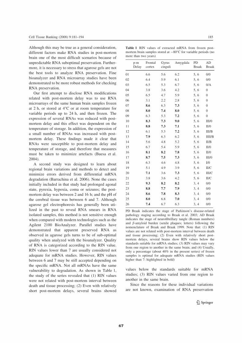
Although this may be true as a general consideration,
different factors make RNA studies in post-mortem
brain one of the most difficult scenarios because of
unpredictable RNA suboptimal preservation. Further-
more, it is necessary to stress that agarose gels are not
the best tools to analyze RNA preservation. Fine
bioanalyzer and RNA microarray studies have been
demonstrated to be more robust methods for checking
RNA preservation.
Our first attempt to disclose RNA modifications
related with post-mortem delay was to use RNA
microarrays of the same human brain samples frozen
at 2 h, or stored at 4�C or at room temperature for
variable periods up to 24 h, and then frozen. The
expression of several RNAs was reduced with post-
mortem delay and this effect was dependent on the
temperature of storage. In addition, the expression of
a small number of RNAs was increased with post-
mortem delay. These findings made it clear that
RNAs were susceptible to post-mortem delay and
temperature of storage, and therefore that measures
must be taken to minimize artefacts (Buesa et al.
2004).
A second study was designed to learn about
regional brain variations and methods to detect and
minimize errors derived from differential mRNA
degradation (Barrachina et al. 2006). None the cases
initially included in that study had prolonged agonal
state, pyrexia, hypoxia, coma or seizures; the post-
mortem delay was between 2 and 16 h; and the pH of
the cerebral tissue was between 6 and 7. Although
agarose gel electrophoresis has generally been uti-
lized in the past to reveal RNA smears in RNA
isolated samples, this method is not sensitive enough
when compared with modern technologies such as the
Agilent 2100 BioAnalyzer. Parallel studies have
demonstrated that apparent preserved RNA as
observed in agarose gels turns to be of sub-optimal
quality when analyzed with the bioanalyzer. Quality
of RNA is categorized according to the RIN value.
RIN values lower than 7 are usually considered not
adequate for mRNA studies. However, RIN values
between 6 and 7 may be still accepted depending on
the specific mRNA. Not all mRNAs have the same
vulnerability to degradation. As shown in Table 1,
the study of the series revealed that (1) RIN values
were not related with post-mortem interval between
death and tissue processing; (2) Even with relatively
short post-mortem delays, several brains showed
values below the standards suitable for mRNA
studies; (3) RIN values varied from one region to
another in the same brain.
Since the reasons for these individual variations
are not known, examination of RNA preservation
Table 1 RIN values of extracted mRNA from frozen post-
mortem brain samples stored at -80�C for variable periods (no
more than two years)
p-m
Delay
Frontal
cortex
Gyrus
cinguli
Amygdala PD
Braak
AD
Braak
01 6.6 5.6 6.2 5, 6 0/0
02 6.4 5.9 6.1 5, 6 0/0
03 6.5 5.3 6.7 5, 6 0/A
04 3.8 3.6 4.2 5, 6 0
05 6.5 4.7 5.9 5, 6 0
06 3.1 2.2 2.8 5, 6 0
07 8.6 6.3 7.3 5, 6 0
08 8.0 7.4 8.0 5, 6 0
09 6.3 5.3 7.2 5, 6 0
10 8.3 7.3 9.0 5, 6 III/0
11 8.8 7.3 7.1 5, 6 II/0
12 6.1 5.3 7.2 5, 6 III/B
13 7.9 6.3 6.2 5, 6 IIII/B
14 5.6 4.8 5.2 5, 6 II/B
15 6.7 5.4 5.9 5, 6 II/0
16 8.1 8.2 7.8 5, 6 II/0
17 8.7 7.5 7.3 5, 6 III/0
18 6.3 4.6 4.8 5, 6 I/0
19 5.1 4.9 3.9 5, 6 II/C
20 7.1 3.6 7.3 5, 6 III/C
21 3.8 3.6 4.2 5, 6 II/C
22 9.3 8.2 8.2 3, 4 0/0
23 8.8 7.7 7.9 3, 4 0/0
24 8.6 7.8 8.3 3, 4 0/0
25 8.0 6.8 7.0 3, 4 0/0
26 7.4 6.7 6.3 3, 4 0/0
PD Braak indicates the stage of Parkinson’s disease-related
pathology staging according to Braak et al. 2003; AD Braak
indicates the stage of neurofibrillary tangle (Roman numbers)
and b-amyloid burden (senile plaques; letters) following the
nomenclature of Braak and Braak 1999. Note that: (1) RIN
values are not related with post-mortem interval between death
and tissue processing; (2) Even with relatively short post-
mortem delays, several brains show RIN values below the
standards suitable for mRNA studies; (3) RIN values may vary
from one region to another in the same brain; and (4) Usually,
only a percentage (about 40% in the present series) of frozen
samples is optimal for adequate mRNA studies (RIN values
higher than 7: highlighted in bold)
Cell Tissue Banking (2008) 9:181–194 185
123
67

must be carried out in every individual subject of
RNA expression studies.
Commonly used endogenous controls belong to the
group of so-called ‘‘housekeeping’’ genes which
ideally are constitutively expressed by all cell types
and which are not affected by disease. For example,
there is a certain concern regarding glyceraldehyde-3-
phosphate dehydrogenase (GAPDH) mRNA expres-
sion as a suitable endogenous control in the study of
degenerative diseases of the nervous system because
GAPDH protein is implicated in apoptosis and neuro-
degeneration (Tatton et al. 2000). In the same line,
mRNA expression levels of b-actin and other genes
regulating the expression of cytoskeletal proteins
a- and b-tubulins might be considered sub-optimal
controls, as b-actin and a- and b-tubulins might be
down-regulated in degenerative diseases of the ner-
vous system. Yet, mRNAs of b-actin, cyclophylin andGAPDH show little fluctuation among several CNS
tissues and disease models (Medhurst et al. 2000).
b-glucuronidase and b-actin have proved to be good
endogenous controls because their expression levels
show a small variation across a representative number
of control and disease cases (Barrachina et al. 2006).
However, RNA samples with RIN\ 6 show reduce
b-actin mRNA levels, whereas b-glucuronidasemRNA levels are not affected by RNA degradation
(Barrachina et al. 2006).
Normalisation to total RNA concentration has
been proposed when comparing tissue biopsies
obtained from different individuals (Tricarico et al.
2002). However, human post-mortem brain presents a
more complex scenario, as the individual study of
each sample for RNA degradation is essential in
control and diseased cases. Finally, normalisation
with a particular control must also be monitored
because RNAs are differentially degraded with post-
mortem delay. For example, levels of superoxide
dismutase 1 (SOD1) and metalloproteinase domain
22 (ADAM22) were apparently not modified when
normalised with b-actin. Yet their expression levels
were reduced with post-mortem delay when values
were normalised with b-glucuronidase (Barrachina
et al. 2006). For practical purposes, several putative
housekeeping genes should be examined in parallel in
order to obtain the correct normalization in the very
specific scenario of study.
A third approach to increase our understanding on
RNA preservation in the post-mortem brain has been
the study of brain RNA in experimental animals. Not
only the post-mortem delay is reduced, as the samples
are immediately processed for RNA preservation, but
there is also no agonal state in this particular
condition. RNA preservation is optimal under these
conditions.
microRNA preservation
The study of microRNAs in diseases of the nervous
system is very recent and only a few studies have been
published, focused on schizophrenia (Burmistrova
et al. 2007; Perkins et al. 2007; Hansen et al. 2007),
Alzheimer’s disease (Lukiw 2007) and Parkinson’s
disease (Kim et al. 2007). Study of microRNAs as
regulators of RNA transcription is crucial in neuro-
degenerative diseases (Rogaev 2005; Hebert and
de Stropper 2007; Nelson and Keller 2007). In
contrast to RNAs, microRNAs are small molecules
thought to be resistant to post-mortem artefacts and
even to fixation. However, the in-vivo half-lives of
specific microRNAs are not known, and their high
uridine plus adenine content and their single-stranded
structure when not complexed to target mRNA make
them potentially labile to degradation (Lukiw 2007).
Therefore, further studies are needed to clarify
specific stability of microRNAs in post-mortem
brain.
Brain protein preservation largely depends on the
post-mortem delay and the post-mortem
temperature of storage
The effects of post-mortem delay on protein preser-
vation largely depend on the temperature of storage.
This aspect has been studied by processing the same
original brain sample with artificial accumulative
post-mortem delay at various temperatures. Samples
of the frontal cortex of cases were obtained at autopsy
two hours after death. Part of the sample was
immediately frozen at -80�C, whereas additional
parts of the same sample were stored at room
temperature, at 4�C or at 1�C for variable periods
up to 48 h, and then frozen at -80�C. Consecutivesamples of the same case were processed for gel
electrophoresis and Western blotting using different
186 Cell Tissue Banking (2008) 9:181–194
123
68

antibodies to selected proteins. Similar samples were
also processed for bidimensional gel electrophoresis,
in-gel digestion and mass spectrometry to pick up
proteins which were differentially expressed over
time Ferrer et al. 2007b).
Results in the first paradigm showed variable
vulnerability among proteins. Some were vulnerable
to degradation whereas others were relatively resis-
tant. Among resistant proteins were subunits of the
20S and 19S proteasome, b-actin and active p38
kinase. Among vulnerable proteins were synaptic
proteins (rab3a, rabphilin, a-synuclein), kinases
(mitogen-activated protein kinases, stress activated
protein kinases, cyclin-dependent kinase 5), trophic
factors and trophic factor receptors, and b-tubulin.The effect of the temperature of storage was
remarkable. The majority of proteins analyzed in
samples stored at room temperature showed marked
protein degradation at 24 h. In contrast, most proteins
in samples stored at 1�C were preserved at 48 h.
Since corpses are rarely or never stored at 1�C, itmay be assumed that, even in the best scenarios of
body storage at 4�C, cumulative protein degradation
with post-mortem delay will be the rule. Preservation
is worse with increased post-mortem delay at room
temperature.
These observations have important implications.
Gel electrophoresis and Western blotting applied to
human post-mortem brains may produce tricky
results depending on multiple factors: protein degra-
dation might be not the same in all the samples
examined, degradation may vary depending on the
protein.
In addition, certain results have to be interpreted
with caution. For example, degradation of soluble,
normal a-synuclein is rather visible with post-mortem
delay. However, a-synuclein aggregates are very
resistant to degradation related with post-mortem
delay. This may explain why Lewy bodies and
neurites in Parkinson’s disease and related disorders
and in multiple system atrophy can be visualized by
immunohistochemistry in tissue sections with poor
tissue preservation albeit in the absence of the typical
synaptic pattern of a-synuclein in the neuropil in
contrast with optimally-preserved tissue sections. The
present findings also confirm previous observations
on the variable vulnerability of synaptic proteins by
immunohistochemistry (Liu and Brun 1995) and
Western blotting (Siew et al. 2004). Synaptophysin
is a resistant protein whereas synucleins, rabphilin,
syntaxins and rab3a are not.
Similarly, tau is rapidly dephosphorylated in
normal conditions, and dephosphorylation of normal
tau occurs a few minutes after death because of the
activity of phosphatases and calpain (Sorimachi et al.
1996). However, hyper-phosphorylated tau, as in
Alzheimer’s disease and tauopathies, is resistant to
degradation. Therefore, neurofibrillary tangles,
grains, astrocytic inclusions and coiled bodies can
still be visualized in suboptimally preserved post-
mortem tissue sections.
Bidimensional gel electrophoresis and mass spec-
trometry permit the discovery of variations in their
expression levels of unexpected proteins. By using
this approach, additional proteins were identified as
vulnerable to post-mortem delay. Several proteins are
vulnerable to post-mortem delay, including a-synuc-lein, b-synuclein, peroxiredoxin, ATP synthase and
superoxide dismutase 1. Others are resistant, includ-
ing several related with the energy metabolism such
as glycogen 3-phosphate dehydrogenase, malate
dehydrogenase and aldolase A.
The use of bidimensional gels has confirmed the
degradation of certain proteins with post-mortem
delay, but has also permitted the identification of
spots whose density increases with post-mortem
delay (Crecelius et al. 2008). This is an unexpected
and important finding as it might imply increased
synthesis, reduced normal degradation or appearance
of fragmented proteins. It must be admitted that little
is known about these putative settings.
The reasons for selective preservation are not
known and we have not found groups of specific
proteins clustering in relation to families or functions,
perhaps with the exception of members of the
proteasome complex, proteases and cathepsine (Com-
paine et al. 1995).
In the same line, the effects of post-mortem delay
(between 5 and 21 h) in the expression levels of G-
proteins in human prefrontal cortex are variable
depending on the protein. Under the same conditions,
Ga-i1, Ga-i2, Ga-S and Gb are stable whereas Ga-qand Ga-o are vulnerable to post-mortem delay (Li
et al. 1996). Similarly, delay in tissue processing
after death results in variable modifications in the
expression levels of microtubule-associated proteins
tau, MAP2, MAP1B and MAP5 in rat hippocampus
(Schwab et al. 1994; Irving et al. 1997).
Cell Tissue Banking (2008) 9:181–194 187
123
69
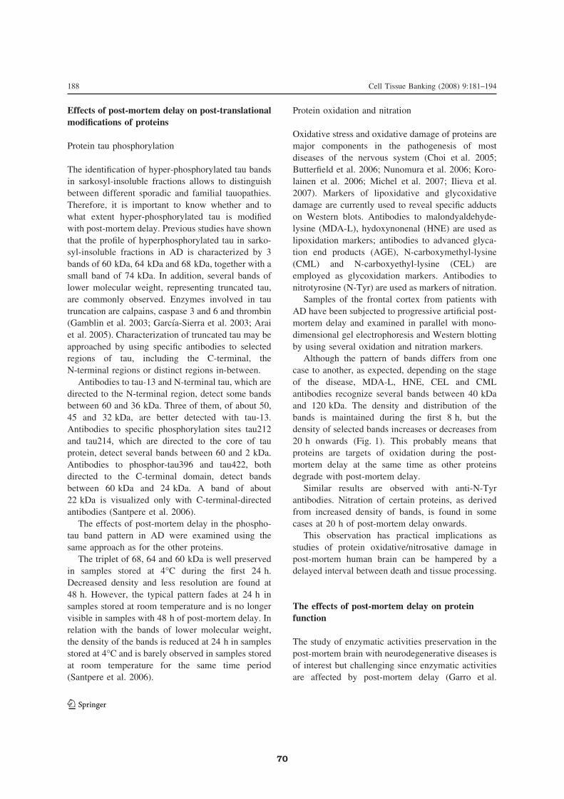
Effects of post-mortem delay on post-translational
modifications of proteins
Protein tau phosphorylation
The identification of hyper-phosphorylated tau bands
in sarkosyl-insoluble fractions allows to distinguish
between different sporadic and familial tauopathies.
Therefore, it is important to know whether and to
what extent hyper-phosphorylated tau is modified
with post-mortem delay. Previous studies have shown
that the profile of hyperphosphorylated tau in sarko-
syl-insoluble fractions in AD is characterized by 3
bands of 60 kDa, 64 kDa and 68 kDa, together with a
small band of 74 kDa. In addition, several bands of
lower molecular weight, representing truncated tau,
are commonly observed. Enzymes involved in tau
truncation are calpains, caspase 3 and 6 and thrombin
(Gamblin et al. 2003; Garcıa-Sierra et al. 2003; Arai
et al. 2005). Characterization of truncated tau may be
approached by using specific antibodies to selected
regions of tau, including the C-terminal, the
N-terminal regions or distinct regions in-between.
Antibodies to tau-13 and N-terminal tau, which are
directed to the N-terminal region, detect some bands
between 60 and 36 kDa. Three of them, of about 50,
45 and 32 kDa, are better detected with tau-13.
Antibodies to specific phosphorylation sites tau212
and tau214, which are directed to the core of tau
protein, detect several bands between 60 and 2 kDa.
Antibodies to phosphor-tau396 and tau422, both
directed to the C-terminal domain, detect bands
between 60 kDa and 24 kDa. A band of about
22 kDa is visualized only with C-terminal-directed
antibodies (Santpere et al. 2006).
The effects of post-mortem delay in the phospho-
tau band pattern in AD were examined using the
same approach as for the other proteins.
The triplet of 68, 64 and 60 kDa is well preserved
in samples stored at 4�C during the first 24 h.
Decreased density and less resolution are found at
48 h. However, the typical pattern fades at 24 h in
samples stored at room temperature and is no longer
visible in samples with 48 h of post-mortem delay. In
relation with the bands of lower molecular weight,
the density of the bands is reduced at 24 h in samples
stored at 4�C and is barely observed in samples stored
at room temperature for the same time period
(Santpere et al. 2006).
Protein oxidation and nitration
Oxidative stress and oxidative damage of proteins are
major components in the pathogenesis of most
diseases of the nervous system (Choi et al. 2005;
Butterfield et al. 2006; Nunomura et al. 2006; Koro-
lainen et al. 2006; Michel et al. 2007; Ilieva et al.
2007). Markers of lipoxidative and glycoxidative
damage are currently used to reveal specific adducts
on Western blots. Antibodies to malondyaldehyde-
lysine (MDA-L), hydoxynonenal (HNE) are used as
lipoxidation markers; antibodies to advanced glyca-
tion end products (AGE), N-carboxymethyl-lysine
(CML) and N-carboxyethyl-lysine (CEL) are
employed as glycoxidation markers. Antibodies to
nitrotyrosine (N-Tyr) are used as markers of nitration.
Samples of the frontal cortex from patients with
AD have been subjected to progressive artificial post-
mortem delay and examined in parallel with mono-
dimensional gel electrophoresis and Western blotting
by using several oxidation and nitration markers.
Although the pattern of bands differs from one
case to another, as expected, depending on the stage
of the disease, MDA-L, HNE, CEL and CML
antibodies recognize several bands between 40 kDa
and 120 kDa. The density and distribution of the
bands is maintained during the first 8 h, but the
density of selected bands increases or decreases from
20 h onwards (Fig. 1). This probably means that
proteins are targets of oxidation during the post-
mortem delay at the same time as other proteins
degrade with post-mortem delay.
Similar results are observed with anti-N-Tyr
antibodies. Nitration of certain proteins, as derived
from increased density of bands, is found in some
cases at 20 h of post-mortem delay onwards.
This observation has practical implications as
studies of protein oxidative/nitrosative damage in
post-mortem human brain can be hampered by a
delayed interval between death and tissue processing.
The effects of post-mortem delay on protein
function
The study of enzymatic activities preservation in the
post-mortem brain with neurodegenerative diseases is
of interest but challenging since enzymatic activities
are affected by post-mortem delay (Garro et al.
188 Cell Tissue Banking (2008) 9:181–194
123
70

2004). Metabolic functions are influenced by a
number of factors, but mainly depend on the preser-
vation of protein involved in a particular pathway.
Thus, it makes sense to first study the profile of
degradation with post-mortem delay of the proteins
under study. Once it is established that protein levels
are stable within a particular range of post-mortem
delay, metabolic studies can be carried out with
certain confidence in tissue samples keeping such
strict conditions.
Proteasomal activities have been analyzed in the
brains of cases with Huntington’s disease (Diaz-
Hernandez et al. 2003). Activation of the RAS-MAP
kinase pathway which is implicated in tau hyper-
phosphorylation has been shown in Alzheimer’s
disease (Ferrer et al. 2001). Immunoprecipitated
active stress-activated protein kinase (SAPK/JNK),
p38 kinase and glycogen synthase kinase-3b from
sarkosyl-insoluble fractions of the frontal cortex in
Alzheimer’s disease and Pick’s disease have the
capacity to phosphorylate recombinant tau in vitro,
thus indicating the presence of active tau kinases in
AD and other tauopathies (Puig et al. 2004; Ferrer
et al. 2005). Combined studies of receptor binding,
RNA and protein expression and enzymatic activities
allowed the demonstration of impaired metabotropic
glutamate receptor/phospholipase C signalling path-
way in the frontal cortex in Dementia with Lewy
bodies, AD and Pick’s disease (Dalfo et al. 2004a, b, c;
Albasanz et al. 2005; Dalfo et al. 2005). A similar
MDA-L
50
70
120
40
30
20
85
2h 5h 8h 20hCEL
46
50
70
120
40
30
20
85
2h 5h 8h 20h
46
50
70
120
40
30
20
85
CML
46
2h 5h 8h 20h
50
70
120
40
30
20
85
AGE
46
2h 5h 8h 20h
50
70
120
40
30
20
85
N-Tyr
46
2h 5h 8h 20h
Fig. 1 Gel electrophoresis and Western blots to oxidative
(MDA-L, CEL, CML, AGE) and nitrosative (N-Tyr) markers
of the same sample from the frontal cortex frozen at 2 h after
death or stored at room temperature after 5, 8 and 20 h and
then frozen at -80�C until use. Modification of the band profile
patterns are seen at 20 h. These changes are represented either
by reduced expression or by increased expression of selected
bands (arrows)
Cell Tissue Banking (2008) 9:181–194 189
123
71

approach has identified up-regulation and activation
of adenosine receptor 1 and adenosine receptor 2A in
the cerebral cortex in Creutzfeldt-Jakob disease and
Pick’s disease, respectively (Albasanz et al. 2006;
Rodrıguez et al. 2006).
Similar levels of a particular protein do not
necessarily mean, however, that there are similar
properties in normal and diseased conditions. Pro-
NGF extracted from the brain of AD cases induces
neuronal apoptosis mediated by p75NTR (Pedraza
et al. 2005). Yet pro-NGF fromAD and normal human
brain displays distinctive abilities to induce processing
and nuclear translocation of intracellular domain of
p75NTR and apoptosis (Podlesniy et al. 2006).
Protein interactions
Several studies have shown abnormal protein inter-
actions in disease states. These abnormalities may
have functional implications. As an example, abnor-
mal a-synuclein interaction with the synaptic protein
rab3a is found in vulnerable regions in Lewy body
diseases and multiple system atrophy (Dalfo et al.
2004b; Dalfo and Ferrer 2005). Similar abnormal
interaction occurs in A30P a-synuclein transgenic
mice bearing the 30 mutation (Dalfo et al. 2004c).
This abnormal interaction may interfere with synaptic
vesicle traffic and docking. Functional studies have
shown retarded synaptic transmission in the hippo-
campus in the same transgenic mice (Steidl et al.
2003).
In spite of these findings, little is known about the
effects of pre-mortem and post-mortem events on
protein-protein interactions, and studies are necessary
to elucidate these points. Not only may protein
interactions be modified with post-mortem delay,
but also transcription factors may translocate from the
nucleus to the cytoplasm in the setting of delayed
post-mortem tissue processing (Boutillier et al. 2007).
Concluding comments
Human post-mortem brain tissue is invaluable for the
study of human diseases of the nervous system. Brain
banks are facilities providing an interface for gener-
ous donation of nervous tissues and research in an
attempt to increase our understanding of diseases of
the nervous system as well as to discover new
diagnostic targets for the diagnosis of diseases and to
procure new strategies to prevent, delay the course of
and cure neurological and mental diseases. Correct
management of brain banks is mandatory in all this
process (Bell et al. 2008). Although every donated
brain is a unique piece of information and subject of
study, strict measures should be taken to understand
the potentialities and limitations of post-mortem
brain tissue for specific studies. Pre-mortem and
post-mortem factors may interfere with molecular
studies, and erroneous conclusions might be drawn as
a consequence of disregarding possible sources of
pitfalls and artefacts. Not all post-mortem tissues are
suitable for DNA, RNA and protein studies; nor are
all tissues appropriate for determinate functional or
enzymatic assays. Special care must be taken to
consider these circumstances when dealing with
human post-mortem brain tissue for research.
Acknowledgements This work was supported by the
European Commission under the Sixth Framework Programme
(BrainNet Europe II, LSHM-CT-2004-503039; and INDABIP).
We thank T. Yohannan for editorial help. Brain samples were
obtained from the Institute of Neuropathology and Brain Bank
following the guidelines and approval of the local ethics
committees.
References
Akbarian S, Ruehl MG, Bliven E, Luiz LA, Peranelli AC, Baker
SP et al (2005) Chromatin alterations associated with
down-regulated metabolic gene expression in the pre-
frontal cortex of subjects with schizophrenia. Arch Gen
Psychiatry 62:829–840. doi:10.1001/archpsyc.62.8.829
Albasanz JL, Dalfo E, Ferrer I, Martın M (2005) Impaired
metabotropic glutamate receptor/phospholipase C signal-
ing pathway in the cerebral cortex in Alzheimer’s disease
and dementia with Lewy bodies correlates with stage of
Alzheimer’s disease-related changes. Neurobiol Dis
20:685–693. doi:10.1016/j.nbd.2005.05.001
Albasanz JL, Rodriguez A, Ferrer I, Martin M (2006) Aden-
osine A2A receptors are up-regulated in Pick’s disease
frontal cortex. Brain Pathol 16:249–255. doi:
10.1111/j.1750-3639.2006.00026.x
Arai T, Guo J, McGeer P (2005) Proteolysis of non-phos-
phorylated and phosphorylated tau by thrombin. J Biol
Chem 280:5145–5153. doi:10.1074/jbc.M409234200
Barrachina M, Castano E, Ferrer I (2006) TaqMan PCR assay
in the control of RNA normalization in human post-
mortem brain tissue. Neurochem Int 49:276–284. doi:
10.1016/j.neuint.2006.01.018
Bazinet RP, Lee HJ, Felder CC, Porter AC, Rapoport SI, Ro-
senberger TA (2005) Rapid high-energy microwave
190 Cell Tissue Banking (2008) 9:181–194
123
72

fixation is required to determine the anandamine (N-
arachidonoylethanolamine) concentration of rat brain.
Neurochem Res 30:597–601. doi:10.1007/s11064-005-
2746-5
Bell JE, Alafuzoff I, Al-Sarraj S, Arzberger T, Bogdanovic N,
Budka H, Dexter DT, Falkai P, Ferrer I, Gelpi E, Gen-
tleman SM, Giaconne G, Huitinga I, Ironside JW,
Klioueva N, Kovacs GG, Meyronet D, Palkovits M, Par-
chi P, Patsouris E, Reynols R, Riederer P, Roggendorf W,
Seilhean D, Schmitt A, Schmitz P, Streichenberger N,
Schwakber A, Kretzschmar H (2008) Management of a
21st century brain bank: experience in the BrainNet
Europe Consortium. Acta Neuropathol 115:497–507
Bird ED, Vonsattel JP (1993) The development of a brain
bank. J Neural Transm 39:17–23
Boutillier S, Lannes B, Buee L, DElacourte A, Rouaux C, Mohr
M, Bellocq JP, Sellal F, Larmet Y, Boutillier AL, Loeffler
JP (2007) Sp3 and Sp4 transcription factor levels are
increased in brains of patients with Alzheimer’s disease.
Neurodegener Dis 4:413–423. doi:10.1159/000107701
Braak H, Braak E (1999) Temporal sequence of Alzheimer’s
disease-related pathology. In: Peters A, Morrison JH
(eds) Cerebral cortex, vol 14: neurodegenerative and
age-related changes in structure and function of cerebral
cortex. Kluwer Academic/Plenum Press, New York, pp
475–512
Braak H, Del Tredici K, Rub U, de Vos RA, Jansen Steur EN,
Braak E (2003) Staging of brain pathology related to
sporadic Parkinson’s disease. Neurobiol Aging 24:
197–211. doi:10.1016/S0197-4580(02)00065-9
Buesa C, Maes T, Subirada F, Barrachina M, Ferrer I (2004)
DNA chip technology in brain banks: confronting
a degrading world. J Neuropathol Exp Neurol 63:
1003–1014
Burmistrova OA, Golstov AY, Abramova LI, Kaleda VG,
Orlova VA, Rogaev EI (2007) MicroRNA in schizo-
phrenia: genetic and expression analysis of miR-130b
(22q11). Biochemistry 72:578–582
Butterfield DA, Poon HF, Clair D, Keller JN, Pierce WM,
Klein JB et al (2006) Redox proteomics identification of
oxidatively modified hippocampal proteins in mild cog-
nitive impairment: insights into the development of
Alzheimer’s disease. Neurobiol Dis 22:223–232. doi:
10.1016/j.nbd.2005.11.002
Calacal GC, Delfin FC, Tan MM, Roewer L, Magtanong DL,
Lara MC et al (2005) Identification of exhumed remains
of fire tragedy victims using conventional methods and
autosomal/Y-chromosomal short tandem repeat DNA
profiling. Am J Forensic Med Pathol 26:285–291. doi:
10.1097/01.paf.0000177338.21951.82
Cavallaro RA, Fuso A, D’Anselmi F, Seminara L, Scarpa S
(2006) The effect of S-adenosylmethionine on CNS gene
expression studied by cDNA microarray analysis. J Alz-
heimers Dis 9:415–419
Choi J, Rees HD, Weintraub ST, Levey AI, Chin LS, Li L
(2005) Oxidative modifications and aggregation of Cu,
Zn-superoxide dismutase associated with Alzheimer and
Parkinson diseases. J Biol Chem 280:11648–11655. doi:
10.1074/jbc.M414327200
Compaine A, Schein JD, Tabbs JS, Mohan PS, Nixon RA
(1995) Limited proteolytic processing of the mature form
of cathepsin D in human and mouse brain: postmortem
stability of enzyme structure and activity. Neurochem Int
27:385–396. doi:10.1016/0197-0186(95)00020-9
Coura R, Prolla JC, Meurer L, Ashton-Prolla P (2005) An
alternative protocol for DNA extraction from formalin
fixed and paraffin wax embedded tissue. J Clin Pathol
58:894–895. doi:10.1136/jcp. 2004.021352
Crecelius A, Gotz A, Arzberger T, Frohlich T, Arnold GJ,
Ferrer I et al (2008) Assessing quantitative post-mortem
changes in the grey white matter of the human frontal
cortex proteome by 2D-DIGE. Proteomics 8:1276–1291.
doi:10.1002/pmic.200700728
Cummings TJ, Strum JC, Yoon LW, Szymanski MH, Hulette
CM (2001) Recovery and expression of messenger RNA
from post-mortem human brain tissue. Mod Pathol
14:1157–1161. doi:10.1038/modpathol.3880451
Dalfo E, Ferrer I (2005) Alpha-synuclein binding to rab-3a in
multiple system atrophy. Neurosci Lett 380:170–175. doi:
10.1016/j.neulet.2005.01.034
Dalfo E, Albasanz JL, Martın M, Ferrer I (2004a) Abnormal
metabotropic glutamate receptor expression and signaling
in the cerebral cortex in Diffuse Lewy body disease is
associated with irregular alpha-synuclein/phospholipase C
interactions. Brain Pathol 14:388–398
Dalfo E, Barrachina M, Rosa JL, Ambrosio S, Ferrer I (2004b)
Abnormal alpha-synuclein interactions with rab3a and
rabphilin in diffuse Lewy body disease. Neurobiol Dis
16:92–97. doi:10.1016/j.nbd.2004.01.001
Dalfo E, Gomez-Isla T, Rosa JL, Nieto Bodelon M, Cuadrado
Tejedor M, Barrachina M et al (2004c) Abnormal alpha-
synuclein interactions with Rab proteins in alpha-synuc-
lein A30P transgenic mice. J Neuropathol Exp Neurol
63:302–313
Dalfo E, Albasanz JL, Rodrıguez A, Martın M, Ferrer I (2005)Abnormal group I metabotropic glutamate receptor
expression and signaling in the frontal cortex in Pick
disease. J Neuropathol Exp Neurol 64:638–647
Daniel SE, Lees AJ (1993) Parkinson’s Disease Society Brain
Bank, London: overview and research. J Neural Transm
39:165–172
Diaz-Hernandez M, Hernadez F, Martin-Aparicio E, Gomez-
Ramos P, Moran MA, Castano JG et al (2003) Neuronal
induction of the immunoproteasome in Huntington’s dis-
ease. J Neurosci 23:11653–11661
Ervin JF, Heinzen EL, Cronin KD, Goldstein D, SzymanskiMH,
Bohmer KA et al (2007) Postmortem delay has minimal
effect on brain RNA integrity. J Neuropathol Exp Neurol
66:1093–1099. doi:10.1097/nen.0b013e31815c196a
Esiri MM (1993) Brain banks: the Oxford experience. J Neural
Transm 39:25–30
Faraco G, Pancani T, Formentini L, Mascagni P, Fossati G,
Leoni F et al (2006) Pharmacological inhibition of histone
deacetylases by suberoylanilide hydroxamic acid specifi-
cally alters gene expression and reduces ischemic injury
in the mouse brain. Mol Pharmacol 70:1876–1884. doi:
10.1124/mol.106.027912
Ferrer I, Blanco R, Carmona M, Ribera R, Goutan E, Puig B
et al (2001) Phosphorylated MAP kinase (ERK1,
ERK2) expression is associated with early tau deposi-
tion in neurones and glial cells, but not with increased
nuclear DNA vulnerability and cell death, in
Cell Tissue Banking (2008) 9:181–194 191
123
73
Alzheimer’s disease, Pick’s disease, progressive supra-
nuclear palsy and corticobasal degeneration. Brain
Pathol 11:144–158
Ferrer I, Gomez-Isla T, Puig B, Freixes M, Ribe E, Dalfo E
et al (2005) Current advances on different kinases
involved in tau phosphorylation, and implications in
Alzheimer’s disease and tauopathies. Curr Alzheimer Res
2:3–18. doi:10.2174/1567205052772713
Ferrer I, Armstrong J, Capellari S, Parchi P, Arzberger T, Bell J
et al (2007a) Effects of formalin fixation, paraffin
embedding, and time of storage on DNA preservation in
brain tissue: a BrainNet Europe study. Brain Pathol
17:297–303. doi:10.1111/j.1750-3639.2007.00073.x
Ferrer I, Santpere G, Arzberger T, Bell J, Blanco R, Boluda S
et al (2007b) Brain protein preservation largely depends
on the postmortem storage temperature: implications for
study of proteins in human neurologic diseases and
management of brain banks: a BrainNet Europe Study. J
Neuropathol Exp Neurol 66:35–46
Fuso A, Seminara L, Cavallaro RA, D’Anselmi F, Scarpa S
(2005) S-adenosylmethionine/homocysteine cycle altera-
tions modify DNA methylation status with consequent
deregulation of PS1 and BACE and beta-amyloid pro-
duction. Mol Cell Neurosci 28:195–204. doi:
10.1016/j.mcn.2004.09.007
Gamblin TC, Chen F, Abraha A, Miller R, Fu Y, Garcia-Sierra
F et al (2003) Caspase cleavage of tau: linking amyloid
and neurofibrillary tangles in Alzheimer’s disease. Proc
Natl Acad Sci USA 100:10032–10037. doi:10.1073/pnas.
1630428100
Garcıa-Sierra F, Ghoshal N, Quinn B, Berry R, Binder LI
(2003) Conformational changes and truncation of tau
protein during tangle evolution in Alzheimer’s disease. J
Alzheimers Dis 5:65–77
Garro MA, Lopez de Jesus M, Ruiz de Azua I, Callado LF,
Meana JJ, Salles J (2004) Differential postmortem delay
effect on agonist-mediated phospholipase Cb activity in
human cortical crude and synaptosomal brain membranes.
Neurochem Res 29:1461–1465. doi:10.1023/B:NERE.000
0026412.66508.14
Hansen T, Olsen L, Lindow M, Jakobsen KD, Ullum H,
Jonsson E et al (2007) Brain expressed microRNAs
implicated in schizophrenia etiology. PLoS ONE 9:1–7
Haroutunian V, Davis KL (2002) Issues and perspectives on
brain tissue banking. Curr Psychiatry Rep 4:233–244. doi:
10.1007/s11920-996-0039-6
Harrison PJ, Heath PR, Eastwood SL, Burnet PW, McDonald
B, Pearson RC (1995) The relative importance of pre-
mortem acidosis and post-mortem interval for human
brain gene expression studies: selective mRNA vulnera-
bility and comparison with their encoded proteins.
Neurosci Lett 200:151–154. doi:10.1016/0304-3940
(95)12102-A
Hebert SS, De Strooper B (2007) Molecular biology miRNAs
in neurodegeneration. Science 317:1179–1180. doi:
10.1126/science.1148530
Hilbig H, Bidmon HJ, Oppermann OT, Remmerbach T (2004)
Influence of post-mortem delay and storage temperature
on the immunohistochemical detection of antigens in the
CNS of mice. Exp Toxicol Pathol 56:159–171. doi:
10.1016/j.etp. 2004.08.002
Huang HS, Matevossian A, Jiang Y, Akbarian S (2006)
Chromatin immunoprecipitation in postmortem brain. J
Neurosci Methods 156:284–292. doi:10.1016/j.jneumeth.
2006.02.018
Hulette CM (2003) Brain banking in the United States. J
Neuropathol Exp Neurol 62:715–722
Hulette CM, Welsh-Bohmer KA, Crain B, Szymanski MH,
Sinclaire NO, Roses AD (1997) Rapid brain autopsy.
The Joseph and Kathleen Bryan Alzheimer’s Disease
Research Center experience. Arch Pathol Lab Med 121:
615–618
Ilieva EV, Ayala V, Jove M, Dalfo E, Cacabelos D, Povedano
M et al (2007) Oxidative and endoplasmic reticulum
stress interplay in sporadic amyotrophic lateral sclerosis.
Brain 130:3111–3123. doi:10.1093/brain/awm190
Irving EA, McCulloch J, Dewar D (1997) The effect of post-
mortem delay on the distribution of microtubule-associated
proteins tau, MAP2, and MAP5 in the rat. Mol Chem
Neuropathol 30:253–271. doi:10.1007/BF02815102
Kim J, Inoue K, Ishii J, Vanti WB, Voronov SV, Murchison
Hannon G et al (2007) A microRNA feedback circuit in
midbrain dopamine neurons. Science 317:1179–1180. doi:
10.1126/science.1148530
Kline MC, Duewer DL, Redman JW, Butler JM (2005) Results
from the NIST 2004 DNA quantitation study. J Forensic
Sci 50:570–578
Konomi N, Lebwohl E, Zhang D (2002) Comparison of DNA
and RNA extraction methods for mummified tissues. Mol
Cell Probes 16:445–451. doi:10.1006/mcpr.2002.0441
Koppelstaetter C, Jennings P, Hochegger K, Perco P, Ischia R,
Karkoszka H et al (2005) Effect of tissue fixatives on telo-
mere length determination by qualitative PCR.MechAgeing
Dev 126:1331–1333. doi:10.1016/j.mad.2005.08.003
Korolainen MA, Goldsteins G, Nyman TA, Alafuzoff I, Ko-
istinaho J, Pirttila T (2006) Oxidative modification of
proteins in the frontal cortex of Alzheimer’s disease brain.
Neurobiol Aging 27:42–53. doi:10.1016/j.neurobiolaging.
2004.11.010
Kovacs Z, Kekesi KT, Bobest M, Torok T, Szilagyi N, Szidra
T et al (2005) Post mortem degradation of nucleosides in
the brain: comparison of human and rat brains for esti-
mation of in vivo concentrations of nucleosides. J
Neurosci Methods 148:88–93. doi:10.1016/j.jneumeth.
2005.04.012
Krajick K (2002) Glacial research melting glaciers release
ancient relics. Science 296:2142. doi:10.1126/science.
296.5567.454
Kunkle RA, Miller JM, Alt DP, Cutlip RC, Cockett NE, Wang
S et al (2006) Determination of sheep prion gene poly-
morphisms from paraffin-embedded tissues. J Vet Diagn
Invest 18:443–447
Ladd-Acosta C, Pevsner J, Sabunciyan S, Yolken RH, Webster
MJ, Dinkins T et al (2007) DNA methylation signatures
within the human brain. Am J Hum Genet 81:1304–1315.
doi:10.1086/524110
Leonard S, Logel J, Luthman D, Casanova M, Kirch D,
Freedman R (1993) Biological stability of mRNA isolated
from human post-mortem brain collections. Biol Psychi-
atry 33:456–466. doi:10.1016/0006-3223(93)90174-C
Li X, Greenwood AF, Powers R, Jope RS (1996) Effects of
postmortem interval, age, and Alzheimer’s disease on
192 Cell Tissue Banking (2008) 9:181–194
123
74
G-proteins in human brain. Neurobiol Aging 17:115–122.
doi:10.1016/0197-4580(95)02023-3
Li JZ, Vawter MP, Walsh DM, Tomita H, Evans SJ, Choudary
PV et al (2004) Systematic changes in gene expression in
post-mortem human brains associated with tissue pH and
terminal medical conditions. Hum Mol Genet 13:609–
616. doi:10.1093/hmg/ddh065
Liu X, Brun A (1995) Synaptophysin immunoreactivity is
stable 36 h postmortem. Dementia 6:211–217. doi:
10.1159/000106949
Luciani S, Fornaciari G, Rickards O, Labarga CM, Rollo F
(2006) Molecular characterization of a pre-Columbian
mummy and in situ coprolite. Am J Phys Anthropol
129:620–629. doi:10.1002/ajpa.20314
Lukiw WJ (2007) Micro-RNA speciation in fetal, adult and
Alzheimer’s disease. Neuroreport 18:297–300. doi:
10.1097/WNR.0b013e3280148e8b
Ma JK, Zhu HE, Piletz JE (2003) Effect of postmortem delay
on imidazoline receptor-binding proteins in human and
mouse brain. Ann N Y Acad Sci 1009:341–346. doi:
10.1196/annals.1304.043
Marota I, Rollo F (2002) Molecular paleontology. Cell Mol
Life Sci 59:97–111. doi:10.1007/s00018-002-8408-8
Medhurst AD, Harrison DC, Read SJ, Campbell CA, Robbins
MJ, Pangalos MN (2000) The use of TaqMan RT-PCR
assays for semiquantitative analysis of gene expression in
CNS tissues and disease models. J Neurosci Methods
98:9–20. doi:10.1016/S0165-0270(00)00178-3
Michel TM, Frangou S, Thiemeyer D, Camara S, Jecel J, Nara
K et al (2007) Evidence for oxidative stress in the frontal
cortex in patients with recurrent depressive disorder-a
postmortem study. Psychiatry Res 151:145–150. doi:
10.1016/j.psychres.2006.04.013
Miething F, Hering S, Hanschke B, Dressler J (2006) Effect of
fixation for the degradation of nuclear and mitochondrial
DNA in different tissues. J Histochem Cytochem 54:371–
374. doi:10.1369/jhc.5B6726.2005
Miller CL, Diglisic S, Leister F, Webster M, Yolken RH (2004)
Evaluating RNA status for RT-PCR in extracts of post-
mortem human brain tissue. Biotechniques 36:628–633
Morgello S, Gelman BB, Kozlowski PB, Vinters HV, Masliah
E, Cornford M et al (2001) The national NeuroAIDS
tissue consortium. Neuropathol Appl Neurobiol 27:326–
335. doi:10.1046/j.0305-1846.2001.00334.x
Murphy DD, Ravina B (2003) Brain banking for neurodegen-
erative diseases. Curr Opin Neurol 16:459–463. doi:
10.1097/00019052-200308000-00003
Nagatsu T, Sawada M (2007) Biochemistry of post-mortem
brains in Parkinson’s disease: historical overview and
future prospects. J Neural Transm 72:113–120
Nelson PT,Keller JN (2007)RNA in brain disease: no longer just
‘‘the messenger in the middle’’. J Neuropathol Exp Neurol
66:461–468. doi:10.1097/01.jnen.0000240474.27791.f3
Newcombe J, Cuzner ML (1993) Organization and research
applications of the UK Multiple Sclerosis Society Tissue
Bank. J Neural Transm 39:155–163
Niwa S (2002) Establishment of the systematic Brain Bank
Network for studies of mental disorders. Seishin Shin-
keigaku Zasshi 104:152–157
Nunomura A, Castrellani RJ, Zhu X, Moreira PI, Perry G,
Smith MA (2006) Involvement of oxidative stress in
Alzheimer disease. J Neuropathol Exp Neurol 65:631–
641. doi:10.1097/01.jnen.0000228136.58062.bf
Paabo S, Poinar H, Serre D, Jaenicke-Despres V, Hebler J,
Rohland N et al (2004) Genetic analyses from ancient
DNA. Annu Rev Genet 38:645–679. doi:10.1146/annurev.
genet.37.110801.143214
Panaro NJ, Yuen PK, Sakazume T, Fortina P, Kricka LJ,
Wilding P (2000) Evaluation of DNA fragment sizing and
quantification by the agilent 2100 bioanalyzer. Clin Chem
46:1851–1853
Pedraza CE, Podlesniy P, Vidal N, Arevalo JC, Lee R,
Hempstead B et al (2005) Pro-NGF isolated from the
human brain affected by Alzheimer’s disease induces
neuronal apoptosis mediated by p75NTR. Am J Pathol
166:533–543
Perkins DO, Jeffries CD, Jarskog LF, Thomson JM, Woods K,
Newman MA et al (2007) microRNA expression in the
prefrontal cortex of individuals with schizophrenia and
schizoaffective disorders. Genome Biol 8(R27):1–11. doi:
10.1186/gb-2007-8-2-r27
Podlesniy P, Kichev A, Pedraza C, Saurat J, Encinas M, Perez
B et al (2006) Pro-NGF from Alzheimer’s disease and
normal human brain displays distinctive abilities to induce
processing and nuclear translocation of intracellular
domain of p75NTR and apoptosis. Am J Pathol 169:119–
131. doi:10.2353/ajpath.2006.050787
Preece P, Cairns NJ (2003) Quantifying mRNA in post-mortem
human brain: influence of gender, age at death, post-
mortem interval, brain pH, agonal state and inter-lobe
mRNA variance. Brain Res Mol Brain Res 118:60–71.
doi:10.1016/S0169-328X(03)00337-1
Puig B, Vinals F, Ferrer I (2004) Active stress kinase p38
enhances and perpetuates abnormal tau phosphorylation
and deposition in Pick’s disease. Acta Neuropathol
107:185–189. doi:10.1007/s00401-003-0793-z
Ravid R (2008) Standard operating procedures, ethical and
legal regulations in BTB (Brain/Tissue/Bio) banking:
what is still missing? Cell Tissue Bank 9(2):121–137
Ren M, Jeong M, Leeds PR, Chuang DM (2004) Valproic acid
reduces brain damage induced by transient focal cerebral
ischemia in rats: potential roles of histone deacetylase
inhibition and heat shock protein induction. J Neurochem
89:1358–1367. doi:10.1111/j.1471-4159.2004.02406.x
Rivero ER, Neves AC, Siklva-Valenzuela MG, Sousa SO,
Nunes FD (2006) Simple salting-out method for DNA
extraction from formalin-fixed, paraffin-embedded tis-
sues. Pathol Res Pract 202:523–529. doi:10.1016/
j.prp. 2006.02.007
Rodrıguez A, Martin M, Albasanz JL, Barrachina M, Espinosa
JC, Torres JM et al (2006) Adenosine A1 receptor protein
levels and activity is increased in the cerebral cortex in
Creutzfeldt-Jakob disease and in Bovine spongiform
encephalopathy-infected bovine-PrP mice. J Neuropathol
Exp Neurol 65:964–975
Rogaev EI (2005) Small RNAs in human brain development
and disorders. Biochemistry 70:1404–1407
Rogaev EI, Lukiw WJ, Lavrushina O, Rogaeva EA, George-
Hyslop PH (1994) The upstream promoter of the beta-
amyloid precursor protein gene (APP) shows differential
patterns of methylation in human brain. Genomics
22:340–347. doi:10.1006/geno.1994.1393
Cell Tissue Banking (2008) 9:181–194 193
123
75
Rollo F, Ermini L, Luciani S, Marota I, Olivieri C, Luiselli D
(2006) Fine characterization of the Iceman’s mtDNA
haplogroup. Am J Phys Anthropol 130:557–564. doi:
10.1002/ajpa.20384
Ross BM, Knowler JT, McCulloch J (1992) On the stability of
messenger RNA and ribosomal RNA in the brains of
control human subjects and patients with Alzheimer’s
disease. J Neurochem 58:1810–1819. doi:10.1111/
j.1471-4159.1992.tb10057.x
Santpere G, Puig B, Ferrer I (2006) Low molecular weight
species of tau in Alzheimer’s disease are dependent on tau
phosphorylation sites but not on delayed post-mortem
delay in tissue processing. Neurosci Lett 399:106–110.
doi:10.1016/j.neulet.2006.01.036
Sarris M, Garrick TM, Sheedy D, Harper CG (2002) Banking
for the future: an Australian experience in brain banking.
Pathology 34:225–229. doi:10.1080/00313020220131260
Scarpa S, Cavallaro RA, D’Anselmi F, Fuso A (2006) Gene
silencing through methylation: an epigenetic intervention
on Alzheimer disease. J Alzheimers Dis 9:407–414
Schmidt A, BauerM,HeinsenH, FeidenW, Falkai P, Alafuzoff I
et al (2007) How a neuropsychiatric brain bank should be
run: a consensus paper of Brainnet Europe II. J Neural
Transm 114:527–537. doi:10.1007/s00702-006-0601-8
Schwab C, Bondada V, Sparks DL, Cahan LD, Geddes JW
(1994) Postmortem changes in the levels and localization
of microtubule-associated proteins (tau, MAP2 and
MAP1B) in the rat and human hippocampus. Hippocam-
pus 4:210–225. doi:10.1002/hipo.450040212
Siew LK, Love S, Dawbarn D, Wilcock GK, Allen SJ (2004)
Measurement of pre- and post-synaptic proteins in cerebral
cortex: effects of post-mortem delay. J Neurosci Methods
139:153–159. doi:10.1016/j.jneumeth.2004.04.020
Sorimachi Y, Harada K, Yoshida K (1996) Involvement of
calpain in postmortem proteolysis in the rat brain.
Forensic Sci Int 81:165–174. doi:10.1016/S0379-0738
(96)01981-0
Stadler F, Kolb G, Rubusch L, Baker SP, Jones EG, Akbarian S
(2005) Histone methylation at gene promoters is associ-
ated with developmental regulation and region-specific
expression of ionotropic and metabotropic glutamate
receptors in human brain. J Neurochem 94:324–336. doi:
10.1111/j.1471-4159.2005.03190.x
Stanta G, Mucelli SP, Petrera F, Bonin S, Bussolati G (2006) A
novel fixative improves opportunities of nucleic acids and
proteomic analysis in human archive’s tissues. Diagn Mol
Pathol 15:115–123. doi:10.1097/00019606-200606000-
00009
Steidl JV, Gomez-Isla T, Mariash A, Ashe KH, Boland LM
(2003) Altered short-term hippocampal plasticity in
mutant synuclein transgenic mice. Neuroreport 14:219–
223. doi:10.1097/00001756-200302100-00012
Tatton WG, Chalmers-Redman RM, Elstner M, Leesch W,
Jagodzinski FB, Stupak DP et al (2000) Glyceraldehyde-
3-phosphate dehydrogenase in neurodegeneration and
apoptosis signaling. J Neural Transm 60:77–100
Tohgi H, Utsugisawa K, Nagane Y, Yoshimura M, Genda Y,
Ukitsu M (1999a) Reduction with age in methylcytosine
in the promoter region -224 approximately -101 of the
amyloid precursor protein gene in autopsy human cortex.
Brain Res Mol Brain Res 70:288–292. doi:10.1016/
S0169-328X(99)00163-1
Tohgi H, Utsugisawa K, Nagane Y, Yoshimura M, Ukitsu M,
Genda Y (1999b) The methylation status of cytosines in a
tau gene promoter region alters with age to downregulate
transcriptional activity in human cerebral cortex. Neurosci
Lett 275:89–92. doi:10.1016/S0304-3940(99)00731-4
Tomita H, Vawter MP, Walsh DM, Evans SJ, Choudary PV, Li
J et al (2004) Effect of agonal and post-mortem factors on
gene expression profile: quality control in microarray
analyses of post-mortem human brain. Biol Psychiatry
55:346–352. doi:10.1016/j.biopsych.2003.10.013
Tourtellotte WW, Rosario IP, Conrad A, Syndulko K (1993)
Human neuro-specimen banking 1961–1992. The
National Neurological Research Specimen Bank (a donor
program of pre- and post-mortem tissues and cerebrospi-
nal fluid/blood; and a collection of cryopreserved human
neurological specimens for neuroscientists). J Neural
Transm 39:5–15
Tricarico C, Pinzani P, Bianchi S, Paglierani M, Distante V,
Pazzagli M et al (2002) Quantitative real-time reverse
transcription polymerase chain reaction: normalization to
rRNA or single housekeeping genes is inappropriate for
human tissue biopsies. Anal Biochem 309:293–300. doi:
10.1016/S0003-2697(02)00311-1
Webster MJ (2006) Tissue preparation and banking. Prog Brain
Res 158:3–14. doi:10.1016/S0079-6123(06)58001-X
West RL, Lee JM, Maroun LE (1995) Hypomethylation of the
amyloid precursor protein gene in the brain of an Alz-
heimer’s disease patient. J Mol Neurosci 6:141–146. doi:
10.1007/BF02736773
Witchell J, Varshney D, Gajjar T, Wangoo A, Goyal M (2008)
RNA isolation and quantitative PCR from HOPE- and
formalin-fixed bovine lymph node tissues. Pathol Res
Pract 204(2):105–111
Wu KH, Penfold PL, Billson FA (2002) Effects of post-mortem
delay and storage duration on the expression of GFAP in
normal human adult retinae. Clin Exp Ophthalmol
309:200–207. doi:10.1046/j.1442-9071.2002.00515.x
Yasojima K, McGeer EG, McGeer PL (2001) High stability of
mRNAs post-mortem and protocols for their assessment
by RT-PCR. Brain Res Prot 8:212–218. doi:
10.1016/S1385-299X(01)00119-2
Yates CM, Butterworth J, Tennant MC, Gordon A (1990)
Enzyme activities in relation to pH and lactate in post-
mortem brain in Alzheimer-type and other dementias. J
Neurochem 55:1624–1630. doi:10.1111/j.1471-4159.1990.tb04948.x
Zhao TC, Cheng G, Zhang LX, Tseng YT, Padbury JF (2007)
Inhibition of histone deacetylases triggers pharmacologic
preconditioning effects againstmyocardial ischemic injury.
Cardiovasc Res 76:473–481. doi:10.1016/j.cardiores.
2007.08.010
194 Cell Tissue Banking (2008) 9:181–194
123
76

V.RESULTATS
2. Glycolitic enzymes are targets of oxidation in aged human frontal cortex and oxidative damage of these proteins is increased in progressive supranuclear
palsy
A. Martínez, E. Dalfó, G. Muntané, I. Ferrer.
J Neural Transm. 2008;115(1):59-66. Epub 2007 Aug 21.
La paràlisi supranuclear progressiva (PSP) és una patologia neurodegenerativa
caracteritzada per pèrdua neuronal i gliosi, principalment en els nuclis subcorticals
però també en l’escorça cerebral. A més a més de la pèrdua de neurones aquestes
presenten dipòsits de tau hiperfosforilat, com també els astròcits i els cossos en
espiral. S’havia descrit en treballs anteriors, un increment de productes d’oxidació en
el nucli subtalàmic i també en l’escorça frontal de mostres de pacients de PSP.
En el present estudi es va examinar la lesió oxidativa en l’escorça frontal de 7
casos de PSP comparats amb 8 casos controls d’edats semblants. Gràcies a l’anàlisi
amb gels en electroforesi i western blot es va observar un increment de la
immunoreactivitat de 4-hidroxi-2-nonenal (HNE) en unes bandes d’entre 40 i 50 kDa
en l’escorça frontal de les mostres de PSP, comparant amb els respectius controls. La
identificació d’aquestes proteïnes, per gels bidimensionals i western blot junt amb
l’anàlisi d’espectometria de masses, van mostrar que les proteïnes modificades per
l’HNE eren la fosfoglicerat cinasa-1 (FGC-1) i la fructosa difosfat aldolasa A (aldolasa
A). La FGC-1 es va detectar en tots els casos patològics i l’aldolasa A en 4 dels 7 casos
de PSP, a la vegada es va veure la presència d’aquests dos enzims modificats en 3 dels
8 casos controls analitzats. Per immunohistoquímica es va veure la localització de
l’aldolasa A en les neurones i en els astròcits i, en el cas de la FGC-1, principalment
en els astròcits.
Aquests resultats mostren que la FGC-1 i l’aldolasa A són dianes d’oxidació en
l’escorça frontal en mostres de cervells humans envellits i que aquesta lesió oxidativa
està considerablement més augmentada en l’escorça frontal de casos diagnosticats
amb PSP.
77


J Neural Transm (2008) 115: 59–66
DOI 10.1007/s00702-007-0800-y
Printed in The Netherlands
Glycolitic enzymes are targets of oxidation in aged humanfrontal cortex and oxidative damage of these proteinsis increased in progressive supranuclear palsy
A. Martınez, E. Dalfo, G. Muntane, I. Ferrer
Institut de Neuropatologia, Servei Anatomia Patol�oogica, IDIBELL-Hospital Universitari de Bellvitge,
Universitat de Barcelona, Hospitalet de Llobregat, Barcelona, Spain
Received 13 February 2007; Accepted 28 July 2007; Published online 21 August 2007
# Springer-Verlag 2007
Summary. Progressive supranuclear palsy (PSP) is a neurodegenerative
disorder pathologically characterized by neuronal loss and gliosis mainly
in specific subcortical nuclei, but also in the cerebral cortex. In addition to
neuron loss, hyperphosphorylated tau deposition is found in neurons, astro-
cytes and coiled bodies. Limited studies have shown that certain oxidative
products are increased in the PSP brain. The present study examines oxida-
tive damage in the frontal cortex in 7 PSP compared with 8 age-matched
controls. Western blotting of the frontal cortex showed increased 4-hydroxy-
2-nonenal (HNE)-immunoreactive bands between 40 and 50kDa in PSP
cases. Bi-dimensional gel electrophoresis and Western blotting, together
with mass spectometry, were used to identify HNE-modified proteins.
Oxidized phosphoglycerate kinase 1 (PGK-1) and fructose bisphosphate
aldolase A (aldolase A) were identified in all cases and 4 of 7 PSP cases,
respectively. In contrast, PGK-1 and aldolase A were oxidized in 3 of 8
controls. Immunohistochemistry revealed the localization of aldolase A in
neurons and astrocytes, and PGK-1 mainly in astrocytes. These findings
show that PGK-1 and aldolase A are targets of oxidation in the frontal
cortex in the aged human cerebral cortex and that oxidative damage of these
proteins is markedly increased in the frontal cortex in PSP.
Keywords: Progressive supranuclear palsy; oxidative stress; phospho-
glycerate kinase 1; fructose bisphosphate aldolase A; astrocytes
Introduction
Progressive supranuclear palsy (PSP) is a degenerative dis-
ease manifested with rigidity, loss of balance and unsteady
gait, frequent falls, bradykinesia, abnormal eye move-
ments, pseudobulbar palsy, mood and personality disorders,
and dementia (Morris et al. 2002; Litvan 2004; Rampello
et al. 2005; Scaravilli et al. 2005). The disease is patholog-
ically characterized by atrophy of the midbrain, including
superior colliculi, de-pigmentation of the substantia nigra
and locus ceruleus, and atrophy of the pallidum, subtha-
lamic nucleus, thalamus, and frontal cortex. Neurofibrillary
tangles (NFTs) and neurons with pre-tangles, as well as
neuropil threads, are typically encountered in the striatum,
globus pallidus, subthalamic nucleus, nucleus basalis of
Meynert, several nuclei of the midbrain, pons and dentate
nuclei, together with variable NFTs and pre-tangles in the
hippocampus, para-hippocampus and isocortex. In addi-
tion, tufted astrocytes and coiled bodies in oligodendro-
cytes are markers of glial involvement in PSP (Litvan et al.
1996; Ikeda et al. 1998; Komori 1999; Dickson 2001; Haw
and Agid 2001; Tawana and Ramsden 2001). Hyper-phos-
phorylated tau, predominantly composed of 4R isoforms, is
accumulated in neurons, astrocytes and oligodendrocytes
in PSP, and this biochemical hallmark characterizes PSP
as a tauopathy (Spillantini and Goedert 1998; Buee et al.
2000; Delacourte and Buee 2000; Lee et al. 2001; Puig
et al. 2005).
Oxidative stress has been proposed as playing an im-
portant role in the pathogenesis of PSP (Litvan 2004;
Rampello et al. 2005). Early studies disclosed high levels
of malondialdehyde (MDA), a well-established marker
of lipid peroxidation, in the subthalamic nucleus in PSP
(Albers et al. 1999). Later, increased MDA levels, and
increased levels of 4-hydoxinonenal (HNE), another mark-
er of lipid peroxidation, were reported in the frontal cortex
and midbrain tissues, respectively, in PSP cases (Albers
Correspondence: Isidre Ferrer, Institut de Neuropatologia, Servei Anatomia
Patol�oogica, IDIBELL-Hospital Universitari de Bellvitge, carrer Feixa
Llarga sn, Hospitalet de Llobregat, 08907 Barcelona, Spain
e-mail: [email protected]
79
et al. 2000; Odetti et al. 2000). Increased levels of the an-
tioxidant superoxide dismutase 1 (SOD1) were found in
several brain regions, whereas increased levels of SOD2
were only encountered in the subthalamic nucleus in PSP
cases (Cantuti-Castelvetri et al. 2002).
The present study is intended to increase our understand-
ing of oxidative damage and to identify possible targets of
oxidation in the frontal cortex in PSP. For this purpose
mono- and bi-dimensional gel electrophoresis and Western
blotting, followed by in-gel digestion of selected spots and
mass spectrometry, were used in 7 cases of PSP and 8 age-
matched controls. HNE adducts occurred in phosphoglyc-
erate kinase 1 (PGK 1) and fructose bisphosphate aldolase
A in aged human cerebral cortex, and were increased in
PSP cases.
Materials and methods
Tissue samples
Brain tissue was obtained as a result of a generous donation for research to
the University of Barcelona and Institute of Neuropathology Brain Banks,
and following strict criteria of full disclosure and approval designed by
the ethics committees of the University of Barcelona and the Hospital
Universitari de Bellvitge. Seven sporadic PSP cases were selected for this
study. In addition to typical clinical data including parkinsonism, frequent
falls and vertical palsy of the gaze, all of them have language deficits and
moderate frontosubcortical dysfunction accompanied by grasping and fron-
tal reflexes, and emotional lability. Frontosubcortical dysfunction was neu-
ropsychologically analyzed and characterized by altered attention, preserved
orientation, impaired verbal, visual, recent and remote memory, mild or
moderate loss of constructive and symbolic praxis, preservation of gnosias,
and moderate impairment of calculation. Dependence on others for daily
activities and moderate instrumental incapacity (as tested with the RDRS,
and Lawton and Brody tests) occurred in all cases. Cortical atrophy, as
revealed with CT and MRI, and=or reduced perfusion predominating in the
frontal lobes, as revealed with SPECT, were recorded in every case. The
duration of the disease was between 4 and 7 years, and the post-mortem
delay between death and processing between 4 and 13h. At autopsy, half of
each brain was fixed in 10% formalin, while the other half was cut in
coronal sections 1 cm thick, frozen on dry ice and stored at �80�C until
use. In addition, samples of the frontal cortex (area 8) were fixed in buffered
4% paraformaldehyde for 48h, then cryoprotected in 30% saccharose, and
frozen at �80�C. Clinical and neuropathological details of these cases are
summarized in Table 1. Frozen material was stored for 6–12 months at
�80�C, depending on the case. This interval did not have an apparent
influence on tissue preservation for biochemical studies (unpublished obser-
vations). In addition, eight aged-matched controls were processed in par-
allel. No neurological signs and symptoms, evidence of metabolic or
infectious disease were observed during life. The post-mortem delay was
between 2 and 18h. As in PSP cases, the pH of the brain tissue was between
6.8 and 7. The neuropathological study did not reveal abnormalities follow-
ing the examination of the same regions and using the same battery of
techniques as those utilized in diseased cases.
Gel electrophoresis and western blotting
Brain samples (0.1 g) of the frontal cortex from PSP and control cases were
homogenized in 1ml of lysis buffer (10mM Tris-HCl pH 7.4, 100mM
NaCl, 10mM EDTA, 0.5% sodium deoxycholate, 0.5% NP-40, 1mM
sodium orthovanadate and Complete protease inhibitor cocktail (Roche
Molecular Systems, Barcelona, Spain) and centrifuged at 5000 rpm
(2600 g) for 10min at 4�C. The pellets were discarded and protein concen-
trations of the supernatants were determined by BCA (bicinchoninic acid)
method with bovine serum albumin (BSA) (Sigma, Barcelona, Spain) as a
standard.
Samples containing 30mg of protein were loaded onto 10% acrylamide
gels and proteins were separated by SDS-PAGE (sodium dodecyl sulfate
polyacrylamide gel electroforesis) and then transferred to nitrocellulose
membranes (200mA=gel for 90min). After transfer, the membranes were
treated with NaBH4; they were washed for 5min in Tris-buffer saline (TBS)
three times and incubated for 30min with NaBH4 10mM. Then the mem-
branes were blocked with 5% skimmed milk in TBS-T buffer (100mM
Tris-buffered saline, 140mM NaCl and 0.1% Tween 20, pH 7.4) for 1 h at
room temperature and incubated with the primary antibody in TBS-T-3%
bovine serum albumin (BSA) at 4�C overnight. The membranes were in-
cubated with rabbit polyclonal anti-HNE antibody (Calbiochem, Barcelona,
Spain). Later, the membranes were washed for five minutes in TBS-T three
times and then incubated for 45min at room temperature with the corre-
sponding secondary antibody labeled with horseradish peroxidase (Dako,
Barcelona, Spain) at a dilution of 1:1000, and then washed in TBS-T for
30min. Immunoreactivity was detected with the chemiluminescence ECL
method (Amersham, Barcelona, Spain). The monoclonal antibody to b-actin(Sigma, Madrid, Spain) was used at a dilution of 1:10,000 as a control of
protein loading.
Densitometry and statistical processing of data
Protein expression levels were determined by densitometry of the bands using
Total laboratory v2.01 software. This software detects the bands obtained by
Western blot and gives individual values which are dependent on the light
quantification of the corresponding band. Measurements are expressed as
arbitrary units. The results were normalized for b-actin. The numerical data
obtained from PSP and the corresponding controls were statistically analyzed
using the Student t-test. Asterisk indicates p values <0.05.
2D gel electrophoresis
In the first dimension electrophoresis, samples were applied onto 7 cm pH
3–10 nonlinear gradient ReadyStrip IPG strips (Bio-Rad) for isoelectric
Table 1. Summary of the cases studied in the present series
Case Age Gender Post-mortem delay Diagnosis
1 71 M 4 PSP
2 65 F 9 PSP
3 69 M 9 PSP
4 67 M 9 PSP
5 65 F 6 PSP
6 68 F 13 PSP
7 72 F 4 PSP
8 58 M 4 C
9 79 M 7 C
10 85 M 6 C
11 73 F 5 C
12 66 M 2 C
13 70 M 13 C
14 65 M 4 C
15 18 F 18 C
Age in years; Post-mortem delay in hours; diagnosis neurological and
neuropathological diagnosis, PSP progressive supranuclear palsy; C no
neurological and neuropathological lesions.
60 A. Martınez et al.
80
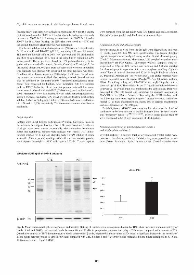
focusing (IEF). The strips were actively re-hydrated at 50V for 16h and the
proteins were focused at 300V for 2 h, after which the voltage was gradually
increased to 500V for 2 h. Focusing was continued at 1000V for 2 h and at
8000V for 18h. After focusing, the IEF strips were stored at �80�C until
the second dimension electrophoresis was performed.
For the second dimension electrophoresis, IPG strips were equilibrated
for 10min in 50mM Tris-HCl (pH 6.8) containing 6M urea, 1% (wt=v)
SDS (sodium dodecyl sulfate), 30% (v=v) glycerol, and 2% dithiotreitol,
and then re-equilibrated for 10min in the same buffer containing 2.5%
iodacetamide. The strips were placed on 10% polyacrilamide gels, to-
gether with standards (Fermentas, Ontario, Canada) at 20mA=gel 2. For
the second dimension, two gels from the same case were run in parallel.
One replicate was stained with silver and the other replicate was trans-
ferred to a nitrocellulose membrane (200mA=gel for 90min). For gel stain-
ing, a mass spectometry-modified silver staining method (Amersham) was
used as described by the manufacturer. Transferred nitrocellulose mem-
branes were processed for blotting. After incubation with 5% skimmed
milk in TBS-T buffer for 1 h at room temperature, nitrocellulose mem-
branes were incubated with anti-HNE (Calbiochem), used at dilution of 1,
1000. Membranes were also incubated with rabbit anti-phosphoglycerate
kinase 1 (Abgent, San Diego, CA, USA) or goat anti-fructose bisphosphate
aldolase A (Novus Biologicals, Littleton, USA) antibodies used at dilutions
of 1:150 and 1:10,000, respectively. The immunoreaction was visualized as
previously.
In-gel digestion
Proteins were in-gel digested with trypsin (Promega, Barcelona, Spain) in
the automatic Investigator ProGest robot of Genomic Solutions. Briefly, ex-
cised gel spots were washed sequentially with ammonium bicarbonate
buffer and acetonitrile. Proteins were reduced with 10mM DTT (dithio-
threitol) solution for 30min and alkylated with 100mM solution of iodine
acetamide. After sequential washings with buffer and acetonitrile, proteins
were digested overnight at 37�C with trypsin 0.27 nM. Tryptic peptides
were extracted from the gel matrix with 10% formic acid and acetonitrile.
The extracts were pooled and dried in a vacuum centrifuge.
Acquisition of MS and MS=MS spectra
Proteins manually excised from the 2D gels were digested and analyzed
by CapLC-nano-ESI-MS-MS mass spectrometry. The tryptic digested
peptide samples were analyzed using on-line liquid chromatography
(CapLC, Micromass-Waters, Manchester, UK) coupled to tandem mass
spectrometry (Q-TOF Global, Micromass-Waters). Samples were re-
suspended in 12 ml of 10% formic acid solution and 4 ml was injected
for chromatographic separation into a reverse-phase capillary C18 col-
umn (75 mm of internal diameter and 15 cm in length, PepMap column,
LC Packings, Amsterdam, The Netherlands). The eluted peptides were
ionized via coated nano-ES needles (PicoTipTM, New Objective, Woburn,
USA). A capillary voltage of 1800–2200V was applied together with a
cone voltage of 80V. The collision in the CID (collision-induced dissocia-
tion) was 25–35 eVand argon was employed as the collision gas. Data were
generated in PKL file format and submitted for database searching in
MASCOT server (Matrix Science, USA) using the NCBI database with
the following parameters: trypsin enzyme, 1 missed cleavage, carbamido-
methyl (C) as fixed modification and oxized (M) as variable modification,
and mass tolerance of 150–250ppm.
Probability-based MOWSE score was used to determine the level of
confidence in the identification of specific isoforms from the mass spectra.
This probability equals 10(-Mowse score=10). Mowse scores greater than 50
were considered to be of high confidence of identification.
Immunohistochemistry to phosphoglycerate kinase 1
and bisphosphate aldolase A
Cryostat sections 14 microns thick of cryoprotected frontal cortex were
processed free-floating with the EnVisionþ system peroxidase proce-
dure (Dako, Barcelona, Spain) in every case. Control samples were
Fig. 1. Mono-dimensional gel electrophoresis and Western blotting of frontal cortex homogenates blotted for HNE show increased immunoreactivity of
bands of 60 and 70 kDa and several bands between 40 and 50 kDa in progressive supranuclear palsy (PSP) when compared with controls (CTL).
Quantitative analysis of HNE-immunoreactive bands, corrected for b-actin, expressed as mean values � SD, reveal a significant increase in the intensity of
all the bands between 40 and 70 kDa in PSP cases compared with CTL. Student T test: � p<0.05. Cases represented in the figure correspond to 8, 15 and
10 (controls), and 1, 2 and 4 (PSP)
Glycolitic enzymes are targets of oxidation in aged human frontal cortex 61
81
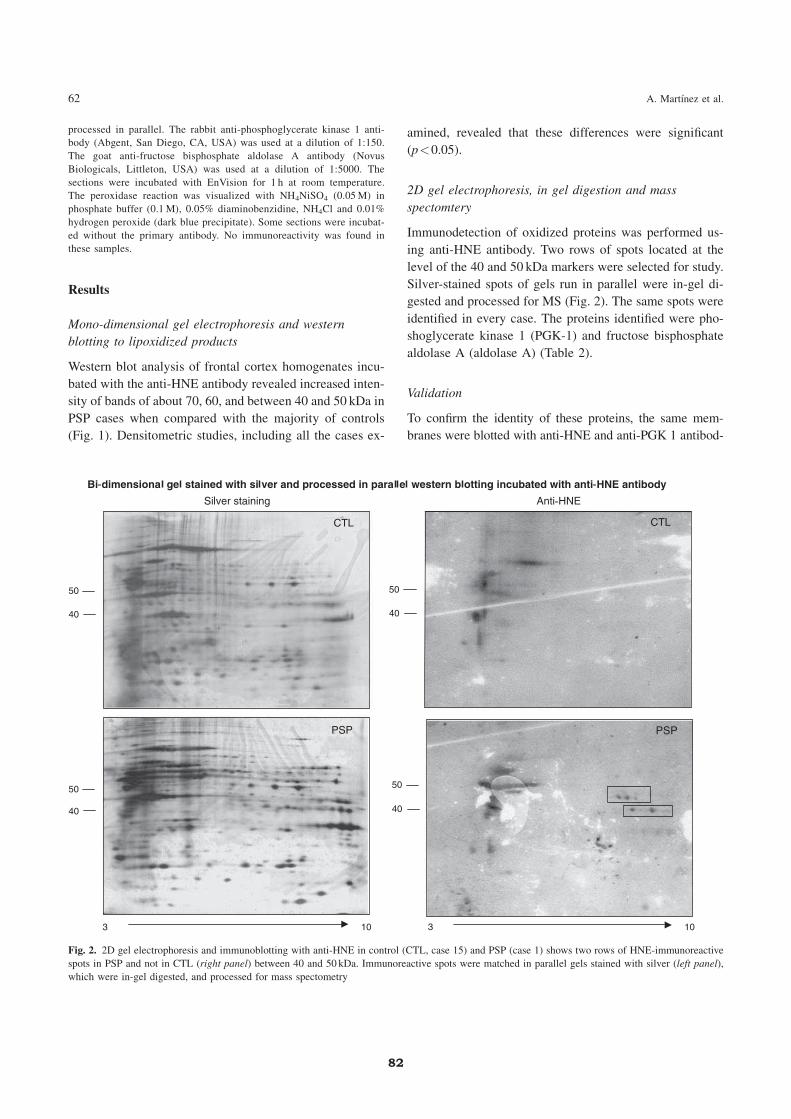
processed in parallel. The rabbit anti-phosphoglycerate kinase 1 anti-
body (Abgent, San Diego, CA, USA) was used at a dilution of 1:150.
The goat anti-fructose bisphosphate aldolase A antibody (Novus
Biologicals, Littleton, USA) was used at a dilution of 1:5000. The
sections were incubated with EnVision for 1 h at room temperature.
The peroxidase reaction was visualized with NH4NiSO4 (0.05M) in
phosphate buffer (0.1M), 0.05% diaminobenzidine, NH4Cl and 0.01%
hydrogen peroxide (dark blue precipitate). Some sections were incubat-
ed without the primary antibody. No immunoreactivity was found in
these samples.
Results
Mono-dimensional gel electrophoresis and western
blotting to lipoxidized products
Western blot analysis of frontal cortex homogenates incu-
bated with the anti-HNE antibody revealed increased inten-
sity of bands of about 70, 60, and between 40 and 50 kDa in
PSP cases when compared with the majority of controls
(Fig. 1). Densitometric studies, including all the cases ex-
amined, revealed that these differences were significant
(p<0.05).
2D gel electrophoresis, in gel digestion and mass
spectomtery
Immunodetection of oxidized proteins was performed us-
ing anti-HNE antibody. Two rows of spots located at the
level of the 40 and 50 kDa markers were selected for study.
Silver-stained spots of gels run in parallel were in-gel di-
gested and processed for MS (Fig. 2). The same spots were
identified in every case. The proteins identified were pho-
shoglycerate kinase 1 (PGK-1) and fructose bisphosphate
aldolase A (aldolase A) (Table 2).
Validation
To confirm the identity of these proteins, the same mem-
branes were blotted with anti-HNE and anti-PGK 1 antibod-
Fig. 2. 2D gel electrophoresis and immunoblotting with anti-HNE in control (CTL, case 15) and PSP (case 1) shows two rows of HNE-immunoreactive
spots in PSP and not in CTL (right panel) between 40 and 50 kDa. Immunoreactive spots were matched in parallel gels stained with silver (left panel),
which were in-gel digested, and processed for mass spectometry
62 A. Martınez et al.
82
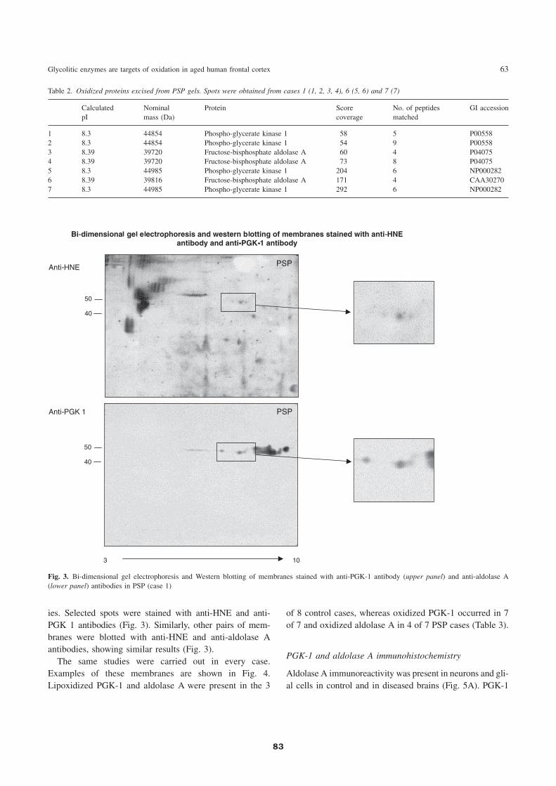
ies. Selected spots were stained with anti-HNE and anti-
PGK 1 antibodies (Fig. 3). Similarly, other pairs of mem-
branes were blotted with anti-HNE and anti-aldolase A
antibodies, showing similar results (Fig. 3).
The same studies were carried out in every case.
Examples of these membranes are shown in Fig. 4.
Lipoxidized PGK-1 and aldolase A were present in the 3
of 8 control cases, whereas oxidized PGK-1 occurred in 7
of 7 and oxidized aldolase A in 4 of 7 PSP cases (Table 3).
PGK-1 and aldolase A immunohistochemistry
Aldolase A immunoreactivity was present in neurons and gli-
al cells in control and in diseased brains (Fig. 5A). PGK-1
Table 2. Oxidized proteins excised from PSP gels. Spots were obtained from cases 1 (1, 2, 3, 4), 6 (5, 6) and 7 (7)
Calculated
pI
Nominal
mass (Da)
Protein Score
coverage
No. of peptides
matched
GI accession
1 8.3 44854 Phospho-glycerate kinase 1 58 5 P00558
2 8.3 44854 Phospho-glycerate kinase 1 54 9 P00558
3 8.39 39720 Fructose-bisphosphate aldolase A 60 4 P04075
4 8.39 39720 Fructose-bisphosphate aldolase A 73 8 P04075
5 8.3 44985 Phospho-glycerate kinase 1 204 6 NP000282
6 8.39 39816 Fructose-bisphosphate aldolase A 171 4 CAA30270
7 8.3 44985 Phospho-glycerate kinase 1 292 6 NP000282
Fig. 3. Bi-dimensional gel electrophoresis and Western blotting of membranes stained with anti-PGK-1 antibody (upper panel) and anti-aldolase A
(lower panel) antibodies in PSP (case 1)
Glycolitic enzymes are targets of oxidation in aged human frontal cortex 63
83
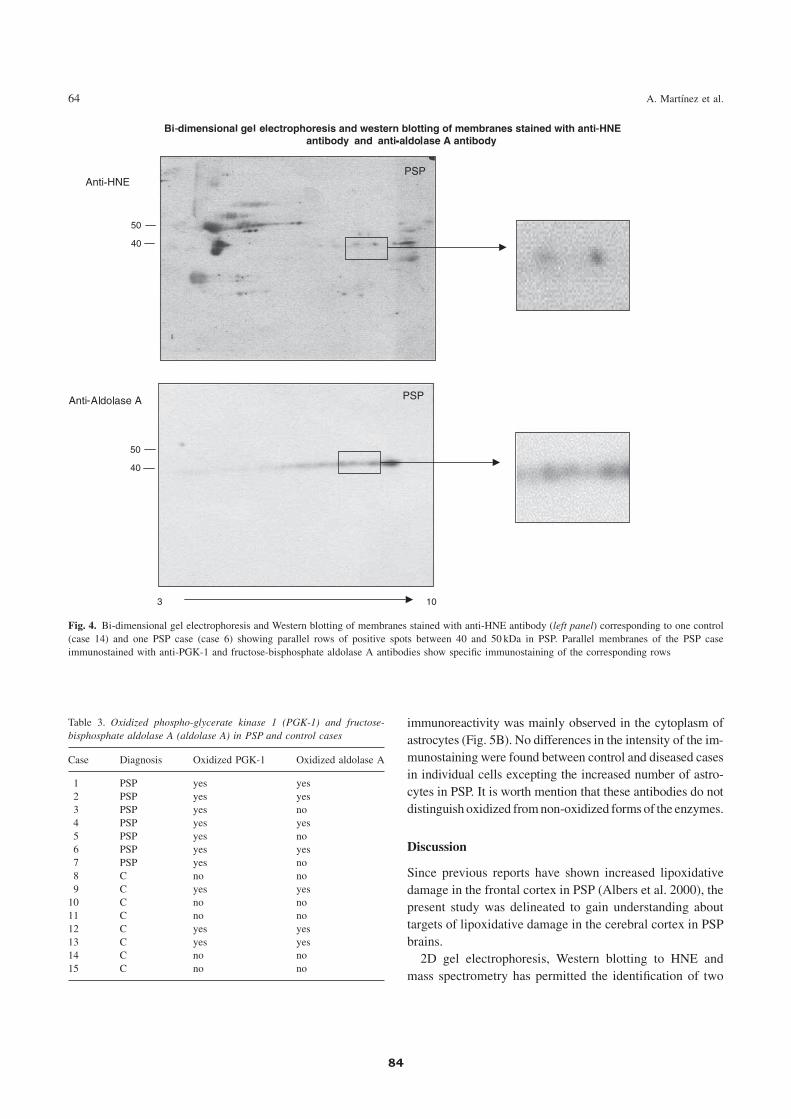
immunoreactivity was mainly observed in the cytoplasm of
astrocytes (Fig. 5B). No differences in the intensity of the im-
munostaining were found between control and diseased cases
in individual cells excepting the increased number of astro-
cytes in PSP. It is worth mention that these antibodies do not
distinguish oxidized fromnon-oxidized forms of the enzymes.
Discussion
Since previous reports have shown increased lipoxidative
damage in the frontal cortex in PSP (Albers et al. 2000), the
present study was delineated to gain understanding about
targets of lipoxidative damage in the cerebral cortex in PSP
brains.
2D gel electrophoresis, Western blotting to HNE and
mass spectrometry has permitted the identification of two
Table 3. Oxidized phospho-glycerate kinase 1 (PGK-1) and fructose-
bisphosphate aldolase A (aldolase A) in PSP and control cases
Case Diagnosis Oxidized PGK-1 Oxidized aldolase A
1 PSP yes yes
2 PSP yes yes
3 PSP yes no
4 PSP yes yes
5 PSP yes no
6 PSP yes yes
7 PSP yes no
8 C no no
9 C yes yes
10 C no no
11 C no no
12 C yes yes
13 C yes yes
14 C no no
15 C no no
Fig. 4. Bi-dimensional gel electrophoresis and Western blotting of membranes stained with anti-HNE antibody (left panel) corresponding to one control
(case 14) and one PSP case (case 6) showing parallel rows of positive spots between 40 and 50 kDa in PSP. Parallel membranes of the PSP case
immunostained with anti-PGK-1 and fructose-bisphosphate aldolase A antibodies show specific immunostaining of the corresponding rows
64 A. Martınez et al.
84

proteins that are oxidized in the frontal cortex in PSP cases:
fructose bisphosphate aldolase A (aldolase A) and phos-
phoglycerate kinase 1 (PGK-1). Both enzymes are impli-
cated in glycolysis. Fructose 1,6-bisphosphate aldolase
A converts fructose-1,6-bisphosphate into glyceraldehyde-
3-phosphate and dihidroxyacetone phosphate. PGK-1
catalyzes the conversion of 1,3-diphosphoglycerate to 3-
phosphoglycerate, leading to the production of the first
ATP in glycolysis. Loss of function of PGK-1 may impair
glucose metabolism and result in the accumulation of gly-
colytic intermediates.
PGK-1 has been described as being oxidized in the fron-
tal cortex in Alzheimer’s disease (AD) (Castegna et al.
2002; Korolainen et al. 2006) and in transgenic Tg2576
mice with Alzheimer plaque pathology (Bigl et al. 2003).
Several proteins related to glycogenesis and glycolysis
have also been reported to be oxidized in experimental
models of AD following injection of amyloid b peptide
(1–42) into rat brain (Boyd-Kimball et al. 2005) and in
rat primary neural cells following amyloid b1–42-inducedoxidative damage (Sultana et al. 2006). Therefore, oxida-
tion of proteins linked with the glycolysis may account for
impaired energy metabolism in the AD brain.
No antecedents of HNE-modified glycolytic enzymes
have been reported so far in PSP. Yet previous studies using
(18F)-2-fluoro-2-deoxy-D-glucose (FDG) positron emis-
sion tomography (PET) have shown a significant alteration
of cerebral glucose metabolism in the frontal cortex in PSP
cases (Karbe et al. 1992). The present findings suggest that
oxidation of aldolase A and PGK-1 may also account for
impaired energy metabolism in PSP, and they provide a
biochemical substrate for impaired glucose metabolism in
PSP. Since aldolase A is present in neurons and astrocytes,
and PGK-1 mainly in astrocytes, the present findings sup-
port neurons and astrocytes as targets of oxidative damage
in PSP.
Fig. 5. Immunohistochemistry to aldolase A (A) and PGK-1 (B) in the PSP frontal cortex (case 1). Aldolase A immunoreactivity is observed in the
cytoplasm of neurons (arrows) and astrocytes (arrowheads). PGK-1 immunoreactivity is mainly found in the cytoplasm of astrocytes (arrowheads).
Cryostat section processed free-floating with no counterstaining. Bar¼ 25 mm
Glycolitic enzymes are targets of oxidation in aged human frontal cortex 65
85

Although PGK-1 has been found to be oxidized in 7 of 7
cases and aldolase A in 4 of 7 PSP cases, oxidation of these
proteins has also been observed in certain control cases.
Thus PGK-1 and aldolase A were oxidized in the frontal
cortex of 3 of 8 control cases. No apparent clinical and
neuropathological differences were noticed between these
three cases and the remaining five controls with no evi-
dence of PGK-1 and aldolase A oxidation, excepting the
young age in a single case.
In summary, the present study has shown that aldolase A
and PGK-1 as targets of lipoxidation in the aged human
cerebral cortex, and that increased lipoxidation of aldolase
A and, particularly, of PGK-1 occurs in the frontal cortex in
PSP. The present findings together with previous observa-
tions in AD support the idea that increased oxidation of
proteins linked with glycolysis is a common feature in
advanced stages of certain neurodegenerative diseases in-
cluding AD and PSP.
Acknowledgements
This work was funded by grants from the Spanish Ministry of Health,
Instituto de Salud Carlos III (PI040184 and PI05=1570), and supported
by the European Commission under the Sixth Framework Programme
(BrainNet Europe II, LSHM-CT-2004-503039). We thank T. Yohannan
for editorial help.
References
Albers DS, Augood SJ, Martin DM, Standaert DG, Vonsattel JP, Beal MF
(1999) Evidence for oxidative stress in the subthalamic nucleus in
progressive supranuclear palsy. J Neurochem 73: 881–884
Albers DS, Augood SJ, Park LC, Browne SE,Martin DM, Adamson J, Hutton
M, Standaert DG, Vonsattel JP, Gibson GE, Beal MF (2000) Frontal
lobe dysfunction in progressive supranuclear palsy: evidence for oxida-
tive stress and mitochondrial impairment. J Neurochem 74: 878–881
Bigl ML, Apelt J, Eschrich K, Schliebs R (2003) Cortical glucose metabo-
lism is altered in aged transgenic Tg2576 mice that demonstrate
Alzheimer plaque pathology. J Neural Transm 110: 77–94
Boyd-Kimball D, Sultana R, Poon HF, Lynn BC, Casamenti F, Pepeu G,
Klein JB, Butterfield DA (2005) Proteomic identification of proteins
specifically oxidized by intracerebral injection of amyloid peptide (1–
42) into rat brain: implications for Alzheimer’s disease. Neuroscience
132: 313–324
Buee L, Bussiere T, Buee-Scherrer V, Delacourte A, Hof PR (2000)
Tau protein isoforms, phosphorylation and role in neurodegenerative
disorders. Brain Res Rev 33: 95–130
Cantuti-Castelvetri I, Keller-McGandy CE, Albers DS, Beal MF, Vonsattel
JP, Standaert DG, Augood SJ (2002) Expression and activity of an-
tioxidants in the brain in progressive supranuclear palsy. Brain Res
930: 170–181
Castegna A, AksenovM, Thonboonkerd V, Klein JB, PierceWM, Booze R,
Markesbery W, Butterfield DA (2002) Proteomic identification of
oxidatively modified proteins in Alzheimer’s disease brain. Part II:
Dihydropyrimidinase-related protein 2, a-enolase and heat shock
cognate 71. J Neurochem 82: 1524–1532
Delacourte A, Buee L (2000) Tau pathology: a marker of neurodegenerative
disorders. Curr Opin Neurol 13: 371–376
Dickson DW (2001) Progressive supranuclear palsy and corticobasal
degeneration. In: Hof PR, Mobbs ChV (eds) Functional neurobiology
of aging. Academic Press, San Diego, pp 155–171
Hauw JJ, Agid Y (2003) Progressive supranuclear palsy (PSP) or Steele-
Richardson-Olszewski disease. In: Dickson D (ed) Neurodegeneration:
the molecular pathology of dementia and movement disorders. ISN
Neuropath Press, Basel, pp 103–114
Ikeda K, Akiyama H, Arai T, Nishimura T (1998) Glial tau pathology in
neurodegenerative diseases: their nature and comparison with neuronal
tangles. Neurobiol Aging 19: S85–S91
Karbe H, Grond M, Huber M, Herholz K, Kessler J, Heiss WD (1992)
Subcortical damage and cortical dysfunction in progressive supra-
nuclear palsy demonstrated by positron emission tomography. J Neurol
239: 98–102
Komori T (1999) Tau-positive glial inclusions in progressive supranuclear
palsy, corticobasal degeneration and Pick’s disease. Brain Pathol 9:
663–679
Korolainen MA, Goldsteins G, Nyman TA, Alafuzoff I, Koistinaho J,
Pirttila T (2006) Oxidative modification of proteins in the fron-
tal cortex of Alzheimer’s disease brain. Neurobiol Aging 27:
42–53
Lee WM, Goedert M, Trojanowski JQ (2001) Neurodegenerative tau-
opathies. Annu Rev Neurosci 24: 1121–1159
Litvan I (2004) Update on progressive supranuclear palsy. Curr Neurol
Neurosci Rep 4: 296–302
Litvan I, Hauw JJ, Bartko JJ, Lantos PL, Daniel SE, Horoupian DS,
McKee A, Dickson D, Bancher C, Tabaton M, Jellinger K, Anderson
DW (1996) Validity and reliability of the preliminary NINDS neuro-
pathologic criteria for progressive supranuclear palsy and related
disorders. J Neuropathol Exp Neurol 55: 97–105
Morris HR, Gibb G, Katzenschlager R, Wood NW, Hanger DP, Strand C,
Lashley T, Daniel SE, Lees AJ, Anderton BH, Revesz T (2002)
Pathological, clinical and genetic heterogeneity in progressive supra-
nuclear palsy. Brain 125: 969–975
Odetti P, Garibaldi S, Norese R, Angelini G, Marinelli L, Valentini S,
Menini S, Traverso N, Zaccheo D, Siedlak S, Perry G, Smith MA,
Tabaton M (2000) Lipoperoxidation is selectively involved in progres-
sive supranuclear palsy. J Neuropathol Exp Neurol 59: 393–397
Puig B, Rey MJ, Ferrer I (2005) Individual and regional variations of
phospho-tau species in progressive supranuclear palsy. Acta Neuro-
pathol 110: 261–268
Rampello l, Butt�aa V, Raffaele R, Vecchio I, Battaglia G, Cormaci G,
Alvano A (2005) Progressive supranuclear palsy: a systematic review.
Neurobiol Dis 20: 179–186
Scaravilli T, Tolosa E, Ferrer I (2005) Progressive supranuclear palsy and
corticobasal degeneration: lumping versus splitting. Mov Disord 20
(Suppl 12): S21–S28
Spillantini MG, Goedert M (1998) Tau protein pathology in neurodegener-
ative diseases. Trends Neurosci 21: 428-433
Sultana R, Newman SF, Abdul HM, Cai J, Pierce WM, Klein JB, Merchant
M, Butterfield DA (2006) Protective effect of D609 against amyloid-
beta1–42-induced oxidative modifications on neuronal proteins: redox
proteomics studies. J Neurosci Res 84: 409–417
Tawana K, Ramsden DB (2001) Progressive supranuclear palsy. Mol Pathol
54: 427–434
66 A. Martınez et al.: Glycolitic enzymes are targets of oxidation in aged human frontal cortex
86
V.RESULTATS
3. Type-Dependent Oxidative Damage in Frontotemporal Lobar Degeneration: Cortical Astrocytes Are Targets of Oxidative Damage
Anna Martínez, Margarita Carmona, Manuel Portero-Otin, Alba Naudí, Reinald
Pamplona, Isidre Ferrer.
J Neuropathol Exp Neurol. 2008 Dec;67(12):1122-36. Erratum in: J Neuropathol
Exp Neurol. 2009 Jan;68(1):112.
En el grup de la degeneració frontotemporal lobar (DFTL) s’engloben diferents
patologies com la DFTL amb la mutació P301L en el gen de la tau (DFTL-tau), la DFTL
amb inclusions negatives per la tau però immunoreactives per la ubiqüitina (DFTL-U) i
la DFTL associada a l’esclerosi lateral amiotròfica (DFTL-ELA). La lesió oxidativa i les
repostes a l’estrès són trets comuns de diferents malalties neurodegeneratives però,
pel que fa a les malalties esmentades, no hi han gaires treballs que descriguin còm
afecta l’estrès oxidatiu a les proteïnes.
L’anàlisi per gels en electroforesi i western blot, van mostrar un increment de
4-hidroxi-2-nonenal (HNE) en l’escorça frontal de 6 de 6 casos de DFTL-tau, en 3 dels
10 casos de DFTL-U i en 2 de les 3 mostres de DFTL-ELA. L’augment del dany proteïc
derivat de la lipoxidació es va veure associat amb una alteració de les membranes
insaturades i del perfil dels àcids grassos en les mostres de DFTL-tau i DFTL-ELA.
Tots els casos de DFTL-tau, i la majoria que presentaven un increment en la
imunoreactivitat de les bandes per western blot amb l’HNE, mostraven una important
gliosi. Aquesta gliosi es va determinar per la immunohistoquímica de la glial fibrillary
acidic protein (GFAP) i per l’increment d’aquesta proteïna per western blot. Gràcies a
les tècniques dels gels bidimensionals, western blot i espectometria de masses es va
identificar la GFAP com la proteïna diana majoritàriament lipoxidada. La microscopia
confocal va mostrar colocalització de l’HNE amb la GFAP en els astròcits corticals, la
superòxid dismutasa 1 (SOD-1) en els astròcits i la superòxid dismutasa 2 (SOD-2) en
els astròcits i les neurones de tots els grups de DFTL.
Per tant, en la DFTL hi ha una variabilitat de la lesió oxidativa depenent de la
malaltia, trobant-se més accentuada en la DFTL-tau. Els astròcits són dianes de dany
lipoxidatiu sent elements importants en situacions de resposta a l’estrès oxidatiu en la
DFTL.
87


Copyright @ 200 by the American Association of Neuropathologists, Inc. Unauthorized reproduction of this article is prohibited.8
ORIGINAL ARTICLE
Type-Dependent Oxidative Damage in FrontotemporalLobar Degeneration: Cortical Astrocytes Are Targets
of Oxidative Damage
Anna Martınez, Margarita Carmona, Manuel Portero-Otin, PhD, Alba Naudı, PhD,Reinald Pamplona, PhD, and Isidre Ferrer, MD, PhD
AbstractOxidative injury and stress responses are common features of
many neurodegenerative diseases. To assess oxidative stress re-sponses in frontotemporal lobar degeneration (FTLD), we identifiedincreased 4-hydroxynonenal (HNE) adducts using gel electropho-resis and Western blotting in frontal cortex samples in 6 of 6 cases ofFTLD with the P301L mutation in the tau gene (FTLD-tau), in 3 of10 cases with tau-negative ubiquitin-immunoreactive inclusions, andin 2 of 3 cases associated with motor neuron disease. Selectivelyincreased lipoxidation-derived protein damage associated with al-tered membrane unsaturation and fatty acid profiles was verified bymass spectrometry in FTLD-tau and FTLD associated with motorneuron disease. All FTLD-tau and most cases with increased HNE-positive bands had marked astrocytosis as determined by glialfibrillary acidic protein (GFAP) immunohistochemistry andincreased GFAP expression on Western blotting; 2 FTLD cases withtau-negative ubiquitin-immunoreactive inclusions and with increasedGFAP expression did not have increased HNE adducts. Bidimen-sional gel electrophoresis, Western blotting, in-gel digestion, andmass spectrometry identified GFAP as a major target of lipoxidationin all positive cases; confocal microscopy revealed colocalization ofHNE and GFAP in cortical astrocytes, superoxide dismutase 1 inastrocytes, and superoxide dismutase 2 in astrocytes and neurons inall FTLD types. Thus, in FTLD, there is variable disease-dependent
oxidative damage that is prominent in FTLD-tau, astrocytes aretargets of oxidative damage, and GFAP is a target of lipoxidation.Astrocytes are, therefore, crucial elements of oxidative stressresponses in FTLD.
Key Words: Astrocytes, Frontotemporal lobar degeneration,Hydroxynonenal, Malondialdehyde-lysine, Membrane unsaturation,Oxidative damage, Superoxide dismutase.
INTRODUCTIONFrontotemporal lobar degeneration (FTLD) designates
a group of diseases having common progressive changesof personality and behavior, often with language impair-ment (i.e. frontotemporal dementia) associated with bilateralatrophy of the frontal and anterior temporal lobes. A prom-inent neuropathologic feature of FTLD is loss of neurons andmicrovacuolation that is mainly in the upper cortical layersand is accompanied by astrocytic gliosis. In addition to thefrontal and temporal neocortex, the anterior cingulate gyrus,anterior insular cortex, hippocampus, entorhinal cortex,amygdala, and striatum (mainly the head of the caudate andputamen) are commonly affected. Loss of neurons andneuromelanin in macrophages, or free in the neuropil, canbe present in the substantia nigra, even in cases with noapparent parkinsonian features (1Y7). The remaining neu-rons in the upper cortical layers have reduced numbers ofdendrites and dendritic spines, and there is a loss of presyn-aptic markers. Glutamatergic projection neurons and calbindin-immunoreactive local-circuit neurons are equally affected.Because the upper cortical layers are the source of shortcortico-cortical connections, involvement of these areas isdramatic evidence of the devastating effects of FTLD on cor-tical connectivity (8Y10).
The causes of frontotemporal dementia are varied. Threemain groups of FTLD have been categorized, namely 1)tauopathies, including familial FTLD (and parkinsonism)linked to chromosome 17 because of mutations in the taugene (MAPT 17q21-q22; FTLD-tau) (11Y17); 2) FTLD withtau-negative ubiquitin-positive inclusions (FTLD-U); and 3)FTLD with no distinctive neuropathologic features. Thesecond group has been subdivided into different types de-pending on the localization, distribution, and predominanceof ubiquitin inclusions in the nucleus, cytoplasm, and/orneuropil (7, 18Y20). Most FTLD-U cases seem sporadic, but
J Neuropathol Exp Neurol � Volume 67, Number 12, December 20081122
J Neuropathol Exp NeurolCopyright � 2008 by the American Association of Neuropathologists, Inc.
Vol. 67, No. 12December 2008pp. 1122Y1136
From the Institut de Neuropatologia, Servei Anatomia Patologica, IDIBELL-Hospital Universitari de Bellvitge; Universitat de Barcelona; Hospitaletde Llobregat; CIBERNED (AM, MC, IF); and Departament de Medi-cina Experimental, Universitat de Lleida-IRBLLEIDA, Lleida, Spain(MP-O, AN, RP).
Send correspondence and reprint requests to: Isidre Ferrer, MD, PhD, Institutde Neuropatologia, Servei Anatomia Patologica, IDIBELL-HospitalUniversitari de Bellvitge, carrer Feixa Llarga sn, 08907 Hospitalet deLlobregat, Spain; E-mail: [email protected]
This work was supported by Grants PI05/1570 and PI05/2214 from theSpanish Ministry of Health, Instituto de Salud Carlos III, and by GrantLSHM-CT-2004-503039 from the European Commission under the SixthFramework Program (BrainNet Europe II) to Isidre Ferrer; by GrantBFU2006-14495/BFI from the Spanish Ministry of Education andScience, by Grant RD06/0013/0012 from the Spanish Ministry ofHealth (ISCIII, Red de Envejecimiento y Fragilidad), and by Grant2005SGR00101 from the Autonomous Government of Catalonia toReinald Pamplona; and by the Spanish Ministry of Health (GrantsPI04/0355, PI05/2214, and PI05/2241), Spanish Ministry of Educationand Science (Grant AGL2006-12433), BLa Caixa[ Foundation, andCOST B-35 Action to Manuel Portero-Otin.
89
Copyright @ 200 by the American Association of Neuropathologists, Inc. Unauthorized reproduction of this article is prohibited.8
mutations in at least 3 genes may cause FTLD-U. They arethe progranulin gene (PGRN) located 17q21-q22, chargedmultivesicular body protein 2B (CHMP2B) in 3p13-3p12,and valosin-containing protein gene (VCP) in 9q21-q22(21Y28). In addition, FTLD with tau-negative ubiquitin-positive inclusions occurs in association with motor neurondisease and amyotrophic lateral sclerosis (FTLD-MND);linkage to chromosome 9 has been demonstrated in some ofthese cases (29, 30). Frontotemporal lobar degeneration withtau-negative ubiquitin-positive neuronal inclusions (FTLD-U)is more common (50%) than FTLD with tau pathology (FTLD-tau) (31). The amount of new information that has accu-mulated in recent years has made it increasingly difficult toconsider the third category (i.e. FTLD with no distinctive neu-ropathologic features) as a separate entity (32).
Oxidative stress is common in degenerative diseasesof the nervous system, including Alzheimer disease (33Y40).Oxidative damage has also been reported in several tauopa-thies (41Y43), but very little is known about oxidative stressin FTLD. Iron, in a redox-active state, is, however, increasedin cases of frontotemporal dementia (44).
In the present study, we investigate the presence ofoxidative stress and its impact on common predominantproteins in FTLD-tau, FTLD-U, and FTLD-MND. For thispurpose, the combination of single and double labeling im-munofluorescence and confocal microscopy, monodimen-sional and bidimensional gel electrophoresis, Western blotanalysis, in-gel digestion, and mass-spectrometry was used forthe first time to analyze the frontal cortex in 19 FTLD cases(FTLD-tau, n = 6; FTLD-U, n = 10; FTLD-MND, n = 3) andcompared with 10 age-matched controls. Subsequently, gaschromatography/mass spectrometry (GC/MS) analyses wereperformed to further dissect the molecular oxidative path-ways by detecting and quantifying specific oxidation, gly-coxidation, and lipoxidation-derived protein damage markers.Finally, to assess the role of structural components that canact as substrate targets in determining the cellular vulner-ability, membrane fatty acid profiles and their degree of un-saturation were also measured. Because the first part of thestudy showed marked individual variations among FTLD-Ucases, further attempts to delineate oxidative damage and lipidprofiles were examined only in FTLD-U and FTLD-MNDcases and controls.
MATERIALS AND METHODS
CasesPatients were clinically categorized as having FTLD
(16 cases) or FTLD + MND (3 cases). Clinical features ofthese patients are detailed elsewhere (45). Age-matched con-trols (n = 10) had not had neurological or metabolic diseases.
The postmortem delay between death and tissue pro-cessing was between 3 and 15 hours; the bodies weremaintained at 4-C. All cases were processed in the sameway following the same sampling and staining protocols.At autopsy, half of each brain was fixed in 10% bufferedformalin, whereas the other half was cut in 1-cm-thick coronalsections, frozen on dry ice, and stored at j80-C until use.
The neuropathologic study permitted the identificationof 6 FTLD-tau (Cases 4Y6, 27Y29), 10 FTLD-U (Cases 7Y12,23Y26), and 3 FTLD-MND (Cases 13Y15). Genetic studiesdemonstrated the P301L mutation in the 6 FTLD-tau cases,and the c908delC and P357HfsX3 mutations in the pro-granulin gene in 2 FTLD-U cases (Cases 10 and 26, respec-tively). The neuropathologic examination was normal in the10 control cases, including lack of neurofibrillary tangles andamyloid deposits in any region, except a few diffuse plaquesin the temporal and orbital cortex in two. The cases are sum-marized in Table 1.
Double Labeling Immunofluorescence andConfocal Microscopy
Dewaxed 5-Km-thick sections were stained with a sat-urated solution of Sudan black B (Merck, Barcelona, Spain)for 10 minutes to block the autofluorescence of lipofuscingranules present in nerve cell bodies; they were then rinsedin 70% ethanol and washed in distilled water. The sections
TABLE 1. Case Summaries*
CaseAge,years Sex
PostmortemDelay Diagnosis
1 65 F 4 hours CTL
2 59 M 7 hours CTL
3 58 F 4 hours CTL
4 52 M 14 hours FTLD-tau (P301L)
5 58 M 13 hours FTLD-tau (P301L)
6 58 M 10 hours FTLD-tau (P301L)
7 64 M 12 hours FTLD-U
8 61 M 8 hours FTLD-U
9 74 F 5 hours FTLD-U
10 74 F 15 hours FTD-U-PGRN (c908delC)
11 77 F 8 hours FTLD-U
12 69 F 10 hours FTLD-U
13 68 M 3 hours FTLD-MND
14 70 F 13 hours FTLD-MND
15 69 F 6 hours FTLD-MND
16 75 F 3 hours CTL
17 78 F 4 hours CTL
18 78 M 2 hours CTL
19 85 M 6 hours CTL
20 73 F 7 hours CTL
21 67 M 5 hours CTL
22 62 M 3 hours CTL
23 74 F 15 hours FTLD-U
24 72 M 6 hours FTLD-U
25 62 F 4 hours FTLD-U
26 51 M 8 hours FTD-U-PGRN (P357HfsX3)
27 62 M 5 hours FTLD-tau (P301L)
28 73 M 10 hours FTLD-tau (P301L)
29 78 M 7 hours FTLD-tau (P301L)
Mutations in MAPT (tau gene) and PGRN (progranulin gene) are indicated.*Diagnoses are based on clinical, neuropathologic, and genetic data.CTL, control; F, female; FTLD-MND, frontotemporal lobar dementia with tau-
negative ubiquitin-positive inclusions that occurs in association with motor neurondisease and amyotrophic lateral sclerosis; FTLD-tau, frontotemporal lobar dementiawith tau pathology; FTLD-U, frontotemporal lobar degeneration with tau-negativeubiquitin-positive neuronal inclusions; M, male.
J Neuropathol Exp Neurol � Volume 67, Number 12, December 2008 Oxidative Damage in FTLD
� 2008 American Association of Neuropathologists, Inc. 1123
90
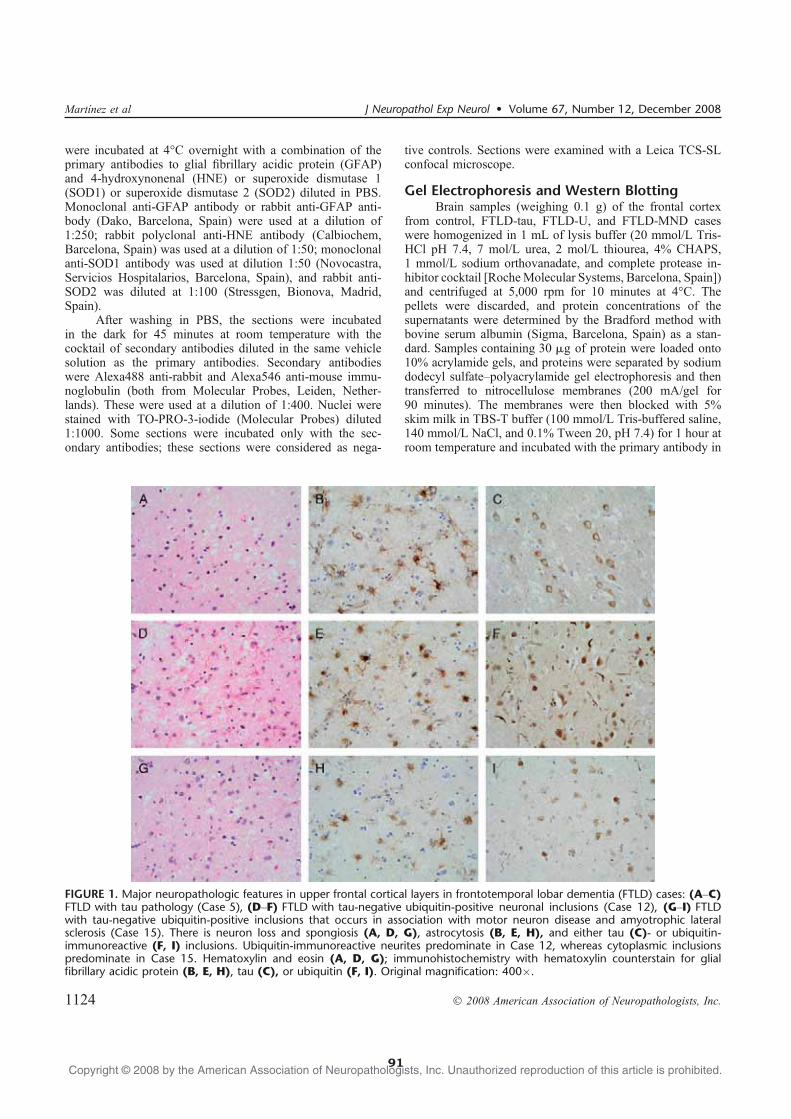
Copyright @ 200 by the American Association of Neuropathologists, Inc. Unauthorized reproduction of this article is prohibited.8
were incubated at 4-C overnight with a combination of theprimary antibodies to glial fibrillary acidic protein (GFAP)and 4-hydroxynonenal (HNE) or superoxide dismutase 1(SOD1) or superoxide dismutase 2 (SOD2) diluted in PBS.Monoclonal anti-GFAP antibody or rabbit anti-GFAP anti-body (Dako, Barcelona, Spain) were used at a dilution of1:250; rabbit polyclonal anti-HNE antibody (Calbiochem,Barcelona, Spain) was used at a dilution of 1:50; monoclonalanti-SOD1 antibody was used at dilution 1:50 (Novocastra,Servicios Hospitalarios, Barcelona, Spain), and rabbit anti-SOD2 was diluted at 1:100 (Stressgen, Bionova, Madrid,Spain).
After washing in PBS, the sections were incubatedin the dark for 45 minutes at room temperature with thecocktail of secondary antibodies diluted in the same vehiclesolution as the primary antibodies. Secondary antibodieswere Alexa488 anti-rabbit and Alexa546 anti-mouse immu-noglobulin (both from Molecular Probes, Leiden, Nether-lands). These were used at a dilution of 1:400. Nuclei werestained with TO-PRO-3-iodide (Molecular Probes) diluted1:1000. Some sections were incubated only with the sec-ondary antibodies; these sections were considered as nega-
tive controls. Sections were examined with a Leica TCS-SLconfocal microscope.
Gel Electrophoresis and Western BlottingBrain samples (weighing 0.1 g) of the frontal cortex
from control, FTLD-tau, FTLD-U, and FTLD-MND caseswere homogenized in 1 mL of lysis buffer (20 mmol/L Tris-HCl pH 7.4, 7 mol/L urea, 2 mol/L thiourea, 4% CHAPS,1 mmol/L sodium orthovanadate, and complete protease in-hibitor cocktail [RocheMolecular Systems, Barcelona, Spain])and centrifuged at 5,000 rpm for 10 minutes at 4-C. Thepellets were discarded, and protein concentrations of thesupernatants were determined by the Bradford method withbovine serum albumin (Sigma, Barcelona, Spain) as a stan-dard. Samples containing 30 Kg of protein were loaded onto10% acrylamide gels, and proteins were separated by sodiumdodecyl sulfateYpolyacrylamide gel electrophoresis and thentransferred to nitrocellulose membranes (200 mA/gel for90 minutes). The membranes were then blocked with 5%skim milk in TBS-T buffer (100 mmol/L Tris-buffered saline,140 mmol/L NaCl, and 0.1% Tween 20, pH 7.4) for 1 hour atroom temperature and incubated with the primary antibody in
FIGURE 1. Major neuropathologic features in upper frontal cortical layers in frontotemporal lobar dementia (FTLD) cases: (AYC)FTLD with tau pathology (Case 5), (DYF) FTLD with tau-negative ubiquitin-positive neuronal inclusions (Case 12), (GYI) FTLDwith tau-negative ubiquitin-positive inclusions that occurs in association with motor neuron disease and amyotrophic lateralsclerosis (Case 15). There is neuron loss and spongiosis (A, D, G), astrocytosis (B, E, H), and either tau (C)- or ubiquitin-immunoreactive (F, I) inclusions. Ubiquitin-immunoreactive neurites predominate in Case 12, whereas cytoplasmic inclusionspredominate in Case 15. Hematoxylin and eosin (A, D, G); immunohistochemistry with hematoxylin counterstain for glialfibrillary acidic protein (B, E, H), tau (C), or ubiquitin (F, I). Original magnification: 400�.
Martınez et al J Neuropathol Exp Neurol � Volume 67, Number 12, December 2008
� 2008 American Association of Neuropathologists, Inc.1124
91

Copyright @ 200 by the American Association of Neuropathologists, Inc. Unauthorized reproduction of this article is prohibited.8
TBS-TY3% bovine serum albumin at 4-C overnight. After theincubation and washes with TBS, the membranes wereblocked with 5% skim milk in TBS-T buffer and thenincubated with rabbit polyclonal anti-HNE antibody (Calbio-chem) at a dilution of 1:1000, rabbit polyclonal anti-catalase(Calbiochem) diluted 1:6000, rabbit polyclonal anti-GFAP(Dako) diluted 1:500, or mouse monoclonal anti-GFAPantibody (Dako) used at a dilution of 1:10000. Later, themembranes were washed 3 times in TBS-T for 5 minutes andthen incubated for 45 minutes at room temperature, with thecorresponding secondary antibody labeled with horseradishperoxidase (Dako) at a dilution of 1:1000 and washed inTBS-T for 30 minutes. The immunoreactivity was detectedwith the chemiluminescence ECL method (Amersham,Barcelona, Spain). The monoclonal antibody to A-actin(Sigma) was used at a dilution of 1:10000 as a control ofprotein loading.
Two-Dimensional Gel ElectrophoresisIn the first dimension electrophoresis, samples were
applied onto a 7-cm 4 to 7 pH nonlinear gradient ReadyStripimmobilized pH gradient strip (Bio-Rad, Barcelona, Spain)for isoelectric focusing. The strips were actively rehydratedat 50 V for 12 hours, and the proteins were focused at 500 Vfor 1 hour, after which the voltage was gradually increased to1000 V for 1 hour. Focusing was increased to 8,000 V for1 hour more, and the voltage was continued at 8,000 V for10 hours. After focusing, the isoelectric focusing strips werestored at j80-C until the second dimension electrophoresiswas performed.
For the second dimension electrophoresis, immobilizedpH gradient strips were equilibrated for 10 minutes in50 mmol/L Tris-HCl (pH 6.8) containing 6 mol/L urea,1% (wt/vol) sodium dodecyl sulfate, 30% (vol/vol) glycerol,and 2% dithiothreitol, and next re-equilibrated for 10 minutesin the same buffer containing 2.5% iodoacetamide. The stripswere placed on 10% polyacrylamide gels together with stan-dards (Fermentas, Ontario, Canada). For the second dimen-sion, 2 gels from the same case were run in parallel. Onereplicate was stained with Coomassie blue (Bio-Rad), and theother replicate was transferred to a nitrocellulose membrane(200 mA/gel for 90 minutes). Transferred nitrocellulose mem-branes were processed for blotting. After incubation with5% skim milk in TBS-T buffer for 1 hour at room tempera-ture, nitrocellulose membranes were incubated with rabbitanti-HNE (Calbiochem), previously incubated with NaBH410 mmol/L in TBS, used at dilution of 1:1000. Membraneswere also incubated with monoclonal anti-GFAP antibody(Dako) used at dilution of 1:10000. The immunoreactionswere visualized as previously described (39).
In-Gel DigestionProteins were in-gel digested with trypsin (Promega,
Barcelona, Spain) in the automatic Investigator ProGest robotof Genomic Solutions (Isogen Life Science, Barcelona,Spain). Briefly, excised gel spots were washed sequentiallywith ammonium bicarbonate buffer and acetonitrile. Proteinswere reduced with 10 mmol/L dithiothreitol solution during
30 minutes and alkylated with 100 mmol/L solution of io-dine acetamide. After sequential washings with buffer andacetonitrile, proteins were digested overnight at 37-C withtrypsin 0.27 nmol/L. Tryptic peptides were extracted from thegel matrix with 10% formic acid and acetonitrile. The ex-tracts were pooled and dried in a vacuum centrifuge.
Acquisition of MS and MS/MS SpectraProteins manually excised from the 2-dimensional (2-D)
gels were digested and analyzed by CapLC-nano-ESI-MS-MS. The tryptic digested peptide samples were analyzedusing an online liquid chromatography (CapLC, Micromass-Waters, Manchester, United Kingdom) coupled to a tandemMS (Q-TOF Global, Micromass-Waters) (43). Data were
TABLE 2. Summary of Neuropathologic andBiochemical ChangesCase Diagnosis Neuron Loss Astrocytes GFAP HNE
1 CTL + + T
2 CTL +/++ +/++ ++
3 CTL + + T
4 FTLD-tau ++ +++ +++ +++
5 FTLD-tau ++ +++ +++ +++
6 FTLD-tau ++ +++ +++ +++
7 FTLD-U + + + +
8 FTLD-U + + + +
9 FTLD-U + + + +
10 FTLD-U ++ ++ +++ +++
11 FTLD-U + ++ ++ +
12 FTLD-U +++ +++ +++ +++
13 FTLD-MND + ++ ++ T
14 FTLD-MND + ++ ++ ++/+++
15 FTLD-MND + ++ ++ ++/+++
16 CTL + + T
17 CTL + + T
18 CTL + + T
19 CTL + + T
20 CTL + Y Y
21 CTL + + T
22 CTL + + T
23 FTLD-U ++ +++ +++ +++
24 FTLD-U + + + T
25 FTLD-U + + Y T
26 FTLD-U + + ++ +
27 FTLD-tau ++ ++ +++ ++
28 FTLD-tau ++ ++ +++ ++
29 FTLD-tau +++ +++ +++ +++
Semiquantitative values for neuron loss and reactive astrocytosis in sections andimmunoreactivities in Western Blots (WBs) for GFAP and HNE were assigned asfollows: +, slight; ++, moderate; +++, severe or strong. +/++ and ++/+++ are used todesignate intermediate values. There is a clear relationship between astrocyte numbers,GFAP protein expression levels, and HNE expression levels in every case, with theexception of Case 11, in which increased GFAP expression is accompanied bymoderate increase in HNE expression levels.
CTL, control; FTLD-MND, frontotemporal lobar dementia with tau-negativeubiquitin-positive inclusions that occurs in association with motor neuron disease andamyotrophic lateral sclerosis; FTLD-tau, frontotemporal lobar dementia with taupathology; FTLD-U, frontotemporal lobar degeneration with tau-negative ubiquitin-positive neuronal inclusions; GFAP, glial fibrillary acidic protein; HNE, 4-hydroxynonenal.
J Neuropathol Exp Neurol � Volume 67, Number 12, December 2008 Oxidative Damage in FTLD
� 2008 American Association of Neuropathologists, Inc. 1125
92
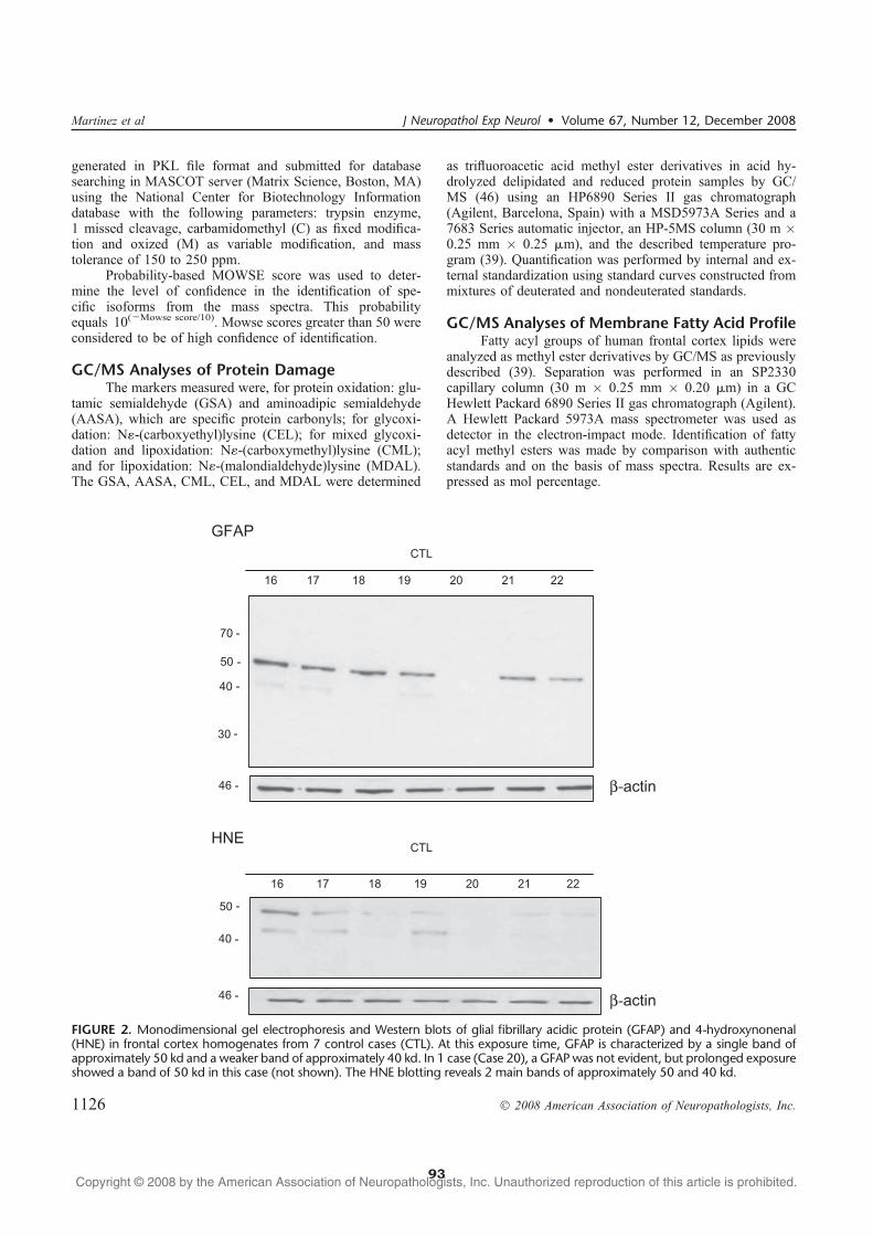
Copyright @ 200 by the American Association of Neuropathologists, Inc. Unauthorized reproduction of this article is prohibited.8
generated in PKL file format and submitted for databasesearching in MASCOT server (Matrix Science, Boston, MA)using the National Center for Biotechnology Informationdatabase with the following parameters: trypsin enzyme,1 missed cleavage, carbamidomethyl (C) as fixed modifica-tion and oxized (M) as variable modification, and masstolerance of 150 to 250 ppm.
Probability-based MOWSE score was used to deter-mine the level of confidence in the identification of spe-cific isoforms from the mass spectra. This probabilityequals 10(jMowse score/10). Mowse scores greater than 50 wereconsidered to be of high confidence of identification.
GC/MS Analyses of Protein DamageThe markers measured were, for protein oxidation: glu-
tamic semialdehyde (GSA) and aminoadipic semialdehyde(AASA), which are specific protein carbonyls; for glycoxi-dation: N?-(carboxyethyl)lysine (CEL); for mixed glycoxi-dation and lipoxidation: N?-(carboxymethyl)lysine (CML);and for lipoxidation: N?-(malondialdehyde)lysine (MDAL).The GSA, AASA, CML, CEL, and MDAL were determined
as trifluoroacetic acid methyl ester derivatives in acid hy-drolyzed delipidated and reduced protein samples by GC/MS (46) using an HP6890 Series II gas chromatograph(Agilent, Barcelona, Spain) with a MSD5973A Series and a7683 Series automatic injector, an HP-5MS column (30 m �0.25 mm � 0.25 Km), and the described temperature pro-gram (39). Quantification was performed by internal and ex-ternal standardization using standard curves constructed frommixtures of deuterated and nondeuterated standards.
GC/MS Analyses of Membrane Fatty Acid ProfileFatty acyl groups of human frontal cortex lipids were
analyzed as methyl ester derivatives by GC/MS as previouslydescribed (39). Separation was performed in an SP2330capillary column (30 m � 0.25 mm � 0.20 Km) in a GCHewlett Packard 6890 Series II gas chromatograph (Agilent).A Hewlett Packard 5973A mass spectrometer was used asdetector in the electron-impact mode. Identification of fattyacyl methyl esters was made by comparison with authenticstandards and on the basis of mass spectra. Results are ex-pressed as mol percentage.
FIGURE 2. Monodimensional gel electrophoresis and Western blots of glial fibrillary acidic protein (GFAP) and 4-hydroxynonenal(HNE) in frontal cortex homogenates from 7 control cases (CTL). At this exposure time, GFAP is characterized by a single band ofapproximately 50 kd and a weaker band of approximately 40 kd. In 1 case (Case 20), a GFAP was not evident, but prolonged exposureshowed a band of 50 kd in this case (not shown). The HNE blotting reveals 2 main bands of approximately 50 and 40 kd.
Martınez et al J Neuropathol Exp Neurol � Volume 67, Number 12, December 2008
� 2008 American Association of Neuropathologists, Inc.1126
93

Copyright @ 200 by the American Association of Neuropathologists, Inc. Unauthorized reproduction of this article is prohibited.8
RESULTS
General Neuropathologic FindingsVariable degrees of neuron loss, spongiosis in the
upper cellular layers, and reactive astrocytosis were presentin every FTLD case, and characteristic inclusions werepresent in every FTLD type (Fig. 1). Cases with FTDL-tau showed tau-immunoreactive neurons and fewer tau-immunoreactive astrocytes (Fig. 1C). Cases with FTLD-Ushowed ubiquitin-immunoreactive intraneuronal inclusionsand neurites (Fig. 1F); cases affected by FTLD-MND hadglobular ubiquitin-immunoreactive neuronal cytoplasmicinclusions (Fig. 1I). The 2 cases with mutation in theprogranulin gene had characteristic cat eyeYshaped intra-nuclear inclusions. All FTLD-U and FTLD-MND cases hadTDP-43Yimmunoreactive inclusions.
The degrees of neuron loss and astrocytosis in thefrontal cortex in FTLD and control cases were assessed in asemiquantitative manner and are summarized in Table 2.
Gel Electrophoresis and Western Blotting toGFAP and HNE in Controls
Monodimensional gel electrophoresis and GFAPWestern blotting of the frontal cortex revealed a band ofapproximately 50 kd and a weaker band of approximately40 kd in most control cases (Fig. 2). Increased GFAP im-munoreactivity was seen in a single case (Case 2; see later).Monodimensional gel electrophoresis to HNE showed 2 bandsof approximately 50 and 40 kd in most cases (Fig. 2), with theexception of moderate HNE immunoreactivity in Case 2.One case showed an apparent absence of GFAP and HNEimmunoreactivity, but typical bands in this case were de-tected with a long time of exposure (data not shown).
Gel Electrophoresis and Western Blotting toGFAP and HNE in Pathological Cases
Monodimensional gel electrophoresis and GFAPWestern blotting of the frontal cortex revealed several bandsof strong GFAP immunoreactivity between 35 and 50 kd in
FIGURE 3. Monodimensional gel electrophoresis and Western blots of glial fibrillary acidic protein ([GFAP] bands between 35 and50 kd) in frontal cortex homogenates showing strong expression levels in the 6 frontotemporal lobar dementia (FTLD) with taupathology cases ([FTLD-tau] P301L; Cases 4Y6, 27Y29), 5 FTLDwith tau-negative ubiquitin-positive neuronal inclusions cases ([FTLD-U]strong 10, 12, and 23; moderate 11, 26), and 2 FTLD with tau-negative ubiquitin-positive inclusions that occurs in association withmotor neuron disease and amyotrophic lateral sclerosis cases ([FTLD-MND] Cases 14, 15). A slight increase is observed in 1 control case([CTL] Case 2). Similar patterns are seen by using mouse monoclonal and rabbit polyclonal anti-GFAP antibodies.
J Neuropathol Exp Neurol � Volume 67, Number 12, December 2008 Oxidative Damage in FTLD
� 2008 American Association of Neuropathologists, Inc. 1127
94

Copyright @ 200 by the American Association of Neuropathologists, Inc. Unauthorized reproduction of this article is prohibited.8
the 6 FTLD-tau (Cases 4Y6, 27Y29), 5 cases with FTLD-U(Cases 10Y12, 23, 26), and 2 cases with FTLD-MND (Cases14, 15) (Fig. 3). One control case (Case 2) had moderateGFAP immunoreactivity. Similar findings were obtainedusing mouse monoclonal and rabbit polyclonal anti-GFAPantibodies (Fig. 3). The intensity of GFAP immunoreactivity,as revealed in Western blot analysis, correlated with theamount of cortical GFAP-immunoreactive astrocytes identi-fied by immunohistochemistry (Table 2).
Monodimensional gel electrophoresis and Western blotanalysis of HNE revealed higher intensities of bands between35 and 50 kd in some pathological cases than in controls.Increased densities of HNE bands were observed in the6 FTLD-tau cases (Cases 4Y6, 27Y29), 3 of 10 FTLD-U cases(Cases 10, 12, 23), and 2 FTLD-MND cases (Cases 14, 15)(Fig. 4). Increased density of the upper band was noticedin another FTLD-U (Case 11). A moderate increase in HNEexpression was present in 1 control (Case 2) (Fig. 4). Theintensity of HNE-immunoreactive bands expressed semi-quantitatively is shown in Table 2 for comparison to othermorphological and biochemical parameters.
2-D Gel Electrophoresis, In-Gel Digestion,and MS
4-Hydroxynonenal antibody was used in Western blotsof 2-D gels. Control (Cases 1Y3) and FTLD cases (Cases4Y6, 10, 12, 14, 15) were processed in parallel on differentdays. Several spots between 65 and 70 kd appeared in someFTLD cases, thus indicating that different proteins could betargets of lipoxidation in FTLD. However, only spots be-
tween 35 and 50 kd that were consistently present in HNE-positive cases were selected for study.
Corresponding Coomassie-stained spots were excisedfrom Cases 4, 10, 12, 14, and 15, in-gel digested, and pro-cessed for MS. All spots were identified as GFAP as primaryor unique option with Mowse scores of high confidence(Table 3). Other spots also appeared in individual cases,suggesting that other proteins were targets of oxidation, butbecause these spots were neither consistent nor related to age,sex, or pathology, no further attempts were made to iden-tify them.
ValidationParallel membranes were blotted with anti-HNE and
anti-GFAP antibodies in control and FTLD cases. Spots ofFTLD-tau, FTLD-U Cases 10 and 12, and FTLD-MNDCases 14 and 15 stained with anti-HNE antibody matchedwith spots incubated with anti-GFAP antibodies (Figs. 5Y7).
Double Labeling Immunofluorescence andConfocal Microscopy
Sections of the frontal cortex in controls and in Cases 4to 6 (FTLD-tau), 10, 12 (FTLD-U), 14, and 15 (FTLD-MND)were double stained with anti-HNE and anti-GFAP. Thisanalysis demonstrated colocalization of these antibodies inreactive astrocytes in FTLD (Fig. 8). Neurons were notstained with anti-HNE antibodies. Double labeling with an-tibodies to GFAP and SOD1 demonstrated the presence ofSOD1 in astrocytes in FTLD-tau and FTLD-U cases (Fig. 9),whereas double labeling immunofluorescence and confocal
FIGURE 4. Monodimensional gel electrophoresis and Western blotting to 4-hydroxynonenal ([HNE] bands between 35 and 50 kd)in frontal cortex homogenates show strong expression levels in the 6 frontotemporal lobar dementia (FTLD) with tau pathologycases ([FTLD-tau] P301L; Cases 4Y6, 27Y29), 3 FTLD with tau-negative ubiquitin-positive neuronal inclusions cases ([FTLD-U] Cases10, 12, 23), and 2 FTLD with tau-negative ubiquitin-positive inclusions that occurs in association with motor neuron disease andamyotrophic lateral sclerosis cases ([FTLD-MND] Cases 14, 15). There is a moderate increase in 1 control case ([CTL] Case 2) and aslight increase in 2 FTLD-U cases (Cases 11, 26). A-Actin is used as a control of protein loading.
Martınez et al J Neuropathol Exp Neurol � Volume 67, Number 12, December 2008
� 2008 American Association of Neuropathologists, Inc.1128
95

Copyright @ 200 by the American Association of Neuropathologists, Inc. Unauthorized reproduction of this article is prohibited.8
FIGURE 5. Bidimensional gel electrophoresis with gels stained with Coomassie (left panels) and membranes immunoblotted withanti<4-hydroxynonenal ([HNE] middle panels) and anti<glial fibrillary acidic protein ([GFAP] right panels) antibodies in control(Case 2) and frontotemporal lobar dementia with tau pathology ([FTLD-tau] Case 4). These cases were processed in parallel.Numbers 1 to 3 are spots selected for in-gel digestion and mass spectrometry.
TABLE 3. Gel Electrophoresis Spot Identification in Selected Cases*
FTLD Case Spot Calculated pINominalMass, d Protein
ScoreCoverage
SequenceCoverage, %
PeptidesMatched
GIAccession
4 1 5.42 49,776 GFAP 91 21 9 38566198
4 2 5.42 49,776 GFAP 136 27 15 38566198
4 3 5.42 49,776 GFAP 275 40 22 38566198
10 1 5.42 49,907 GFAP 978 38 50 4503979
10 2 5.42 49,776 GFAP 404 46 25 38566198
12 1 5.42 49,776 GFAP 158 35 19 38566198
12 2 5.42 49,776 GFAP 442 42 23 38566198
12 3 5.42 49,776 GFAP 359 43 24 38566198
14 (1) 5.42 49,907 GFAP 540 26 12 4503979
14 (2) 5.47 49,835 GFAP 177 12 5 16265836
14 (4) 5.42 49,907 GFAP 502 29 13 4503979
14 (5) 5.42 49,907 GFAP 548 29 13 4503979
15 (3) 5.42 49,907 GFAP 270 15 6 4503979
15 (4) 5.42 49,907 GFAP 591 30 16 4503979
15 (5) 5.42 49,907 GFAP 790 39 23 4503979
*Case 4 is an FTLD-tau case; Cases 10 and 12 are FTLD-U; Cases 14 and 15 are FTLD-MND cases. Spots identified are indicated in Figures 5, 6, and 7 for the correspondingcases.
FTLD-MND, frontotemporal lobar dementia with tau-negative ubiquitin-positive inclusions that occurs in association with motor neuron disease and amyotrophic lateral sclerosis;FTLD-tau, frontotemporal lobar dementia with tau pathology; FTLD-U, frontotemporal lobar degeneration with tau-negative ubiquitin-positive neuronal inclusions; GFAP, glialfibrillary acidic protein; GI, genInfo identification number; pI, isoelectric point.
J Neuropathol Exp Neurol � Volume 67, Number 12, December 2008 Oxidative Damage in FTLD
� 2008 American Association of Neuropathologists, Inc. 1129
96
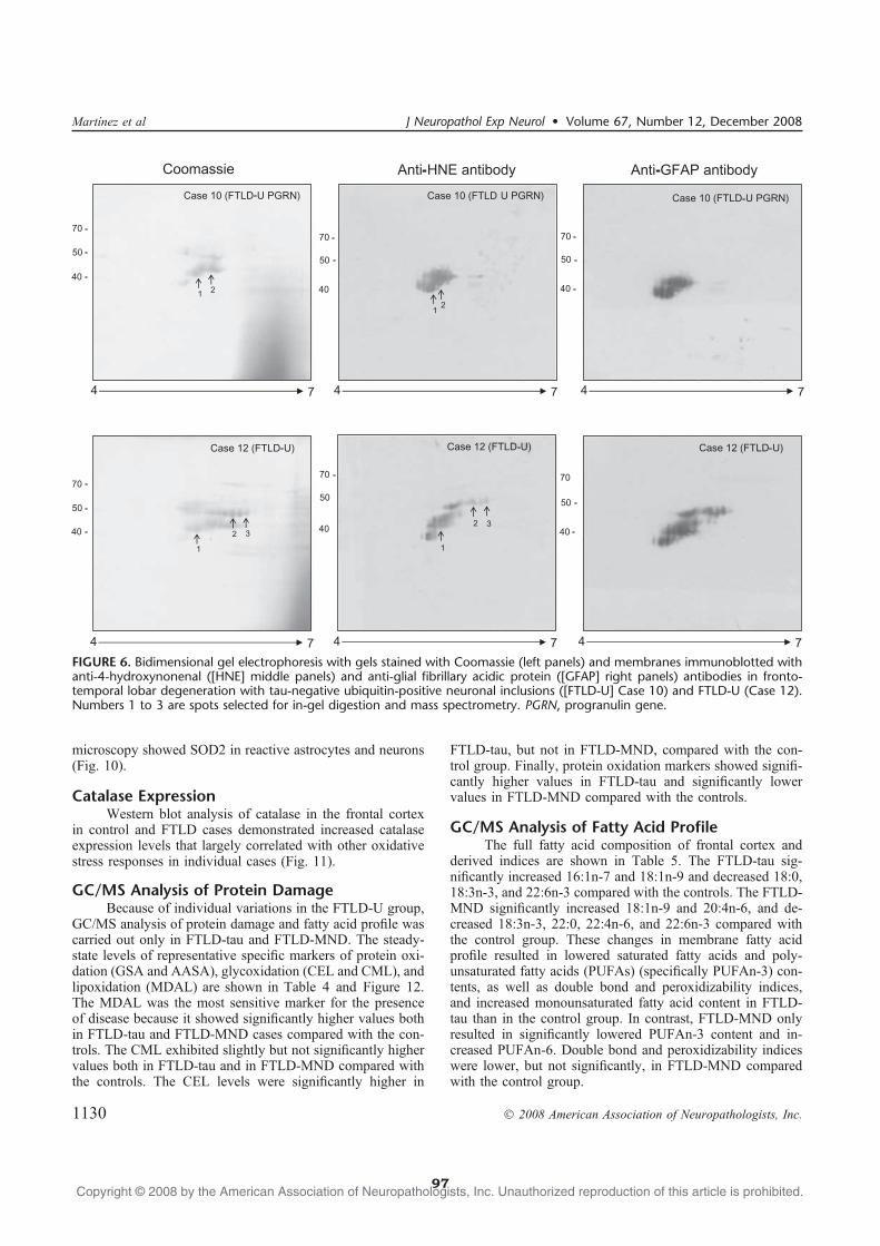
Copyright @ 200 by the American Association of Neuropathologists, Inc. Unauthorized reproduction of this article is prohibited.8
microscopy showed SOD2 in reactive astrocytes and neurons(Fig. 10).
Catalase ExpressionWestern blot analysis of catalase in the frontal cortex
in control and FTLD cases demonstrated increased catalaseexpression levels that largely correlated with other oxidativestress responses in individual cases (Fig. 11).
GC/MS Analysis of Protein DamageBecause of individual variations in the FTLD-U group,
GC/MS analysis of protein damage and fatty acid profile wascarried out only in FTLD-tau and FTLD-MND. The steady-state levels of representative specific markers of protein oxi-dation (GSA and AASA), glycoxidation (CEL and CML), andlipoxidation (MDAL) are shown in Table 4 and Figure 12.The MDAL was the most sensitive marker for the presenceof disease because it showed significantly higher values bothin FTLD-tau and FTLD-MND cases compared with the con-trols. The CML exhibited slightly but not significantly highervalues both in FTLD-tau and in FTLD-MND compared withthe controls. The CEL levels were significantly higher in
FTLD-tau, but not in FTLD-MND, compared with the con-trol group. Finally, protein oxidation markers showed signifi-cantly higher values in FTLD-tau and significantly lowervalues in FTLD-MND compared with the controls.
GC/MS Analysis of Fatty Acid ProfileThe full fatty acid composition of frontal cortex and
derived indices are shown in Table 5. The FTLD-tau sig-nificantly increased 16:1n-7 and 18:1n-9 and decreased 18:0,18:3n-3, and 22:6n-3 compared with the controls. The FTLD-MND significantly increased 18:1n-9 and 20:4n-6, and de-creased 18:3n-3, 22:0, 22:4n-6, and 22:6n-3 compared withthe control group. These changes in membrane fatty acidprofile resulted in lowered saturated fatty acids and poly-unsaturated fatty acids (PUFAs) (specifically PUFAn-3) con-tents, as well as double bond and peroxidizability indices,and increased monounsaturated fatty acid content in FTLD-tau than in the control group. In contrast, FTLD-MND onlyresulted in significantly lowered PUFAn-3 content and in-creased PUFAn-6. Double bond and peroxidizability indiceswere lower, but not significantly, in FTLD-MND comparedwith the control group.
FIGURE 6. Bidimensional gel electrophoresis with gels stained with Coomassie (left panels) and membranes immunoblotted withanti<4<hydroxynonenal ([HNE] middle panels) and anti<glial fibrillary acidic protein ([GFAP] right panels) antibodies in fronto-temporal lobar degeneration with tau-negative ubiquitin-positive neuronal inclusions ([FTLD-U] Case 10) and FTLD-U (Case 12).Numbers 1 to 3 are spots selected for in-gel digestion and mass spectrometry. PGRN, progranulin gene.
Martınez et al J Neuropathol Exp Neurol � Volume 67, Number 12, December 2008
� 2008 American Association of Neuropathologists, Inc.1130
97
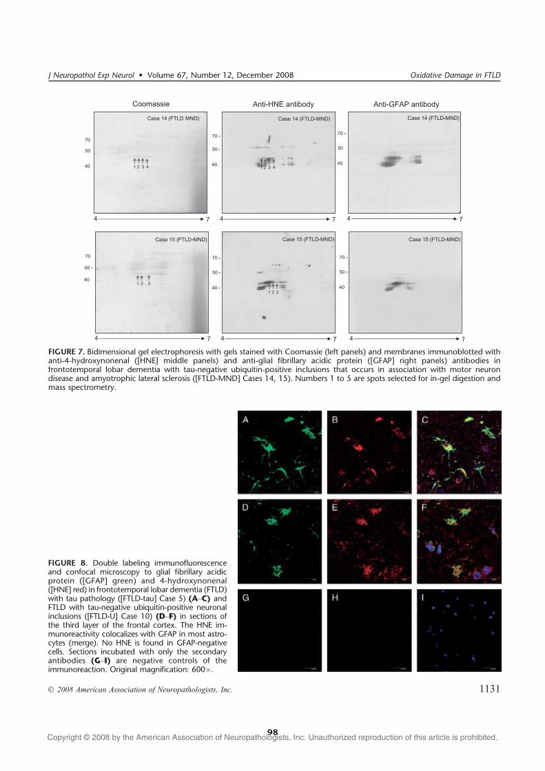
Copyright @ 200 by the American Association of Neuropathologists, Inc. Unauthorized reproduction of this article is prohibited.8
FIGURE 7. Bidimensional gel electrophoresis with gels stained with Coomassie (left panels) and membranes immunoblotted withanti<4-hydroxynonenal ([HNE] middle panels) and anti<glial fibrillary acidic protein ([GFAP] right panels) antibodies infrontotemporal lobar dementia with tau-negative ubiquitin-positive inclusions that occurs in association with motor neurondisease and amyotrophic lateral sclerosis ([FTLD-MND] Cases 14, 15). Numbers 1 to 5 are spots selected for in-gel digestion andmass spectrometry.
FIGURE 8. Double labeling immunofluorescenceand confocal microscopy to glial fibrillary acidicprotein ([GFAP] green) and 4-hydroxynonenal([HNE] red) in frontotemporal lobar dementia (FTLD)with tau pathology ([FTLD-tau] Case 5) (AYC) andFTLD with tau-negative ubiquitin-positive neuronalinclusions ([FTLD-U] Case 10) (DYF) in sections ofthe third layer of the frontal cortex. The HNE im-munoreactivity colocalizes with GFAP in most astro-cytes (merge). No HNE is found in GFAP-negativecells. Sections incubated with only the secondaryantibodies (GYI) are negative controls of theimmunoreaction. Original magnification: 600�.
J Neuropathol Exp Neurol � Volume 67, Number 12, December 2008 Oxidative Damage in FTLD
� 2008 American Association of Neuropathologists, Inc. 1131
98
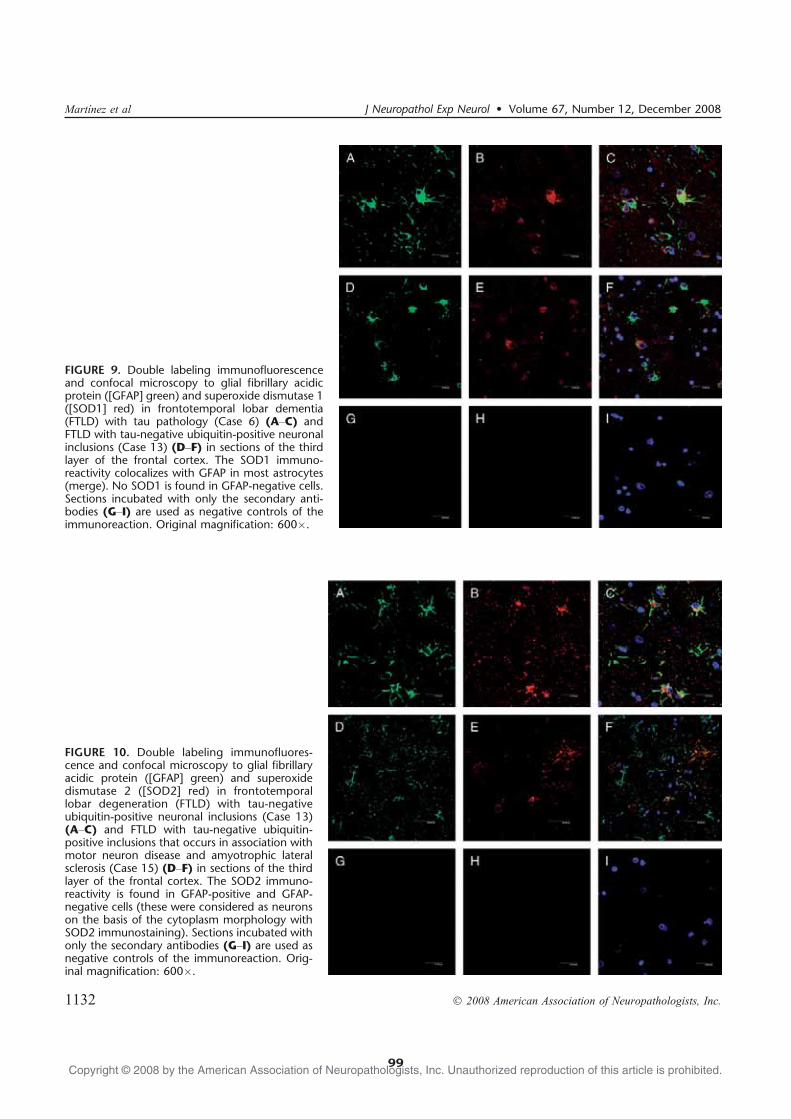
Copyright @ 200 by the American Association of Neuropathologists, Inc. Unauthorized reproduction of this article is prohibited.8
FIGURE 9. Double labeling immunofluorescenceand confocal microscopy to glial fibrillary acidicprotein ([GFAP] green) and superoxide dismutase 1([SOD1] red) in frontotemporal lobar dementia(FTLD) with tau pathology (Case 6) (AYC) andFTLD with tau-negative ubiquitin-positive neuronalinclusions (Case 13) (DYF) in sections of the thirdlayer of the frontal cortex. The SOD1 immuno-reactivity colocalizes with GFAP in most astrocytes(merge). No SOD1 is found in GFAP-negative cells.Sections incubated with only the secondary anti-bodies (GYI) are used as negative controls of theimmunoreaction. Original magnification: 600�.
FIGURE 10. Double labeling immunofluores-cence and confocal microscopy to glial fibrillaryacidic protein ([GFAP] green) and superoxidedismutase 2 ([SOD2] red) in frontotemporallobar degeneration (FTLD) with tau-negativeubiquitin-positive neuronal inclusions (Case 13)(AYC) and FTLD with tau-negative ubiquitin-positive inclusions that occurs in association withmotor neuron disease and amyotrophic lateralsclerosis (Case 15) (DYF) in sections of the thirdlayer of the frontal cortex. The SOD2 immuno-reactivity is found in GFAP-positive and GFAP-negative cells (these were considered as neuronson the basis of the cytoplasm morphology withSOD2 immunostaining). Sections incubated withonly the secondary antibodies (GYI) are used asnegative controls of the immunoreaction. Orig-inal magnification: 600�.
Martınez et al J Neuropathol Exp Neurol � Volume 67, Number 12, December 2008
� 2008 American Association of Neuropathologists, Inc.1132
99
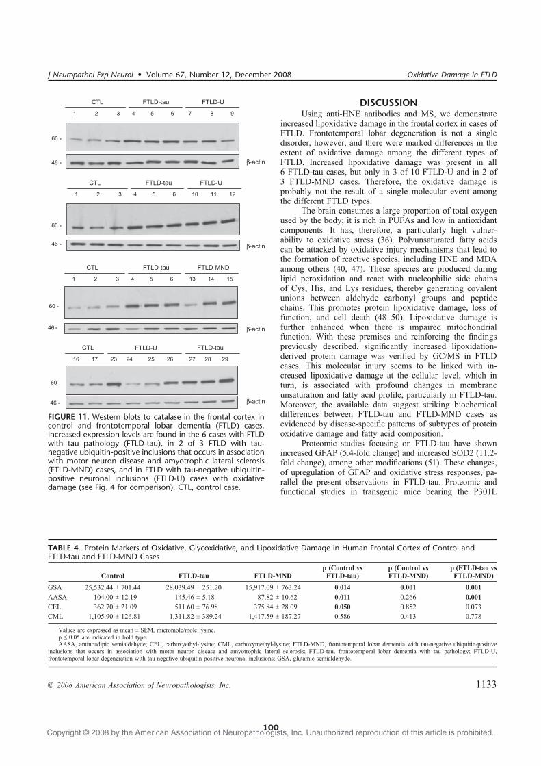
Copyright @ 200 by the American Association of Neuropathologists, Inc. Unauthorized reproduction of this article is prohibited.8
DISCUSSIONUsing anti-HNE antibodies and MS, we demonstrate
increased lipoxidative damage in the frontal cortex in cases ofFTLD. Frontotemporal lobar degeneration is not a singledisorder, however, and there were marked differences in theextent of oxidative damage among the different types ofFTLD. Increased lipoxidative damage was present in all6 FTLD-tau cases, but only in 3 of 10 FTLD-U and in 2 of3 FTLD-MND cases. Therefore, the oxidative damage isprobably not the result of a single molecular event amongthe different FTLD types.
The brain consumes a large proportion of total oxygenused by the body; it is rich in PUFAs and low in antioxidantcomponents. It has, therefore, a particularly high vulner-ability to oxidative stress (36). Polyunsaturated fatty acidscan be attacked by oxidative injury mechanisms that lead tothe formation of reactive species, including HNE and MDAamong others (40, 47). These species are produced duringlipid peroxidation and react with nucleophilic side chainsof Cys, His, and Lys residues, thereby generating covalentunions between aldehyde carbonyl groups and peptidechains. This promotes protein lipoxidative damage, loss offunction, and cell death (48Y50). Lipoxidative damage isfurther enhanced when there is impaired mitochondrialfunction. With these premises and reinforcing the findingspreviously described, significantly increased lipoxidation-derived protein damage was verified by GC/MS in FTLDcases. This molecular injury seems to be linked with in-creased lipoxidative damage at the cellular level, which inturn, is associated with profound changes in membraneunsaturation and fatty acid profile, particularly in FTLD-tau.Moreover, the available data suggest striking biochemicaldifferences between FTLD-tau and FTLD-MND cases asevidenced by disease-specific patterns of subtypes of proteinoxidative damage and fatty acid composition.
Proteomic studies focusing on FTLD-tau have shownincreased GFAP (5.4-fold change) and increased SOD2 (11.2-fold change), among other modifications (51). These changes,of upregulation of GFAP and oxidative stress responses, pa-rallel the present observations in FTLD-tau. Proteomic andfunctional studies in transgenic mice bearing the P301L
FIGURE 11. Western blots to catalase in the frontal cortex incontrol and frontotemporal lobar dementia (FTLD) cases.Increased expression levels are found in the 6 cases with FTLDwith tau pathology (FTLD-tau), in 2 of 3 FTLD with tau-negative ubiquitin-positive inclusions that occurs in associationwith motor neuron disease and amyotrophic lateral sclerosis(FTLD-MND) cases, and in FTLD with tau-negative ubiquitin-positive neuronal inclusions (FTLD-U) cases with oxidativedamage (see Fig. 4 for comparison). CTL, control case.
TABLE 4. Protein Markers of Oxidative, Glycoxidative, and Lipoxidative Damage in Human Frontal Cortex of Control andFTLD-tau and FTLD-MND Cases
Control FTLD-tau FTLD-MNDp (Control vsFTLD-tau)
p (Control vsFTLD-MND)
p (FTLD-tau vsFTLD-MND)
GSA 25,532.44 T 701.44 28,039.49 T 251.20 15,917.09 T 763.24 0.014 0.001 0.001
AASA 104.00 T 12.19 145.46 T 5.18 87.82 T 10.62 0.011 0.266 0.001
CEL 362.70 T 21.09 511.60 T 76.98 375.84 T 28.09 0.050 0.852 0.073
CML 1,105.90 T 126.81 1,311.82 T 389.24 1,417.59 T 187.27 0.586 0.413 0.778
Values are expressed as mean T SEM, micromole/mole lysine.p e 0.05 are indicated in bold type.AASA, aminoadipic semialdehyde; CEL, carboxyethyl-lysine; CML, carboxymethyl-lysine; FTLD-MND, frontotemporal lobar dementia with tau-negative ubiquitin-positive
inclusions that occurs in association with motor neuron disease and amyotrophic lateral sclerosis; FTLD-tau, frontotemporal lobar dementia with tau pathology; FTLD-U,frontotemporal lobar degeneration with tau-negative ubiquitin-positive neuronal inclusions; GSA, glutamic semialdehyde.
J Neuropathol Exp Neurol � Volume 67, Number 12, December 2008 Oxidative Damage in FTLD
� 2008 American Association of Neuropathologists, Inc. 1133
100
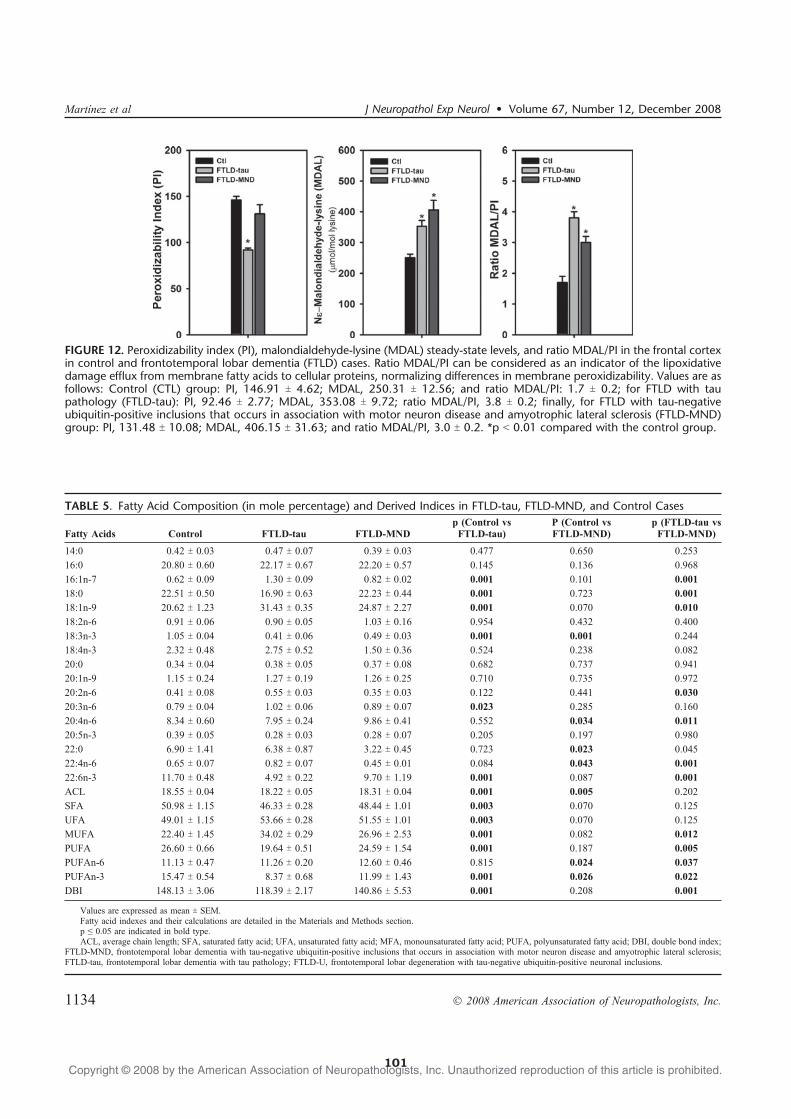
Copyright @ 200 by the American Association of Neuropathologists, Inc. Unauthorized reproduction of this article is prohibited.8
FIGURE 12. Peroxidizability index (PI), malondialdehyde-lysine (MDAL) steady-state levels, and ratio MDAL/PI in the frontal cortexin control and frontotemporal lobar dementia (FTLD) cases. Ratio MDAL/PI can be considered as an indicator of the lipoxidativedamage efflux from membrane fatty acids to cellular proteins, normalizing differences in membrane peroxidizability. Values are asfollows: Control (CTL) group: PI, 146.91 T 4.62; MDAL, 250.31 T 12.56; and ratio MDAL/PI: 1.7 T 0.2; for FTLD with taupathology (FTLD-tau): PI, 92.46 T 2.77; MDAL, 353.08 T 9.72; ratio MDAL/PI, 3.8 T 0.2; finally, for FTLD with tau-negativeubiquitin-positive inclusions that occurs in association with motor neuron disease and amyotrophic lateral sclerosis (FTLD-MND)group: PI, 131.48 T 10.08; MDAL, 406.15 T 31.63; and ratio MDAL/PI, 3.0 T 0.2. *p G 0.01 compared with the control group.
TABLE 5. Fatty Acid Composition (in mole percentage) and Derived Indices in FTLD-tau, FTLD-MND, and Control Cases
Fatty Acids Control FTLD-tau FTLD-MNDp (Control vsFTLD-tau)
P (Control vsFTLD-MND)
p (FTLD-tau vsFTLD-MND)
14:0 0.42 T 0.03 0.47 T 0.07 0.39 T 0.03 0.477 0.650 0.253
16:0 20.80 T 0.60 22.17 T 0.67 22.20 T 0.57 0.145 0.136 0.968
16:1n-7 0.62 T 0.09 1.30 T 0.09 0.82 T 0.02 0.001 0.101 0.001
18:0 22.51 T 0.50 16.90 T 0.63 22.23 T 0.44 0.001 0.723 0.001
18:1n-9 20.62 T 1.23 31.43 T 0.35 24.87 T 2.27 0.001 0.070 0.010
18:2n-6 0.91 T 0.06 0.90 T 0.05 1.03 T 0.16 0.954 0.432 0.400
18:3n-3 1.05 T 0.04 0.41 T 0.06 0.49 T 0.03 0.001 0.001 0.244
18:4n-3 2.32 T 0.48 2.75 T 0.52 1.50 T 0.36 0.524 0.238 0.082
20:0 0.34 T 0.04 0.38 T 0.05 0.37 T 0.08 0.682 0.737 0.941
20:1n-9 1.15 T 0.24 1.27 T 0.19 1.26 T 0.25 0.710 0.735 0.972
20:2n-6 0.41 T 0.08 0.55 T 0.03 0.35 T 0.03 0.122 0.441 0.030
20:3n-6 0.79 T 0.04 1.02 T 0.06 0.89 T 0.07 0.023 0.285 0.160
20:4n-6 8.34 T 0.60 7.95 T 0.24 9.86 T 0.41 0.552 0.034 0.011
20:5n-3 0.39 T 0.05 0.28 T 0.03 0.28 T 0.07 0.205 0.197 0.980
22:0 6.90 T 1.41 6.38 T 0.87 3.22 T 0.45 0.723 0.023 0.045
22:4n-6 0.65 T 0.07 0.82 T 0.07 0.45 T 0.01 0.084 0.043 0.001
22:6n-3 11.70 T 0.48 4.92 T 0.22 9.70 T 1.19 0.001 0.087 0.001
ACL 18.55 T 0.04 18.22 T 0.05 18.31 T 0.04 0.001 0.005 0.202
SFA 50.98 T 1.15 46.33 T 0.28 48.44 T 1.01 0.003 0.070 0.125
UFA 49.01 T 1.15 53.66 T 0.28 51.55 T 1.01 0.003 0.070 0.125
MUFA 22.40 T 1.45 34.02 T 0.29 26.96 T 2.53 0.001 0.082 0.012
PUFA 26.60 T 0.66 19.64 T 0.51 24.59 T 1.54 0.001 0.187 0.005
PUFAn-6 11.13 T 0.47 11.26 T 0.20 12.60 T 0.46 0.815 0.024 0.037
PUFAn-3 15.47 T 0.54 8.37 T 0.68 11.99 T 1.43 0.001 0.026 0.022
DBI 148.13 T 3.06 118.39 T 2.17 140.86 T 5.53 0.001 0.208 0.001
Values are expressed as mean T SEM.Fatty acid indexes and their calculations are detailed in the Materials and Methods section.p e 0.05 are indicated in bold type.ACL, average chain length; SFA, saturated fatty acid; UFA, unsaturated fatty acid; MFA, monounsaturated fatty acid; PUFA, polyunsaturated fatty acid; DBI, double bond index;
FTLD-MND, frontotemporal lobar dementia with tau-negative ubiquitin-positive inclusions that occurs in association with motor neuron disease and amyotrophic lateral sclerosis;FTLD-tau, frontotemporal lobar dementia with tau pathology; FTLD-U, frontotemporal lobar degeneration with tau-negative ubiquitin-positive neuronal inclusions.
Martınez et al J Neuropathol Exp Neurol � Volume 67, Number 12, December 2008
� 2008 American Association of Neuropathologists, Inc.1134
101
Copyright @ 200 by the American Association of Neuropathologists, Inc. Unauthorized reproduction of this article is prohibited.8
mutation in the tau gene have also shown reduced NADH-ubiquinone oxireductase activity, reduced mitochondrial res-piration, and reduced adenosine triphosphate synthase,together with increased reactive oxygen species, elevatedantioxidant enzymes, enhanced SOD and glutathione reduc-tase activities, and lowered in vivo lipoxidation and increasedin vitro lipoxidation, as estimated by MDA levels (52). In lightof the present findings, this last observation is likely caused bya lower membrane unsaturation in P301L mutants. Despitethat the origin of this change in membrane unsaturation isunknown, it is likely caused by the combined effect of dif-ferent factors, including 1) changes in cellular populations(neuronal loss and astrocytosis) with cell-specific fatty acidprofiles, 2) alterations in metabolic pathways where specificfatty acids (particularly 22:6n-3) participate, 3) cellular mem-brane adaptations to the pathological state, and 4) the flux ofoxidative damage directed to the cellular membrane.
No similar studies have been performed in FTLD-U,and no available models of FTLD-U are currently available.Recent studies have, however, shown a link between oxi-dative stress, oxidative damage, mitochondrial abnormalities,and endoplasmic reticulum stress in the spinal cord in patientswith amyotrophic lateral sclerosis (53). For these reasons, de-spite being a neuropathologic variety of FTLD-U, the FTLD-MND cases in the present study have been considered apart.Because of the limited number of cases and considering thatcauses of oxidative damage in FTLD-MND may differ fromthose of other FTLD-U cases, further studies are necessary toprobe specific impairment of metabolic pathways in FTLD-MND compared with other FTLD-U cases.
With the exception of 2 cases in which GFAP expres-sion levels were higher than those corresponding to HNE,the intensity of oxidative damage correlated with astrocy-tosis ascertained by GFAP immunohistochemistry in the ad-jacent cortex and by levels of GFAP protein expression asrevealed by Western blot analysis of total cortex homoge-nates. Bidimensional gel electrophoresis, Western blottingto HNE, in-gel digestion, and MS revealed GFAP as the firstor unique candidate in every individually analyzed spot fromFTLD-tau, FTLD-U, and FTLD-MND cases. Additionalspots were also observed in individual cases, thus suggest-ing that other proteins may be additional targets of lip-oxidation. Because of the extreme variability among cases,however, no further attempts were made to characterize theseadditional spots. Finally, double labeling immunofluores-cence and confocal microscopy revealed the presence ofHNE and SOD1 only in reactive cortical astrocytes in FTLDcases and SOD2 in neurons and astrocytes. Increased ex-pression levels of catalase that correlated with oxidativedamage were also demonstrated in Western blots of totalfrontal cortex homogenates.
Previous studies have shown GFAP to be a target ofoxidation in Alzheimer disease (39, 54) and certain tauopa-thies such as Pick disease (46) and progressive supranu-clear palsy (55). Glial fibrillary acidic protein oxidation has,however, also been reported in conditions not associated withtau pathology, such as aceruloplasminemia (56), diabeticretina (57), and Huntington disease (58). The present ob-servations also indicate that although prominent GFAP lip-
oxidative damage occurs in FTLD-tau, GFAP is alsoinvolved in FTLD cases without tau pathology, includingFTLD-U and FTLD-MND. On the other hand, not all FTLD-Ucases with increased GFAP have similarly increased ex-pression of HNE adducts.
In summary, disease-specific oxidative damage andfatty acid patterns seem to underlie different FTLD subtypes;these are particularly pronounced in FTLD-tau cases.Furthermore, the present findings show astrocytes as targetsof lipoxidative damage, GFAP as a target of lipoxidation, andthat astrocytes are crucial elements of oxidative stressresponses in FTLD. Further work is needed to elucidatecommon and specific pathological metabolic pathways inFTLD-tau, FTLD-U, and FTLD-MND.
ACKNOWLEDGMENTSThe authors thank the anonymous reviewers for
suggestions and T. Yohannan for editorial help.Brain samples were obtained from the Institute of
Neuropathology and University of Barcelona/Hospital ClinicBrain Banks following the guidelines and approval of thelocal ethics committees.
REFERENCES1. Lowe J, Rossor M. Frontotemporal lobar degeneration. In: Dickson D,
ed. Neurodegeneration: The Molecular Pathology of Dementia andMovement Disorders. Basel, Switzerland: ISN Neuropath Press, 2003;342Y48
2. Froelich-Fabre S, Skoglund L, Ostojic J, et al. Clinical and molecularaspects of frontotemporal dementia. Neurodegener Dis 2004;1:218Y24
3. Neary D, Snowden J, Mann D. Frontotemporal dementia. Lancet Neurol2005;4:771Y80
4. Knibb JA, Kipps CM, Hodges JR. Frontotemporal dementia. Curr OpinNeurol 2006;19:565Y71
5. Brun A. Identification and characterization of frontal lobe degeneration:Historical perspective on the development of FTD. Alzheimer Dis AssocDisord 2007;21:S3YS4
6. Kumar-Singh S, van Broeckhoven C. Frontotemporal lobar degenera-tion: Current concepts in the light of recent advances. Brain Pathol 2007;17:104Y13
7. Cairns NJ, Bigio EH, Mackenzie IRA, et al. Neuropathologic diagnosticand nosologic criteria for frontotemporal lobar degeneration: Consensusof the Consortium for Frontotemporal Lobar Degeneration. Acta Neuro-pathol 2007;114:5Y22
8. Ferrer I, Roig C, Espino A, et al. Dementia of frontal lobe type andmotor neuron disease. A Golgi study of the frontal cortex. J NeurolNeurosurg Psychiatr 1991;54:932Y34
9. Ferrer I, Tunon T, Serrano MT, et al. Calbindin D-28k and parval-bumin immunoreactivity in the frontal cortex in patients with frontallobe dementia of non-Alzheimer type associated with amyotrophiclateral sclerosis. J Neurol Neurosurg Psychiatr 1993;56:257Y61
10. Ferrer I. Neurons and their dendrites in frontotemporal dementia.Dement Geriat Cogn Disord 1999;10(suppl 1):55Y60
11. Goedert M, Crowther RA, Spillantini MG. Tau mutations cause fronto-temporal dementias. Neuron 1998;21:955Y58
12. Lee VM, Goedert M, Trojanowski JQ. Neurodegenerative tauopathies.Ann Rev Neurosci 2001;24:1121Y59
13. Ghetti B, Hutton ML, Wszolek ZK. Frontotemporal dementia and par-kinsonism linked to chromosome 17 associated with tau gene mutations(FTDP-17T). In: Dickson D, ed. Neurodegeneration: The MolecularPathology of Dementia and Movement Disorders. Basel, Switzerland:ISN Neuropath Press, 2003;86Y102
14. Alonso A, Mederlyova A, Novak M, et al. Promotion of hyper-phosphorylation by frontotemporal dementia tau mutations. J Biol Chem2004;279:34873Y81
J Neuropathol Exp Neurol � Volume 67, Number 12, December 2008 Oxidative Damage in FTLD
� 2008 American Association of Neuropathologists, Inc. 1135
102

Copyright @ 200 by the American Association of Neuropathologists, Inc. Unauthorized reproduction of this article is prohibited.8
15. Goedert M. Tau gene mutations and their effects. Mov Disord 2005;20(suppl 12):S45YS52
16. Gasparini L, Terni B, Spillantini MG. Frontotemporal dementia with taupathology. Neurodegener Dis 2007;4:236Y53
17. Munoz DG, Ferrer I. Neuropathology of hereditary forms of fronto-temporal dementia and parkinsonism. Handb Clin Neurol 2008;89:393Y414
18. Josephs KA, Holton JL, Rossor MN, et al. Frontotemporal lobar de-generation and ubiquitin immunohistochemistry. Neuropathol ApplNeurobiol 2004;30:369Y73
19. Sampathu DM, Neumann M, Kwong LK, et al. Pathological hetero-geneity of frontotemporal lobar degeneration with ubiquitin-positive in-clusions delineated by ubiquitin immunohistochemistry and novelmonoclonal antibodies. Am J Pathol 2006:169:1343Y52
20. Mackenzie IR, Baborie A, Pickering-Brown S, et al. Heterogeneityof ubiquitin pathology in frontotemporal lobar degeneration: Classifica-tion and relation to clinical phenotype. Acta Neuropathol 2006;112:539Y49
21. Baker M, Mackenzie IR, Pickering-Brown SM, et al. Mutations inprogranulin cause tau-negative frontotemporal dementia linked to chro-mosome 17. Nature 2006;442:916Y19
22. Cruts M, Gijselinck I, van der Zee J, et al. Null mutations in progranulincause ubiquitin-positive frontotemporal dementia linked to chromosome17q21. Nature 2006;442:920Y24
23. Mukherjee O, Pastor P, Cairns NJ, et al. HDDD2 is a familialfrontotemporal lobar degeneration with ubiquitin-positive, tau-negativeinclusions caused by a missense mutation in the signal peptide of pro-granulin. Ann Neurol 2006;60:314Y22
24. Gass J, Cannon A, Mackenzie IR, et al. Mutations in progranulin are amajor cause of ubiquitin-positive frontotemporal lobar degeneration.Hum Mol Genet 2006;15:2988Y3001
25. Watts GD, Wymer J, Kovach MJ, et al. Inclusion body myopathyassociated with Paget disease of bone and frontotemporal dementia iscaused by mutant valosin-containing protein. Nat Genet 2004;36:377Y81
26. Guyant-Marechal L, Laquerriere A, Duyckaerts C, et al. Valosin-con-taining protein gene mutations: clinical and neuropathologic features.Neurology 2006;67:644Y51
27. Skibinski G, Parkinson NJ, Brown JM, et al. Mutations in the endosomalESCRTIII-complex subunit CHMP2B in frontotemporal dementia. NatGenet 2005;37:806Y8
28. Holm IE, Englund E, Mackenzie IR, et al. A reassessment of theneuropathology of frontotemporal dementia linked to chromosome 3. JNeuropathol Exp Neurol 2007;66:884Y91
29. Vance C, Al-Chalabi A, Ruddy D, et al. Familial amyotrophic lateralsclerosis with frontotemporal dementia is linked to a locus on chro-mosome 9p13.2Y21.3. Brain 2006;129:868Y76
30. Talbot K, Ansorge O. Recent advances in the genetics of amyotrophiclateral sclerosis and frontotemporal dementia: Common pathways inneurodegenerative disease. Hum Mol Genet 2006;15:R182Y87
31. Mackenzie IR, Rademakers R. The molecular genetics and neuro-pathology of frontotemporal lobar degeneration: Recent developments.Neurogenetics 2007;8:237Y48
32. Mackenzie IR, Shi J, Shaw CL, et al. Dementia lacking distinctivehistology (DLDH) revisited. Acta Neuropathol 2006;112:551Y59
33. Markesbery WR, Carney JM. Oxidative alterations in Alzheimer’sdisease. Brain Pathol 1999;9:133Y46
34. Varadarajan S, Yatin S, Aksenova M, et al. Review: Alzheimer’s amy-loid peptideYassociated free radical oxidative stress and neurotoxicity.J Struct Biol 2000;130:184Y208
35. Smith MA, Rottkamp CA, Nunomura A, et al. Oxidative stress inAlzheimer’s disease. Biochem Biophys Acta 2000;1502:139Y44
36. Butterfield DA, Kanski J. Brain protein oxidation in age-relatedneurodegenerative disorders that are associated with aggregated proteins.Mech Ageing Dev 2001;122:945Y62
37. Liu Q, Raina AK, Smith MA, et al. Hydroxynonenal, toxic carbonyls,and Alzheimer disease. Mol Aspects Med 2003;24:305Y13
38. Keller JN, Schmitt FA, Scheff SW, et al. Evidence of increased oxidativedamage in subjects with mild cognitive impairment. Neurology 2005;64:1152Y56
39. Pamplona R, Dalfo E, Ayala V, et al. Proteins in human brain cortex aremodified by oxidation, glycoxidation, and lipoxidation. Effects ofAlzheimer disease and identification of lipoxidation targets. J BiolChem 2005;280:21522Y30
40. Pamplona R, Ilieva E, Ayala V, et al. Maillard reaction versus othernon-enzymatic modifications in neurodegenerative processes. Ann N YAcad Sci 2008;1126:315Y19
41. Albers DS, Augood SJ, Park LC, et al. Frontal lobe dysfunction inprogressive supranuclear palsy: Evidence for oxidative stress andmitochondrial impairment. J Neurochem 2000;74:878Y81
42. Odetti P, Garibaldi S, Norese R, et al. Lipoperoxidation is selectivelyinvolved in progressive supranuclear palsy. J Neuropathol Exp Neurol2000;59:393Y97
43. Martinez A, Dalfo E, Muntane G, et al. Glycolitic enzymes are targets ofoxidation in aged human frontal cortex and oxidative damage of theseproteins is increased in progressive supranuclear palsy. J Neural Transm2008;115:59Y66
44. Gerst JL, Siedlak SL, Nunomura A, et al. Role of oxidative stressin frontotemporal dementia. Dement Geriatr Cogn Disord 1999;10(suppl 1):85Y87
45. Llado A, Sanchez-Valle R, Rey MJ, et al. Clinicopathological andgenetic correlates of frontotemporal lobar degeneration and corticobasaldegeneration. J Neurol 2008;114:1051Y54
46. Muntane G, Dalfo E, Martınez A, et al. Glial fibrillary acidic protein is amajor target of glycoxidative and lipoxidative damage in Pick’s disease.J Neurochem 2006;99:177Y85
47. Dalle-Donne I, Aldini G, Carini M, et al. Protein carbonylation, cellu-lar dysfunction, and disease progression. J Cell Mol Med 2006;10:389Y406
48. Kruman I, Bruce-Keller AJ, Bredesen D, et al. Evidence that4-hydroxynonenal mediates oxidative stress-induced neuronal apoptosis.J Neurosci 1997;17:5089Y100
49. Uchida K. 4-Hydroxy-2-nonenal: A product and mediator of oxidativestress. Prog Lipid Res 2003;42:318Y43
50. Zarkovic N. 4-hydroxynonenal as a bioactive marker of pathophysio-logical processes. Mol Aspects Med 2003;24:281Y91
51. Schweitzer K, Decker E, Zhu L, et al. Aberrantly regulated proteins infrontotemporal dementia. Biochem Biophys Res Commun 2006;348:465Y72
52. David DC, Hauptmann S, Scherping I, et al. Proteomic and functionalanalysis reveal a mitochondrial dysfunction in P301L tau transgenicmice. J Biol Chem 2005;280:23802Y14
53. Ilieva EV, Ayala V, Jove M, et al. Oxidative and endoplasmic reticulumstress interplay in sporadic amyotrophic lateral sclerosis. Brain 2007;130:3111Y23
54. Korolainen MA, Auriola S, Nyman TA, et al. Proteomic analysis of glialfibrillary acidic protein in Alzheimer’s disease and aging brain. Neuro-biol Dis 2005;20:858Y70
55. Santpere G, Ferrer I. Delineation of early changes in cases withprogressive supranuclear palsy-like pathology. Astrocytes in striatumare primary targets of tau phosphorylation and GFAP oxidation. BrainPathol 2008; May 6 [Epub ahead of print]
56. Kaneko K, Nakamura A, Yoshida K, et al. Glial fibrillary acidic proteinis greatly modified by oxidative stress in aceruloplasminemia brain. FreeRadic Res 2002;36:303Y6
57. Baydas G, Tuzcu M, Yasar A, Baydas B. Early changes in glial re-activity and lipoid peroxidation in diabetic rat retina: Effects of mel-atonin. Acta Diabetol 2004;41:123Y28
58. Sorolla MA, Reverter-Branchat G, Tamarit J, et al. Proteomic andoxidative stress analysis in human brain samples of Huntington’sdisease. Free Radic Biol Med 2008;45:667Y78
Martınez et al J Neuropathol Exp Neurol � Volume 67, Number 12, December 2008
� 2008 American Association of Neuropathologists, Inc.1136
103

V.RESULTATS
4. Protein Targets of Oxidative Damage in Human Neurodegenerative Diseases with Abnormal Protein Aggregates
Anna Martínez, Manuel Portero-Otin, Reinald Pamplona, Isidre Ferrer.
Brain Pathol. 2009 Aug 6. [Epub ahead of print]
Les malalties humanes neurodegeneratives presenten proteïnes anormalment
agregades, associades amb alteracions en les modificacions post translacionals, la
solubilitat, l’agregació i la formació de fibrilles. A més a més de l’acumulació
d’aquestes proteïnes principals, hi han d’altres mecanismes implicats en la
neurodegeneració que probablement podrien explicar alguns aspectes importants, com
per exemple determinar la seqüència dels diferents processos moleculars, la selectiva
vulnerabilitat cel·lular i la progressió de la malaltia en els diferents casos individuals.
Un d’aquests mecanismes és l’estrès oxidatiu, el qual es troba en la majoria, per no dir
en totes, de malalties degeneratives del sistema nerviós.
La present revisió descriu la majoria de proteïnes dianes que han estat
identificades com a modificades nitro/oxidativament, utilitzant gels d’electroforesi
bidimensionals, western blot i espectometria de masses en la malaltia d’Alzheimer,
d’altres taupaties com la paràlisi supranuclear progressiva, la malaltia de Pick, la
malaltia de grans argidòfils i la degeneració del lòbul frontotemporal lligada a
mutacions de la proteïna tau; la malaltia de Parkinson i �-sinucleopaties relacionades;
la malaltia d’Huntington i l’esclerosi lateral amiotròfica, juntament amb models
animals i cultius cel·lulars. Les proteïnes vulnerables es van agrupar majoritàriament
en: vies metabòliques (incloses la glicòlisi i el metabolisme energètic), citoesquelet,
xaperones, resposta a l’estrès cel·lular i elements del sistema ubiqüitina-proteosoma.
Gràcies al recull d’informació bibliogràfica es va fer patent la importància de les vies
metabòliques que es troben perjudicades per la lesió oxidativa en diverses malalties
degeneratives humanes i que el dany oxidatiu succeeix en estadis primerens de la
malaltia. Es va observar que els estudis funcionals paral·lels a la identificació de la
proteïna oxidada són limitats i que són necessaris d’altres anàlisis per corroborar que
els resultats de l’oxidació de les proteïnes comporten una pèrdua de l’activitat i/o
alteracions en la seva funcionalitat. Un millor coneixement de les proteïnes
susceptibles a l’oxidació i a la nitració permetrà definir la relació de les alteracions
metabòliques en estadiatges inicials de la malaltia, per poder intervenir
terapèuticament atenuant la progressió de la malaltia.
105


R E V I E W A R T I C L E bpa_326 1..17
Protein Targets of Oxidative Damage in HumanNeurodegenerative Diseases with Abnormal ProteinAggregatesAnna Martínez1; Manuel Portero-Otin2; Reinald Pamplona2; Isidre Ferrer1
1 Institut de Neuropatologia, Institut d’Investigacio de Bellvitge-Hospital Universitari de Bellvitge, Universitat de Barcelona, Hospitalet de LLobregat,Centro de Inbvestigación Biomédica en Red de Enfermedades Neurodegenerativas, Spain.2 Departament de Medicina Experimental, Universitat de Lleida-Institut de Recerca Biomedica de Lleida, Lleida, Spain.
AbstractHuman neurodegenerative diseases with abnormal protein aggregates are associated withaberrant post-translational modifications, solubility, aggregation and fibril formation ofselected proteins which cannot be degraded by cytosolic proteases, ubiquitin–protesomesystem and autophagy, and, therefore, accumulate in cells and extracellular compartments asresidual debris. In addition to the accumulation of “primary” proteins, several other mecha-nisms are involved in the degenerative process and probably may explain crucial aspectssuch as the timing, selective cellular vulnerability and progression of the disease in particu-lar individuals. One of these mechanisms is oxidative stress, which occurs in the vastmajority of, if not all, degenerative diseases of the nervous system. The present reviewcovers most of the protein targets that have been recognized as modified proteins mainlyusing bidimensional gel electrophoresis, Western blotting with oxidative and nitrosativemarkers, and identified by mass spectrometry in Alzheimer disease; certain tauopathies suchas progressive supranuclear palsy, Pick disease, argyrophilic grain disease and frontotempo-ral lobar degeneration linked to mutations in tau protein, for example, FTLD-tau, Parkinsondisease and related a-synucleinopathies; Huntington disease; and amyotrophic lateral scle-rosis, together with related animal and cellular models. Vulnerable proteins can be mostlygrouped in defined metabolic pathways covering glycolysis and energy metabolism, cytosk-eletal, chaperoning, cellular stress responses, and members of the ubiquitin–proteasomesystem. Available information points to the fact that vital metabolic pathways are hamperedby protein oxidative damage in several human degenerative diseases and that oxidativedamage occurs at very early stages of the disease. Yet parallel functional studies are limitedand further work is needed to document whether protein oxidation results in loss of activityand impaired performance. A better understanding of proteins susceptible to oxidation andnitration may serve to define damaged metabolic networks at early stages of disease and toadvance therapeutic interventions to attenuate disease progression.
Keywords
Alzheimer disease, amyotrophic lateralsclerosis, cytoskeleton, energy metabolism,Huntington disease, mitochondria,neurodegeneration, nitration, oxidative stress,Parkinson disease, tauopathies.
Correspondence author:
Isidro Ferrer, MD, Institut Neuropatologia,Servei Anatomia Patològica, IDIBELL-HospitalUniversitari de Bellvitge, Carrer Feixa LLargasn, 08907 Hospitalet de LLobregat, Spain(E-mail: [email protected])
Received 15 July 2009; accepted 17 July 2009.
doi:10.1111/j.1750-3639.2009.00326.x
INTRODUCTIONIn most aerobic cell types the mitochondrial respiratory chain isone of the main sources of generation of reactive oxygen species(ROS) under physiologic conditions (6, 45, 46, 68, 83, 112). Inaddition to mitochondria, peroxisomes, endoplasmatic reticulum,microsomes, nucleus and plasma membrane oxidases are potentialsources of ROS. Univalent oxygen reduction by the mitochondrialrespiratory chain, as well as metal-ion-catalyzed reactions, gener-ates a wide diversity of highly reactive metabolites of oxygen andnitrogen. These products mainly include superoxide anion (O-
2),hydrogen peroxide (H2O2), hydroxyl radical (HO·), which can beformed from either the O-
2 and H2O2 (Haber–Weiss reaction) orfrom metal ion (Fe2+, Fe3+) and H2O2 (Fenton reaction), peroxyl
radical (RO·2), alkoxyl radical (RO·), hydroperoxyl radical (HO·2),hypochlorous acid (HOCl), hypobromous acid (HOBr) and singletoxygen (O2) (50).
ROS plays a vital signaling role in physiologic conditions (50,66, 120). However, ROS surpassing antioxidant cellular stressresponses can be considered a significant source of endogenousstructural damage to other cellular macromolecules, includingDNA, RNA, carbohydrates, lipids and proteins, finally producingcytotoxic effects.
Nitric oxide (NO·) is produced by the oxidation of one of theterminal guanidonitrogen atoms of L-arginine catalyzed by differ-ent isoforms of nitric oxide synthase. NO plays a crucial role inphysiologic conditions, such as autoimmunity, muscular relaxationand neurotransmission.
Brain Pathology ISSN 1015-6305
1Brain Pathology (2009)
© 2009 The Authors; Journal Compilation © 2009 International Society of Neuropathology
107

Nevertheless, NO· is also a source of harmful reactive nitrogenspecies (RNS). Main RNS are nitrogen dioxide (·NO2), nitrous acid(HNO2), nitrosyl cation (NO+), nitrosyl anion (NO-), dinitrogentetroxide (N2O4), dinitrogen trioxide (N2O3), peroxynitrite(ONOO-), peroxynitrous acid (ONOOH), alkyl peroxynitrites(ROONO), nitronium cation (NO+2) and nitryl chloride (NO2Cl)(66).
All amino acid residues are susceptible to oxidation, but ion-catalyzed oxidation of some residues may result in the productionof protein carbonyl derivatives (37, 108, 111). Characteristic prod-ucts are glutamic semialdehyde and aminoadipic semialdehyde,which are derived from arginine/proline and lysine, respectively(38, 102). Because the magnitude of protein carbonylation ishigher than any other primary change resulting from oxidation,carbonylation of proteins is currently used as a marker of proteinoxidation in variegated settings (1, 15, 35, 60, 110).
In addition to direct effects, protein oxidative modifications mayalso occur following the reaction of distinct reactive carbonylspecies (RCS) as glyoxal, glycoaldehyde, methylglyoxal, malondi-aldehyde (MDA) and 4-hydroxynonenal (HNE), derived from theoxidation of carbohydrates and lipids. Carbonyl species react withlysine, arginine and cysteine residues leading to the formation ofadvanced glycation and lipoxidation end-products (AGE/ALEs) inproteins. Typical AGEs/ALEs adducts are MDA-lysine (MDAL),carboxymethyl-lysine (CML) and carboxyethyl-lysine (CEL),among many others (82, 85, 122, 124).
Regarding RNS, NO damage to thiols, amines and hydroxylsleads to nitrosative damage. Reactions with RNS lead to the forma-tion of 3-nitrotyrosine (nitration) and to oxidation of distinct sub-strates. As an example, reactive peroxinitrite is able to nitratetyrosine residues and to oxidize methionine residues of proteins(55, 99, 121).
Cells have developed different mechanisms to prevent oxidativemolecular damage. Antioxidant enzymes are superoxide dismu-tases, including cytosolic Cu,Zn-superoxide dismutase (SOD1),matrix mitochondrial Mn-superoxide dismutase (SOD2) and extra-cellular superoxide dismutase 3 (SOD3); catalase; glutathioneperoxidase; peroxiredoxin; and some molecular chaperones. Non-enzymatic systems composed of different proteins such as ferritin(binds iron in the cytoplasm of mammalian cells) and ceruloplas-min (binds copper in plasma) have the capacity to bind transitionmetals in oxidation reactions. Finally, a-tocopherol (vitamin E),ascorbic acid (vitamin C), glutathione (L-g-glutamyl-L-cysteinyl-glycine), flavonoids and carotenoids may act as antioxidants (50).
The concept of oxidative stress has been applied to the imbalancebetween the generation of ROS/RNS/RCS, and the cellular antioxi-dant defense mechanisms (4, 49). This may result in oxidativedamage to varied molecules including DNA, RNA, lipids and pro-teins. Oxidative damage increases in aging (15, 47, 60, 97, 109–111). The nervous system is particularly susceptible to oxidativestress because of the abundance of Polyunsaturated fatty acids(PUFA) content, especially arachidonic and docosahexaenoic acids,the high oxygen consumption rate, and the relatively low levels ofantioxidant pathways (7, 14, 31). The presence of increased oxida-tive stress and oxidative damage in neurodegenerative diseases hasbeen recognized for years, and it has been the subject of hundreds ofpapers and reviews (25, 39, 41, 44, 61–63, 67, 75, 80, 81, 90, 98, 113,127–129). However, little is known about the specific protein targetsof oxidative damage in human neurodegenerative diseases.
The term proteomics is used to define the analysis of the wholeproteins expressed by a genome. Redox proteomics is used to namethe analysis of proteins modified by oxidation and nitration (16,34). The term redox proteomics is instrumental as it serves toidentify proteins that are damaged as a result of oxidation as well asthe methods used to recognize modified proteins (36).
The objectives of the present review are (i) to list proteins modi-fied by oxidation/nitration identified so far in neurodegenerativediseases covering Alzheimer disease (AD), tauopathies, Parkinsondisease (PD) and related a-synucleinopathies, Huntington disease(HD), and amyotrophic lateral sclerosis (ALS), and related animaland cellular models; (ii) to give information about the methodsused to identify those proteins in the different studies; (iii) to iden-tify vulnerable metabolic pathways in individual diseases and vul-nerable proteins common to different neurodegenerative disorders;(iv) to investigate the effects of oxidative stress on protein targets atearly stages of neurodegenerative diseases to learn whether oxida-tive damage to proteins is an early event in degenerative diseasesof the nervous system; (v) to find out whether studies dealingwith protein damage resulting from oxidation/nitration have beenaccompanied by studies focused on associated loss of function; (vi)to clarify whether information is available regarding the involve-ment of particular cell types; (vii) to discuss limitations of redoxproteomics; and (viii) to comment on aspects that may help toimprove the use and results of redox proteomics applied to thestudy of neurodegenerative diseases.
IDENTIFICATION OF OXIDIZED ANDNITRATED PROTEINSSeveral methods are currently used to identify oxidative stress andoxidative damage in tissues (36, 37). Yet the majority of studiesdealing with the identification of proteins modified by oxidation inhuman neurodegenerative diseases are based on bidimensional gelelectrophoresis of paired gels run in parallel—one of them is usedto transfer proteins to membranes to carry out Western blottingwith specific oxidative damage markers, and the other serves topick up selected spots for protein identification by mass spectrom-etry. Common antibodies utilized to identify modified proteins areanti-AGE, anti-CEL, anti-CML, anti-3-NTyr, anti-MDAL and anti-HNE. The recognition of spots of modified proteins in Westernblots is conducted in the parallel gels stained with Coomassie blue,SYPRO Ruby or silver. This is followed by in-gel digestion of theselected spots, analysis of fingerprints by MALDI mass spectrom-etry and identification of proteins using a database. To detectcarbonyls, samples are derivatized to hydrazones with 2,4-dinitrophenylhydrazyne (DNPH) usually before the separation ofproteins.
Several variables are introduced in different studies, includingthe characteristics of buffers of homogenates and the solutionsused for protein loading. These aspects may have implications onthe range and type of proteins finally transferred to membranes forWestern blotting labeling (see section Pitfalls and limitations).
The effects of post-mortem delay in the study of oxidized/nitrated proteins in human brain has been analyzed by freezing partof the sample immediately 1 or 2 h after death, and storing piecesof the remaining sample at -4°C (thus mimicking corpse preserva-tion) and then freezing them at 2, 6, 8, 12, 18, 24 and 48 h at –80°Cuntil use. Monodimensional gel electrophoresis and Western blot-
Targets of Oxidative Damage in Neurodegenarative Diseases Martínez et al
2 Brain Pathology (2009)
© 2009 The Authors; Journal Compilation © 2009 International Society of Neuropathology
108

ting to anti-MDAL, anti-HNE, anti-CEL, anti-CML and anti-3-NTyr antibodies has demonstrated good preservation up to12–18 h. Yet reduction or enhancement of the intensity of previousbands and appearance of new bands occurs from this time onward(43).
The relevant methodological aspects in individual studies,including use of total homogenates or subfractions, buffers, regionsexamined, gel staining, methods employed for mass spectrometryand software characteristics, are shown in Tables S1–S3 (Support-ing Information).
ALZHEIMER DISEASEExcellent reviews of oxidatively damaged proteins in AD andrelated models, identified by researchers of the University ofKentucky, have recently appeared (20, 118). Yet other groups havesignificantly contributed to identify proteins modified by oxidation.For these reasons, the present review updates and complements thelist by adding important observations made in other centers. Tofacilitate understanding, vulnerable proteins have been groupedin defined metabolic pathways covering glycolysis and energymetabolism, mitochondrial electron transport chain and oxidativephosphorylation, structural proteins, chaperones, stress proteins,ubiquitin–proteasome system components, and other proteins.
Glycolysis and energy metabolism
Aldolase A, which catalyzes D-fructose 1,6-bisphosphate to glycer-aldehyde 3-phosphate and dihydroxyacetone phosphate, is modi-fied by oxidation and nitration at middle and advanced stagesof AD (57, 101); oxidative damage to aldolase C has also beendetected in advanced stages of AD (58). Triose phosphateisomerase, which catalyzes the reversible interconversion of glyc-eraldehyde 3-phosphate and dihydroxyacetone phosphate, is alsomodified by oxidation and nitration at middle and advanced stages(24, 101, 115). Glyceraldehyde 3-phosphate dehydrogenase(GAPDH), involved in the two-step reaction that transforms glyc-eradehyde 3-phosphate to D-glycerate 1,3-bisphosphate, is modi-fied by S-glutathionylation (77) and nitration (117) in AD.
Interestingly, GAPDH is carbonylated in the brain of Wistar ratsfollowing intracerebral injection of amyloid-beta1–42 (Ab1–42) (12),and in neuronal cultures treated with Ab1–42 (116).
Phosphoglycerate kinase (PGK) catalyzes the reaction ofD-glycerate 1,3-bisphosphate with adenosine diphosphate (ADP)to form adenosine triphosphate (ATP) and 3-phosphoglycerate.Oxidized PGK, as detected using anti-4-HNE, has been shown atmiddle stages of AD-related pathology to be clinically manifestedas mild cognitive impairment (100).
Phosphoglycerate mutase (PGM) catalyzes an internal transferof a phosphate group from 3-phosphoglycerate to 2-phosphoglycerate. Isoform B (PGM-1) is more oxidized andnitrated in AD cases than in age-matched controls (101, 115).
Similarly, PGM-1 was more carbonylated in 3-month-old maleWistar rats following intracerebral injection of Ab1–42 when com-pared with controls (12).
Enolases, which modulate the reaction of 2-phosphoglycerate tophosphoenolpyruvate in the next-to-last step of glycolysis, aretargets of oxidative and nitrosative damage in AD. a-Enolasehas increased carbonylation, lipoxidation, S-glutathionylation and
nitration levels in advanced stages of AD and in cases of mildcognitive impairment (19, 23, 24, 77, 84, 100, 101, 114, 115).Likewise, increased oxidized a-enolase has been found in the brainof mutant Tg2576 mice which bear the Swedish APP mutationcausative of familial AD (105).
Increased modification of enolase, as revealed with anti-DNPH,anti-MDAL and anti-3NTyr antibodies, has been described in spo-radic AD and in cases with familial AD linked with mutations inpresenilin-1 (18, 24, 84).
The last enzyme of glycolysis is pyruvate kinase (PK), whichcatalyzes the step from phosphoenolpyruvate to pyruvate, thustransferring phosphate to ADP to form ATP. Increased oxidation ofisoform PK-M2 was reported in human brain samples of cases withmild cognitive impairment by using anti-DNPH and anti-HNE anti-bodies (19, 100). In the same line, increased PK oxidation occurredin neuronal cultures from rat fetuses exposed toAb1–42 (116).
Two enzymes of the Krebs cycle appear to be targets of oxidationin AD and related models. Pyruvate dehydrogenase, which cata-lyzes the step from pyruvate to acetyl-CoA, is more carbonylated inthe brain of rats treated with intracerebral injection of Ab1–42 (12).Malate dehydrogenase, which catalyzes the interconversion ofmalate and oxaloacetate using nicotinamide adenine as a coen-zyme, has increased levels of carbonylation in primary rat neuronalcultures treated with Ab1–42 (116). Moreover, similar results havebeen obtained in transgenic Caenorhabditis elegans (13).
In addition to enzymes involved in glycolysis and Krebs cycle,several proteins linked to variegated metabolic reactions have beenshown to be targets of oxidative damage in AD. These includecarbonyl reductase 1, an oxoreductase enzyme related to arachi-donic acid metabolism (100), carbonyl anhydrase (58), carbonicanhydrase II (CA II) (114, 117), and glutamate dehydrogenase,which converts glutamate to a-ketoglutarate (101). Increased car-bonyl levels in GDH have also been discovered in gerbil synapto-somes exposed to Ab1–42 (11).
Related to nitrogen metabolism, glutamine synthetase (GS),which catalyzes glutamate and ammonia to form glutamine, ismore oxidized in AD cases when compared with age-matched con-trols (19, 22). Increased oxidation of glutamate-ammonia ligase, atransferase enzyme, occurs after intracerebral injection of Ab1–42 inrat brain (12). Finally, lactate dehydrogenase B (LDH 2), whichparticipates in the interconversion of pyruvate and lactate, is moreoxidized in cases of AD presenting as mild cognitive impairmentcompared with controls (100).
Electron transport chain, oxidativephosphorylation and other mitochondrialcomponents
Complex V or ATP synthase catalyzes the synthesis of ATP fromADP and inorganic phosphate with a flow of protons from theintermembrane space to the matrix side. Several studies have foundlipoxidized and nitrated ATP synthase in middle and advancedstages of AD (84, 100, 117). ATP synthase oxidative damage is avery early event in AD, as ATP synthase has been found oxidizedand its function reduced in the entorhinal cortex in asymptomaticcases with Braak II AD-related pathology (119).
In the same line, ATP synthase is more carbonylated in stabletransgenic C. elegans strain CL 2337 when compared with wildworms (13).
Martínez et al Targets of Oxidative Damage in Neurodegenarative Diseases
3Brain Pathology (2009)
© 2009 The Authors; Journal Compilation © 2009 International Society of Neuropathology
109
Ubiquinol–cytochrome c reductase complex core protein I is acomponent of the complex III, which helps to link the complexbetween cytochromes c and c1. This protein is more lipoxidated inthe frontal cerebral cortex of advanced AD when compared withcontrol samples (84).
Evidence of increased oxidative damage in creatine kinase BB(CK BB) derives from the observation that specific protein carbo-nyl content is higher in AD cases when compared with controls (2,3, 22), and in AD-related mice models (26).
Voltage-dependent anion-channel protein-1 (VDAC-1) is a porinthat forms a channel through the mitochondrial outer membraneand the plasma membrane. It helps the transport of a variety ofpurine nucleotides (responsible for ATP/ADP exchange) andallows the diffusion of small hydrophilic molecules. VDAC-1 alsohas an important role as a regulator of mitochondrial function.Nitrated VDAC-1 is significantly increased in AD (117).
Structural proteins
Cytoskeletal proteins are targets of oxidative damage in AD.Increased b-actin carbonylation has been found in sporadic AD andin familial AD due to mutations in presenilin-1 (3, 18). Similarchanges occur at earlier stages of AD corresponding with clinicalsymptoms of mild cognitive impairment (100). Transgenic C.elegans expressing human Ab1–42 also show b-actin oxidativedamage (13). Increased b-actin oxidation also occurs in synapto-somes of Mongolian gerbils exposed to Ab1–42 (11, 12).
Oxidative damage to a-tubulin 1, as revealed with anti-MDALantibodies, has been reported in AD (84). Tubulins are also targetsof oxidative damage in the brains of rats following intracerebralinjection of Ab1–42 (12).
By using a different approach, it has been shown that highmolecular neurofilament proteins are substrates of adduction byHNE (123). Carbonyl-related modifications of neurofilamentprotein have been shown in neurofibrillary tangles in AD (106).
Glial fibrillary acidic protein (GFAP) is oxidized in the normalaged brain, but GFAP oxidative damage increases in AD (57, 84)and related animal models (11).
Chaperones, stress proteins andstress responses
There is cumulative evidence of increased oxidation of severalchaperones including HSC-71 (23) and HSP-70 (100) in AD, andHSP-60 in experimental models (12, 26). aB-crystallin is also atarget of S-glutathionylation in AD (77).
Pin-1 is a protein within the peptidyl-prolyl isomerase familywith chaperone activity involved in several cellular functions,including the modulation of assembly and folding of several pro-teins. Increased oxidized Pin-1 level, using anti-DNPH antibodies,has been found in the hippocampus in AD (114).
Regarding oxidative stress responses, SOD1 is oxidativelydamaged in AD (28).
Ubiquitin–proteasome system
Ubiquitin carboxy-terminal hydrolase L-1 (UCHL-1) belongs to afamily of proteases with high specificity for ubiquitinated sub-strates. Increased levels of carbonylated and oxidized UCHL-1
have been reported in AD (22). Oxidative modifications ofUCHL-1 in AD have also been reported by independent groups(27).
Additional targets of oxidative damage in AD
Other proteins are oxidatively damaged or are targets of nitration inAD cases. Most of the descriptions refer to unique reports thatshould be validated by further studies. Moreover, some of themappear as isolated molecules within a particular metabolic pathway.A list of altered proteins in AD is shown in Table 1. In addition toAD, details are also provided for several experimental modelsincluding intracerebral injection of Ab1–42 in rats and gerbils, trans-genic mice, transgenic C. elegans and transfected cell lines.
TAUOPATHIESStudies in tauopathies including progressive supranuclear palsy(PSP), Pick disease (PiD), argyrophilic grain disease (AGD) andfamilial frontotemporal lobar degeneration linked to MAPT muta-tions (FTLD-tau) are still very limited.
Energy metabolism enzymes phosphoglycerate kinase 1(PGK-1) and aldolase A have been shown to be oxidatively modi-fied in frontal cortex in terminal PSP stages (65). In addition, GFAPhas been identified as a major target of oxidative damage in thestriatum in conventional PSP and in cases with PSP-like pathologyconsistent with early pre-symptomatic stages of the disease (103).
Increased oxidation of GFAP is also encountered in theamygdala in AGD (103), and in the cerebral cortex of FTLD-tau(64) and PiD (54, 74). Oxidative damage to GFAP also occurs,although to a lesser extent, in the cerebral cortex in FTLD withubiquitin-positive, tau-negative inclusions (FTLD-U) and in FTLDassociated with motor neuron disease FTLD-MND (64).
Other proteins that are targets of oxidative damage in PiD arelisted as follows: vesicle-fusing ATPase, cathepsin D precursorisoforms, carbonyl reductase NADPH1 isoforms, GAPDH andHSP-7054.
Additional data of studies dealing with protein oxidative damagein selected tauopathies is shown in Table 2.
PARKINSON DISEASE AND RELATEDa-SYNUCLEINOPATHIESSeveral proteins are damaged by oxidation in PD and dementiawith Lewy bodies (DLB). One of them is a-synuclein, which isoxidized in the substantia nigra even at very early stages of PD(32). a-synuclein oxidative damage also occurs in the frontalcortex in PD and DLB, but also at preclinical (pre-motor) stages inwhich neuropathologic features (Lewy bodies) are restricted toselected nuclei of the brain stem at the time of the post-mortemstudy (incidental PD) (32). Other proteins, the mutations of whichare causative of familial or sporadic PD, are also targets of oxida-tive damage in sporadic PD, such as DJ-1, which is modified bycarbonylation (29). Parkin is S-nitrosated in the brain of PD (30,126). These aspects have functional implications as oxidativestress-induced aggregation of parkin is followed by decreasedparkin E3 ligase activity and impaired proteasome function (59).Down-regulation and increased oxidation of UCHL-1 has alsobeen reported in PD (27).
Targets of Oxidative Damage in Neurodegenarative Diseases Martínez et al
4 Brain Pathology (2009)
© 2009 The Authors; Journal Compilation © 2009 International Society of Neuropathology
110

Table 1. Alzheimer disease.
Reference Disease/stage Protein Function Detectionantibody
Degree ofoxidation inrelation toage-matchedcontrols
Enzymaticactivity
Total proteinlevels
Comments
Aksenov et al (2) AD CK BB Energy transduction Anti-DNPHantibody
↑ ↓ ↓ —
Aksenov et al (3) AD b-tubulin Cytoskeleton Anti-DNPHantibody
= NA NA —b-actin Cytoskeleton ↑ NA NA —CK BB Energy transduction ↑ NA NA —
Castegna et al (22) AD CK BB Energy transduction Anti-DNPHantibody
↑ NA NA —UCHL-1 UPS ↑ NA NA —GS Amino acid biosynthesis ↑ NA NA —
Castegna et al (23) AD DRP-2 Pyrimidine metabolism Anti-DNPHantibody
↑ NA NA —a-enolase Glycolysis ↑ NA NA —HSC-71 Chaperone ↑ NA NA —
Castegna et al (24) AD a-Enolase Glycolysis Anti-3-NTyrantibody
↑ NA NA —Triosephosphate isomerase Glycolysis ↑ NA NA —Neuropolypeptide h3 Phospholipid binding
protein↑ NA NA —
b-actin Cytoskeleton = NA NA —LDH Glycolysis = NA NA —g-enolase Glycolysis = NA NA —
Choi et al (27) AD UCH-L1 UPS Anti-DNPHantibody
↑ NA ↓ 50% identificationof oxidationsites
Choi et al (28) AD SOD1 Antioxidant response Anti-DNPHantibody
↑ NA ↑ IHC
Pamplona et al (84) ADStages
V-VI/C
Neurofilament triplet L Cytoskeleton Anti-MDAL-antibody
↑ (3-fold) NA NA —
Vimentin Cytoskeleton = NA NA —b-tubulin 2 Cytoskeleton = NA NA —a-tubulin 1 Cytoskeleton ↑ (9-fold) NA NA —a-tubulin 4 Cytoskeleton = NA NA —a-tubulin 6 Cytoskeleton = NA NA —b-actin Cytoskeleton = NA NA —g-actin Cytoskeleton = NA NA —GFAP Cytoskeleton ↑ (8-fold) NA NA —g-enolase Glycolysis ↑ (2-fold) NA NA —a-enolase Glycolysis = NA NA —Ubiquinol-cytochrome c
reductase complex coreprotein I
Electron transport chain ↑ (4-fold) NA NA —
ATP synthase (b chain) Oxidative phosphorylation ↑ (4-fold) NA NA —CK BB Energy transduction = NA NA —GS Amino acid biosynthesis = NA NA —Glutamate dehydrogenase 1 Amino acid biosynthesis = NA NA —Guanine nucleotide-binding
protein G(I)/G(S)/G(T) bSignal transduction = NA NA —
60-kDa HSP Chaperone = NA NA —Dihydropyrimidinase-related
protein-2Pyrimidine metabolism = NA NA —
Korolainen et al (57) AD GFAP Cytoskeleton Anti-DNPHantibody
↑ NA ↑ —
a-actin Cytoskeleton ↑ NA NA —b-actin Cytoskeleton ↑ NA NA —Enolase Glycolysis ↑ NA NA —CK BB Energy transduction ↑ NA NA —
Korolainen et al (58) ADBraak
stages V-VI
CA II CO2 metabolism Anti-DNPHantibody
↑ NA = Oxp/tp =
MDH1a Kreb’s cycle ↓ NA ↑ Oxp/tp ↓MDH1b Kreb’s cycle ↓ NA = Oxp/tp ↓Aconitase Kreb’s cycle ↓ NA = Oxp/tp =Glutamate dehydrogenase Amino acid biosynthesis = NA = Oxp/tp ↓14-3-3 protein zeta/delta Cell signaling ↓ NA = Oxp/tp =Aldolase C Glycolysis ↓ NA = Oxp/tp =Aldolase A Glycolysis ↓ NA = Oxp/tp =ATP synthase Oxidative phosphorylation = NA ↑ Oxp/tp =
Martínez et al Targets of Oxidative Damage in Neurodegenarative Diseases
5Brain Pathology (2009)
© 2009 The Authors; Journal Compilation © 2009 International Society of Neuropathology
111
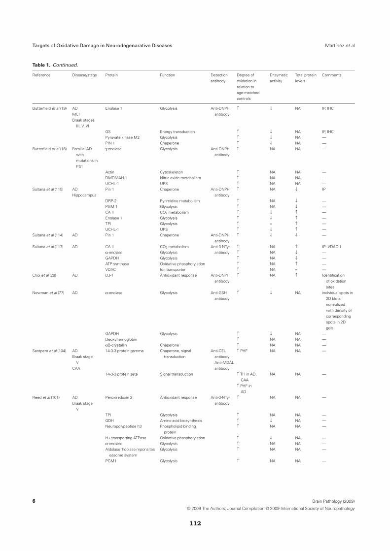
Table 1. Continued.
Reference Disease/stage Protein Function Detectionantibody
Degree ofoxidation inrelation toage-matchedcontrols
Enzymaticactivity
Total proteinlevels
Comments
Butterfield et al (19) ADMCIBraak stages
III, V, VI
Enolase 1 Glycolysis Anti-DNPHantibody
↑ ↓ NA IP, IHC
GS Energy transduction ↑ ↓ NA IP, IHCPyruvate kinase M2 Glycolysis ↑ ↓ NA —PIN 1 Chaperone ↑ ↓ NA —
Butterfield et al (18) Familial ADwithmutations inPS1
g-enolase Glycolysis Anti-DNPHantibody
↑ NA NA —
Actin Cytoskeleton ↑ NA NA —DMDMAH-1 Nitric oxide metabolism ↑ NA NA —UCHL-1 UPS ↑ NA NA —
Sultana et al (115) ADHippocampus
Pin 1 Chaperone Anti-DNPHantibody
↑ NA ↓ IP
DRP-2 Pyrimidine metabolism ↑ NA ↓ —PGM 1 Glycolysis ↑ NA ↓ —CA II CO2 metabolism ↑ ↓ ↑ —Enolase 1 Glycolysis ↑ ↓ ↑ —TPI Glycolysis ↑ = ↑ —UCHL-1 UPS ↑ ↓ ↑ —
Sultana et al (114) AD Pin 1 Chaperone Anti-DNPHantibody
↑ ↓ ↓ —
Sultana et al (117) AD CA II CO2 metabolism Anti-3-NTyrantibody
↑ NA ↑ IP: VDAC-1a-enolase Glycolysis ↑ NA ↓ —GAPDH Glycolysis ↑ NA ↓ —ATP synthase Oxidative phosphorylation ↑ NA ↑ —VDAC Ion transporter ↑ NA = —
Choi et al (29) AD DJ-1 Antioxidant response Anti-DNPHantibody
↑ NA ↑ Identificationof oxidationsites
Newman et al (77) AD a-enolase Glycolysis Anti-GSHantibody
↑ ↓ NA individual spots in2D blotsnormalizedwith density ofcorrespondingspots in 2Dgels
GAPDH Glycolysis ↑ ↓ NA —Deoxyhemoglobin ↑ NA NA —aB-crystallin Chaperone ↑ NA NA —
Santpere et al (104) ADBraak stage
VCAA
14-3-3 protein gamma Chaperone, signaltransduction
Anti-CELantibodyAnti-MDALantibody
↑ PHF NA NA —
14-3-3 protein zeta Signal transduction ↑ TH in AD,CAA
↑ PHF inAD
NA NA —
Reed et al (101) ADBraak stage
V
Peroxiredoxin 2 Antioxidant response Anti-3-NTyrantibody
↑ NA NA —
TPI Glycolysis ↑ NA NA —GDH Amino acid biosynthesis ↑ ↓ NA —Neuropolypeptide h3 Phospholipid binding
protein↑ NA NA —
H+ transporting ATPase Oxidative phosphorylation ↑ ↓ NA —a-enolase Glycolysis ↑ NA NA —Aldolase 1ldolase mponsites
easome systemGlycolysis ↑ NA NA —
PGM1 Glycolysis ↑ NA NA —
Targets of Oxidative Damage in Neurodegenarative Diseases Martínez et al
6 Brain Pathology (2009)
© 2009 The Authors; Journal Compilation © 2009 International Society of Neuropathology
112

Table 1. Continued.
Reference Disease/stage Protein Function Detectionantibody
Degree ofoxidation inrelation toage-matchedcontrols
Enzymaticactivity
Total proteinlevels
Comments
Reed et al (100) ADMCIBraak stages
III, IV, V, VI
Hippocampus Anti-HNEantibody
Neuropolypeptide h3 Phospholipid bindingprotein
↑ NA NA —
Carbonyl reductase 1 Antioxidant response ↑ NA NA —LDH Glycolysis ↑ ↓ NA —PGK Glycolysis ↑ NA NA —HSP70 Chaperone ↑ NA NA —ATP synthase Oxidative phosphorylation ↑ ↓ NA IPa-enolase Glycolysis ↑ NA NA —IPL Anti-HNE
antibodyb-actin Cytoskeleton ↑ NA NA —Pyruvate kinase Glycolysis ↑ ↓ NA —ATP synthase Oxidative phosphorylation ↑ ↓ NA —eIF-a Protein synthesis ↑ NA NA —EF-Tu Protein synthesis ↑ NA NA —
Terni et al (119) ADBraak stages
I/II
ATP synthase Oxidative phosphorylation Anti-HNEantibody
↑ ↓ complexV
= Treatment with10 mMNaBH4 for30 min
Choi et al (26) ApoE-deficientmice
GFAP Cytoskeleton Anti-DNPHantibody
↑ Hippocampus= cortex
NA NA In-strip DNPderivatization
CK BB Energy transduction ↑ Hippocampus= cortex
NA NA —
Glucose regulated protein,Erp61
Chaperone ↑ Hippocampus= cortex
NA NA —
Chaperonin subunit 5 Chaperone ↑ Hippocampus= cortex
NA NA —
Dihydropyrimidinase-relatedprotein 2
Pyrimidine metabolism ↑ Hippocampus= cortex
NA NA —
Mortalin, Grp70 Chaperone ↑ Hippocampus= cortex
NA NA —
Shin et al (105) Tg2576 micebearing theAPP Swedishmutation
Anti-DNPH antibody Anti-DNPHantibodyAnti-3NTyrantibody
—
a-enolase Glycolysis ↑ NA NA —Laminin receptor 1 Neurite growth ↑ NA NA —Anti-3NTyr antibodyAtp5b Oxidative phosphorylation ↑ NA NA —Calpain 12 Cytoskeleton remodeling
processes, celldiferentiation, apoptosis,signal transduction
↑ NA NA —
a-enolase Glycolysis ↑ NA NA —Boyd-Kimball et al
(12)Inracerebral
injection ofAb (1–42) to3-month-oldWistar rats
NBM Anti-DNPHantibody
Sonicationin re-hydrationbuffer on ice
IP of GAPDH
14-3-3 z Signal transduction ↑ NA NA —HSP-60 Chaperone ↑ NA NA —Cortex Anti-DNPH
antibodyGlutamate-ammonia ligase Amino acid biosynthesis ↑ NA NA —Tubulin b chain 15/a-tubulin Cytoskeleton ↑ NA NA —Hippocampus Anti-DNPH
antibodyb-synuclein Regulator of a-synucleinaggregation
↑ NA NA —
14-3-3 z Signal transduction ↑ NA NA —GAPDH Glycolysis ↑ NA NA —Pyruvate dehydrogenase Glycolysis-Kreb’s cycle ↑ NA NA —Phosphoglycerate mutase 1 Glycolysis ↑ NA NA —
Martínez et al Targets of Oxidative Damage in Neurodegenarative Diseases
7Brain Pathology (2009)
© 2009 The Authors; Journal Compilation © 2009 International Society of Neuropathology
113
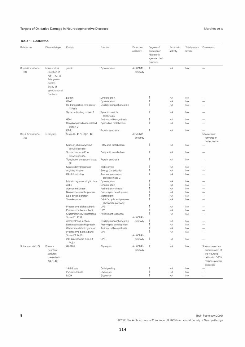
Table 1. Continued.
Reference Disease/stage Protein Function Detectionantibody
Degree ofoxidation inrelation toage-matchedcontrols
Enzymaticactivity
Total proteinlevels
Comments
Boyd-Kimball et al(11)
Intracerebralinjection ofAb (1–42) toMongoliangerbils.Study ofsynaptosomalfractions
g-actin Cytoskeleton Anti-DNPHantibody
↑ NA NA —
b-actin Cytoskeleton ↑ NA NA —GFAP Cytoskeleton ↑ NA NA —H+-transporting two-sector
ATPaseOxidative phosphorylation ↑ NA NA —
Syntaxin binding protein 1 Synaptic vesicleexocytosis
↑ NA NA —
GDH Amino acid biosynthesis ↑ NA NA —Dihydropyrimidinase-related
protein-2Pyrimidine metabolism ↑ NA NA —
EF-Tu Protein synthesis ↑ NA NA —Boyd-Kimball et al
(13)C.elegans Strain CL 4176 (Ab 1–42) Anti-DNPH
antibodySonication in
rehydrationbuffer on ice
Medium-chain acyl-CoAdehydrogenase
Fatty acid metabolism ↑ NA NA —
Short-chain acyl-CoAdehydrogenase
Fatty acid metabolism ↑ NA NA —
Translation elongation factorEF
Protein synthesis ↑ NA NA —
Malate dehydrogenase Kreb’s cycle ↑ NA NA —Arginine kinase Energy transduction ↑ NA NA —RACK1 ortholog Anchoring activated
protein kinase C↑ NA NA —
Myosin regulatory light chain Cytoskeleton ↑ NA NA —Actin Cytoskeleton ↑ NA NA —Adenosine kinase Purine biosynthesis ↑ NA NA —Nematode specific protein Presynaptic development ↑ NA NA —Lipid binding protein Metabolism ↑ NA NA —Transketolase Calvin’s cycle and pentose
phosphate pathway↑ NA NA —
Proteasome alpha subunit UPS ↑ NA NA —Proteasome beta subunit UPS ↑ NA NA —Glutathionine S-transferase Antioxidant response ↑ NA NA —Strain CL 2337 Anti-DNPH
antibodyATP synthase a chain Oxidative phosphorylation ↑ NA NA —Nematode-specific protein Presynaptic development ↑ NA NA —Glutamate dehydrogenase Amino acid biosynthesis ↑ NA NA —Proteasome beta subunit UPS ↑ NA NA —Strain XA 1440 Anti-DNPH
antibody20S proteasome subunitPAS-4
UPS ↑ NA NA —
Sultana et al (116) Primaryneuronalculturestreated withAb (1–42)
GAPDH Glycolysis Anti-DNPHantibody
↑ NA NA Sonication on icepretreatment ofthe neuronalcells with D609reduces proteinoxidation
14-3-3 zeta Cell signaling ↑ NA NA —Pyruvate kinase Glycolysis ↑ NA NA —MDH Glycolysis ↑ NA NA —
Targets of Oxidative Damage in Neurodegenarative Diseases Martínez et al
8 Brain Pathology (2009)
© 2009 The Authors; Journal Compilation © 2009 International Society of Neuropathology
114
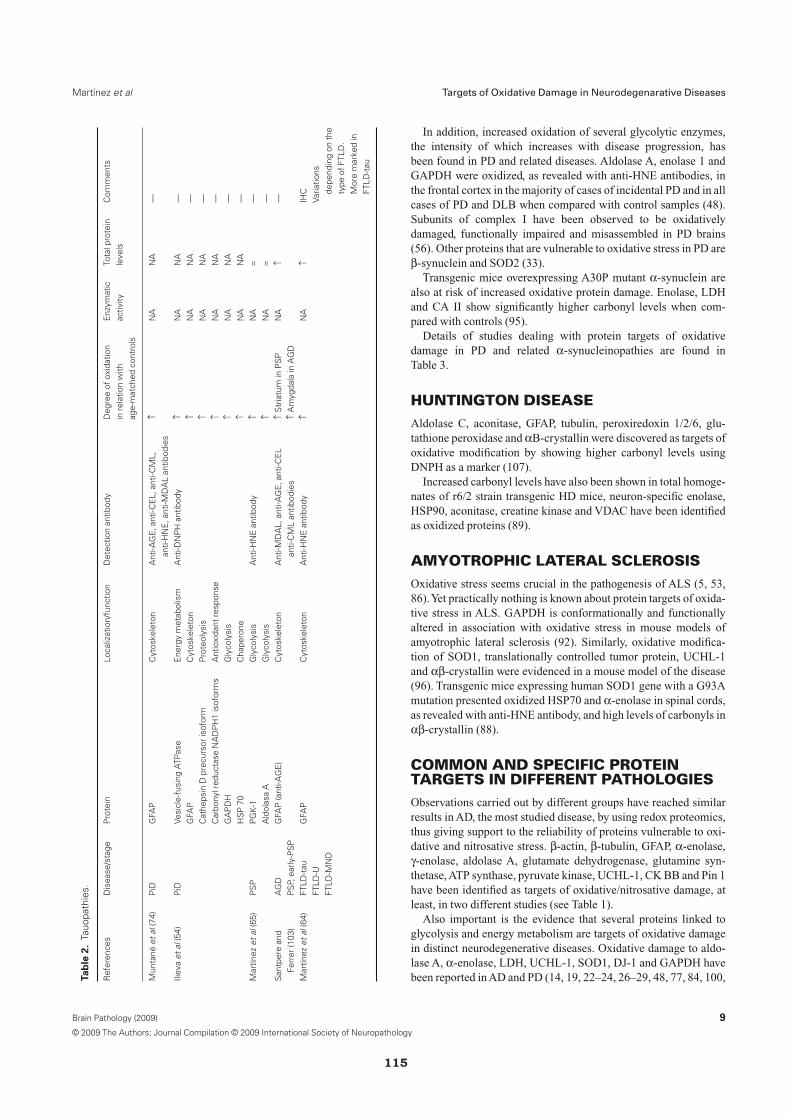
In addition, increased oxidation of several glycolytic enzymes,the intensity of which increases with disease progression, hasbeen found in PD and related diseases. Aldolase A, enolase 1 andGAPDH were oxidized, as revealed with anti-HNE antibodies, inthe frontal cortex in the majority of cases of incidental PD and in allcases of PD and DLB when compared with control samples (48).Subunits of complex I have been observed to be oxidativelydamaged, functionally impaired and misassembled in PD brains(56). Other proteins that are vulnerable to oxidative stress in PD areb-synuclein and SOD2 (33).
Transgenic mice overexpressing A30P mutant a-synuclein arealso at risk of increased oxidative protein damage. Enolase, LDHand CA II show significantly higher carbonyl levels when com-pared with controls (95).
Details of studies dealing with protein targets of oxidativedamage in PD and related a-synucleinopathies are found inTable 3.
HUNTINGTON DISEASEAldolase C, aconitase, GFAP, tubulin, peroxiredoxin 1/2/6, glu-tathione peroxidase and aB-crystallin were discovered as targets ofoxidative modification by showing higher carbonyl levels usingDNPH as a marker (107).
Increased carbonyl levels have also been shown in total homoge-nates of r6/2 strain transgenic HD mice, neuron-specific enolase,HSP90, aconitase, creatine kinase and VDAC have been identifiedas oxidized proteins (89).
AMYOTROPHIC LATERAL SCLEROSISOxidative stress seems crucial in the pathogenesis of ALS (5, 53,86).Yet practically nothing is known about protein targets of oxida-tive stress in ALS. GAPDH is conformationally and functionallyaltered in association with oxidative stress in mouse models ofamyotrophic lateral sclerosis (92). Similarly, oxidative modifica-tion of SOD1, translationally controlled tumor protein, UCHL-1and ab-crystallin were evidenced in a mouse model of the disease(96). Transgenic mice expressing human SOD1 gene with a G93Amutation presented oxidized HSP70 and a-enolase in spinal cords,as revealed with anti-HNE antibody, and high levels of carbonyls inab-crystallin (88).
COMMON AND SPECIFIC PROTEINTARGETS IN DIFFERENT PATHOLOGIESObservations carried out by different groups have reached similarresults in AD, the most studied disease, by using redox proteomics,thus giving support to the reliability of proteins vulnerable to oxi-dative and nitrosative stress. b-actin, b-tubulin, GFAP, a-enolase,g-enolase, aldolase A, glutamate dehydrogenase, glutamine syn-thetase, ATP synthase, pyruvate kinase, UCHL-1, CK BB and Pin 1have been identified as targets of oxidative/nitrosative damage, atleast, in two different studies (see Table 1).
Also important is the evidence that several proteins linked toglycolysis and energy metabolism are targets of oxidative damagein distinct neurodegenerative diseases. Oxidative damage to aldo-lase A, a-enolase, LDH, UCHL-1, SOD1, DJ-1 and GAPDH havebeen reported in AD and PD (14, 19, 22–24, 26–29, 48, 77, 84, 100,T
ab
le2
.Ta
uopa
thie
s.
Ref
eren
ces
Dis
ease
/sta
geP
rote
inLo
caliz
atio
n/fu
nctio
nD
etec
tion
antib
ody
Deg
ree
ofox
idat
ion
inre
latio
nw
ithag
e-m
atch
edco
ntro
ls
Enz
ymat
icac
tivity
Tota
lpro
tein
leve
lsC
omm
ents
Mun
tané
etal
(74)
PiD
GFA
PC
ytos
kele
ton
Ant
i-AG
E,a
nti-C
EL,
anti-
CM
L,an
ti-H
NE
,ant
i-MD
AL
antib
odie
s↑
NA
NA
—
Iliev
aet
al(5
4)P
iDVe
sicl
e-fu
sing
ATP
ase
Ene
rgy
met
abol
ism
Ant
i-DN
PH
antib
ody
↑N
AN
A—
GFA
PC
ytos
kele
ton
↑N
AN
A—
Cat
heps
inD
prec
urso
riso
form
Pro
teol
ysis
↑N
AN
A—
Car
bony
lred
ucta
seN
AD
PH
1is
ofor
ms
Ant
ioxi
dant
resp
onse
↑N
AN
A—
GA
PD
HG
lyco
lysi
s↑
NA
NA
—H
SP
70C
hape
rone
↑N
AN
A—
Mar
tínez
etal
(65)
PS
PP
GK
-1G
lyco
lysi
sA
nti-H
NE
antib
ody
↑N
A=
—A
ldol
asa
AG
lyco
lysi
s↑
NA
=—
San
tper
ean
dFe
rrer
(103
)A
GD
PS
P,ea
rly-P
SP
GFA
P(a
nti-A
GE
)C
ytos
kele
ton
Ant
i-MD
AL,
anti-
AG
E,a
nti-C
EL
anti-
CM
Lan
tibod
ies
↑S
tria
tum
inP
SP
↑A
myg
dala
inA
GD
NA
↑—
Mar
tínez
etal
(64)
FTLD
-tau
FTLD
-UFT
LD-M
ND
GFA
PC
ytos
kele
ton
Ant
i-HN
Ean
tibod
y↑
NA
↑IH
CVa
riatio
nsde
pend
ing
onth
ety
peof
FTLD
.M
ore
mar
ked
inFT
LD-t
au
Martínez et al Targets of Oxidative Damage in Neurodegenarative Diseases
9Brain Pathology (2009)
© 2009 The Authors; Journal Compilation © 2009 International Society of Neuropathology
115

101, 115, 116). Increased oxidative damage to GFAP occurs in AD,tauopathies (PiD, PSP, FTLD-tau and AGD) and HD (54, 57, 65,84, 103, 107).
Mitochondrial proteins are also vulnerable to oxidative/nitrosative stress in different conditions, although vulnerability ofparticular proteins appears to be disease-dependent. ATP synthase(complex V) is a target of oxidative damage in AD (84, 100, 117,119), whereas subunits of complex I have been observed to beoxidatively damaged, functionally impaired and misassembled inPD (56).
These examples will serve to emphasize that certain proteins aredamaged by oxidative stress in different pathologies whereas otherproteins are selectively damaged in one degenerative disease butapparently not in another. Mitochondrial proteins are paradigms ofthis assumption.
Available evidence clearly indicates that only a small fraction ofproteins exhibit discernible oxidative modifications, suggestingselective vulnerability. Obviously, more studies are needed toevaluate structural/functional factors shared by these proteins (ifany) in order to explain this “specificity.” ROS probably act in arandom fashion; however, the sensitivities and proximities ofpotential targets differ. The factors that can affect selectivity ofoxidative damage to proteins could include the presence of a metal-binding site, molecular conformation, rate of proteolysis, relativeabundance of amino acid residues susceptible to metal-catalyzedoxidation, or even protein abundance (91), among others. In thisline, it is clear that modifications present in predominant proteinsare easier to detect than modifications in proteins that are lessabundant. Studies geared to analyze proteins that are represented atlow levels in the brain will improve our understanding of selectivevs. non-selective vulnerability.
However, disease-related specificities in protein vulnerabilityhave been demonstrated as well. This is best exemplified withselective vulnerability of certain subunits of the complexes of therespiratory chain in AD and PD. ATP synthase is consistently oxi-datively damaged in early stages of AD-related pathology, mildcognitive impairment with AD pathology and advanced stages ofAD. In contrast, complex I is consistently altered in PD.
OXIDATIVE CHANGES INPOST-MORTEM BRAIN AREPRINCIPALLY PRIMARYPrimary or secondary oxidative damage in post-mortem brain isdifficult to ascertain as several pre-mortem factors may haveproduce oxidative damage. However, several complementary datasupport a primary origin of the observed protein modifications inpost-mortem human brain. It is important to stress that targets ofoxidative damage are similar in human neurodegenerative diseases,in a case-control approach, and in several animal models coveringintracerebral injection of Ab1–42 in rats and gerbils, and transgenicmice and worm models bearing human mutations of APP. Similarprofiles have been reproduced in primary cortical cultures treatedwith Ab1–42.
Together these data point to the likelihood that at least manyproteins identified as oxidized in the post-mortem human brain arenot modified as a consequence of pre-mortem agonic state but,rather, those modifications are directly linked to the degenerativeprocess.T
ab
le3
.S
ynuc
leop
athi
es.
Ref
eren
ceD
isea
se/s
tage
Pro
tein
Loca
lizat
ion/
func
tion
Det
ectio
nan
tibod
yD
egre
eof
oxid
atio
nin
rela
tion
toag
e-m
atch
edco
ntro
ls
Enz
ymat
icac
tivity
Tota
lpro
tein
leve
lsC
omm
ents
Cho
iet
al(2
6)P
DU
CH
L-1
UP
SA
nti-D
NP
Han
tibod
y↑
(10-
fold
)N
AN
AId
entifi
catio
nof
oxid
atio
nsi
tes
Chu
nget
al(3
0)P
DP
arki
nU
PS
Ant
i-S-n
itro-
syla
ted
park
inan
tibod
y↑
NA
=In
crea
seof
S-n
itros
ylat
edpr
otei
nsm
easu
red
byth
eS
avill
em
etho
dpa
rkin
S-n
itros
ylat
edby
the
biot
insw
itch
met
hod
Yao
etal
(126
)P
DP
arki
nU
PS
Ant
i-S-n
itros
o-pa
rkin
antib
ody
↑N
A=
Det
ectio
nof
S-n
itros
ylat
edpr
otei
nsby
the
biot
insw
itch
met
hod
Cho
iet
al(2
8)P
DS
OD
1A
ntio
xida
ntre
spon
seA
nti-D
NP
Han
tibod
y↑
NA
↑Id
entifi
catio
nof
oxid
atio
nsi
tes
Dal
fóet
al(3
3)iP
Db-
synu
clei
nR
egul
ator
ofa-
synu
clei
nag
greg
atio
nA
nti-M
DA
Lan
tibod
y↑
NA
NA
—S
OD
2A
ntio
xida
ntre
spon
se↑
NA
NA
—C
hoie
tal
(29)
PD
DJ-
1A
ntio
xida
ntre
spon
seA
nti-D
NP
Han
tibod
y↑
NA
↑Id
entifi
catio
nof
oxid
atio
nsi
tes
Kee
ney
etal
(56)
PD
Mito
chon
dria
lco
mpl
exI
Ele
ctro
ntr
ansp
ortc
hain
Ant
i-DN
PH
antib
ody
↑↓
=Im
mun
ocap
ture
tois
olat
eth
eco
mpl
ete
com
plex
Iin
isol
ated
mito
chon
dria
Dal
fóan
dFe
rrer
(32)
iPD
a-sy
nucl
einn
dS
ynap
sis
Ant
i-MD
AL
antib
ody
↑N
AN
AIP
anti-
MD
AL
antib
ody
Góm
ezan
dFe
rrer
(48)
iPD
PD
DLB
Ald
olas
eA
Gly
coly
sis
Ant
i-HN
Ean
tibod
y↑
NA
NA
—
Eno
lase
1G
lyco
lysi
s↑
NA
NA
—G
AD
PH
Gly
coly
sis
↑N
AN
A—
Poo
net
al(9
5)A
30Pa-
synu
clei
ntr
ansg
enic
mic
eLD
HG
lyco
lysi
sA
nti-D
NP
Han
tibod
y↑
↓N
A—
Eno
lase
Gly
coly
sis
↑↓
NA
—C
arbo
nic
anhy
dras
eC
O2
met
abol
ism
↑↓
NA
—
Targets of Oxidative Damage in Neurodegenarative Diseases Martínez et al
10 Brain Pathology (2009)
© 2009 The Authors; Journal Compilation © 2009 International Society of Neuropathology
116
PROTEIN OXIDATION IS AN EARLYEVENT IN NEURODEGENERATIVEDISEASESPioneering studies stressed oxidative damage as an early event inAD (78, 79). In agreement with those predictions, several proteinshave been identified as targets of oxidation and nitration in casesclinically manifested as mild cognitive impairment and pathologi-cally verified as middle (IV) or early advanced (V of Braak) stagesof AD-related pathology (17, 19, 20, 100, 101). More impressive,oxidative damage of ATP synthase and its associated loss of func-tion has been observed in the entorhinal cortex in asymptomaticcases with neuropathologic AD-related pathology restricted to theentorhinal and perirhinal cortices (stage II of Braak), thus repre-senting the earlier oxidative damage to proteins reported in AD(119).
Protein oxidative damage has also been investigated in otherconditions. Protein oxidative damage was increased in brain cortexfrom ALS patients with lumbar debut (53). Increased oxidativedamage of a-synuclein has been found in the substantia nigra atpre-clinical or pre-motor stages of PD (stages II and III of Braak)also known as incidental PD (32). Importantly, increased oxidativedamage of a-synuclein, b-synuclein, SOD2, aldolase A, enolase 1and GADPH has been shown in the cerebral cortex in incidental PD(in addition to PD and DLB) (32, 33, 48). This indicates that thecerebral cortex in PD is involved at very early stages of the diseaseand that oxidative damage to enzymes linked to energy metabolismand glycolysis, oxidative stress responses, and synucleins isalready present at these early stages of the disease and not associ-ated with Lewy pathology (42).
Further studies are needed to unveil oxidative damage at pre-clinical stages in tauopathies and other diseases with abnormalprotein aggregates. However, recent studies have shown increasedGFAP oxidative damage at pre-clinical stages of cases with PSP-like pathology (103).
OXIDATIVE DAMAGE AND LOSS OFFUNCTIONEnzymatic activity decline has been noted in AD with diseaseprogression. Yet loss of activity may be due to reduced numberof cells or specific cellular types, as well to cell redistribution,reduced amount of the enzyme or to modifications in the proteinthat led to protein dysfunction. All these scenarios are probably atwork in advanced stages of neurodegenerative diseases. Amongthese possibilities, it is well known that oxidative damage of pro-teins has consequences in cell function (21).
Unfortunately, the majority of studies dealing with oxidized pro-teins in neurodegenerative diseases are not accompanied by func-tional studies. Perhaps historical events may account for this situa-tion as pioneering works focused on the mere presence of increasedoxidative stress and oxidative damage in aging and degenerativeconditions. This was followed by the identification of DNA, RNA,lipids and proteins as targets of oxidative damage. Subsequentstudies have been centered on the identification of particularproteins.
The enzymatic activities of certain oxidatively damaged proteinshave been analyzed in parallel in a few studies. Increased oxidativedamage accompanied by decreased activity has been shown for
CK BB, enolase 1, glutamine synthetase, Pin-1, CA II, UCHL-1,a-enolase, GAPDH, GDH, H+ transporting ATPase, LDH, ATPsynthase and pyruvate kinase in AD (2, 19, 77, 100, 101, 114, 115,119). However, decreased activity can be related to lower totallevels of the protein; therefore, the value of reduction due to oxida-tion or to the total amount of the particular protein cannot be solvedin those works.
Only a few studies have included the identification of the oxida-tively damaged protein, the quantification of total protein levelsand the reduction of enzymatic activity (2, 114, 115, 119).
No similar data are available in PD, but oxidative damage toLDH, enolase and CA II anhydrase is associated with the corre-sponding decreased enzymatic activities in transgenic mice overex-pressing the human A30P a-synuclein mutation (95).
CLINICAL IMPLICATIONSReduced oxygen uptake and impaired glycolysis have been recog-nized by means of neuroimaging functional studies at relativelyearly stages of AD manifested as mild cognitive impairment, and inadvanced stages of AD. Impaired energy metabolism in the cere-bral cortex has also been reported in pre-clinical stages of individu-als with familial AD (8, 10, 40, 51, 69–73, 93, 94). Unfortunately,no functional neuroimaging and neuropathologic studies have beenperformed in the same cases. As a result, the cause of impairedenergy metabolism in these cases is not known.
However, preserved protein expression levels, together withdecreased enzymatic function associated with oxidative damage ofrelevant energy metabolism enzymes and components of the respi-ratory chain, have been found in a few well-documented studies (2,114, 115, 119). These examples are particularly illuminating asthey demonstrate that reduced oxygen uptake and impaired energymetabolism may be a result of oxidative damage to selected pro-teins rather than a consequence of neuronal loss, at least at earlystages of AD-related pathology.
In the same line, mitochondrial dysfunction and impaired energymetabolism in the cerebral cortex has been demonstrated by severalconvergent neurologic, neuroimaging and biochemical studies inPD (42). These observations reinforce a causal link between oxida-tive modifications of selected proteins and functional impairmentof energy metabolism in PD.
Prognostic implications of these observations are obviousbecause oxidative damage is subject of therapeutic intervention asoxidatively damaged molecules can be substituted by new oneswhereas neuronal loss is not.
CELLULAR LOCALIZATION OFOXIDATIVELY DAMAGED PROTEINSSeminal immunohistochemical studies carried out several yearsago showed that neurons and glial cells were putative sources ofoxidative stress in the nervous system as they were labeled withantibodies that recognize oxidative adducts. Moreover, certain oxi-dative stress responses are particularly robust in astroglia.
In contrast with these observations, it is assumed that oxidativelydamaged proteins, as detected by redox proteomics, are predomi-nantly neuronal proteins. In fact there is little evidence that a par-ticular damaged protein is neuronal or glial, unless the localizationof the protein is known in advance. It is logical to interpret that
Martínez et al Targets of Oxidative Damage in Neurodegenarative Diseases
11Brain Pathology (2009)
© 2009 The Authors; Journal Compilation © 2009 International Society of Neuropathology
117
oxidatively damaged neurofilaments are localized in neurons,whereas oxidized glial fibrillary protein is localized in astrocytes.In some examples, HNE adducts co-localize with GFAP in astro-cytes, as revealed by double-labeling immunofluorescence andconfocal microscopy, at the time that HNE-modified GFAP is iden-tified by bidimensional gel electrophoresis, Western blotting, in-geldigestion and mass spectrometry (64).
It is clear that further studies are needed to elucidate the localiza-tion of damaged molecules in neurons and glial cells to understandthe implications of the abnormalities in definite cellular types. Thisis not only valid considering neurons vs. glial cells, but also amongdifferent neuronal types. The fact that similar molecules are oxida-tively damaged in variegated degenerative diseases does not provethat the same neurons are affected in the different conditions.
PITFALLS AND LIMITATIONSWith no doubt, redox proteomics is a useful tool for detectingdamaged proteins resulting from oxidative/nitrosative damage. Yetavailable studies have also detected limitations and pitfalls.
First of all, information about post-mortem delay is important ascertain proteins might be affected by oxidation/nitration with post-mortem delay. On the other hand, degradation of proteins withpost-mortem delay may minimize the abundance of oxidized pro-teins due to non-specific post-mortem degradation. Several avail-able studies take into consideration this aspect whereas thisinformation is lacking in many others. A caveat derived from thisobservation is that identification of oxidized/nitrated proteins inhuman neurodegenerative diseases is feasible provided that tissuesamples are examined within a time not surpassing thresholds ofprotein degradation and vulnerability to oxidative damage whichare variable from one protein to another.
A second point is the overrepresentation of most abundant pro-teins whereas oxidative damage of minority proteins is probablyunderdetected. This may lead to an oversimplification of damagedmetabolic pathways and, therefore, to the putative neglect ofdamaged crucial components.
A third point is the lack of information regarding cellular typesinvolved, including particular neuronal types in different neurode-generative disorders. We barely understand the reasons for selec-tive cell vulnerability in general terms, and this shortage may alsobe applied to selective vulnerability of individual molecules inparticular cell types.
Another source of possible confusion is based on the conver-gence of abnormalities in particular metabolic pathways that mayobscure the real impact of oxidative damage in determined cellularfunctions. As an example, abnormal mitochondrial function in ADmay be the result of several components, including increasedmitochondrial DNA deletions, abnormal fusion and fission of mito-chondria, and decreased expression of certain complexes of therespiratory chain, such as complex IV (45, 52, 68, 87, 125). Mito-chondrial alterations result in increased oxidative stress. Oxidativedamage to ATP synthase (complex V), VDAC, ubiquinol–cytochrome c reductase complex core protein I, H+ transportingATPase and Atp5b (11, 13, 58, 84, 100, 105, 117, 119) may, in turn,increase mitochondrial dysfunction.
Similar considerations can be applied to the cerebral cortex inPD (42, 76).
REFINING METHODS TO IMPROVEREDOX PROTEOMICSThe large majority of studies in human brain and animal modelshave been carried out by analyzing total homogenates of a par-ticular region. This is with no doubt an adequate approachalthough damage of minority proteins can be easily underrepre-sented. The study of cellular fractions may improve the resolutionof the study by increasing the total amount of a particular protein.In this line, sarkosyl-insoluble fractions have been used torecover proteins in paired-helical filament-enriched fractions(104), mitochondria-enriched fractions to recover proteins princi-pally related with mitochondria (30, 48), or synaptosomal-enriched fractions to reveal abnormalities of synapsis-relatedproteins (11).
Another important point is the use of different protocols andbuffers to increase the capture of different proteins. The combina-tion of different buffers to grind or to focus samples is a strategy toimprove the reproducibility at the acidic or alkaline extremes of theelectrophoresis gel and, likewise, to solubilize different proteinsreceiving a better number of spots and resolution (9). This observa-tion, originally applied to general bidimensional gel electro-phoresis methodology, may be of considerable interest in redoxproteomics to optimize spots detected as oxidized proteins. Weshould also take into account that membrane proteins are difficultto detect by current bidimensional gel electrophoresis, thus pro-bably accounting for the low numbers of membrane proteinsidentified as targets of oxidation.
Finally, detection of specific residues of oxidative modificationsmay increase understanding of specific oxidation sites and theirrelevance to protein function. However, this approach is time-consuming and a combination of different methods is needed. Byusing MALDI-TOF/MS and HPLC-ESI/MS/MS techniques, oxi-dation sites have been identified only in UCH-L1, SOD1 and DJ-1in AD and PD as yet (27, 28).
CONCLUDING COMMENTSNeurodegenerative diseases with abnormal protein aggregates areassociated with modifications of solubility, aggregation and fibrilformation of selected proteins. Mutant proteins resulting fromDNA mutations are causative in familial and certain sporadic set-tings. More commonly, post-translational modifications of pro-teins are involved in the majority of sporadic cases. Understand-ing the mechanisms involved in such modifications is crucialfrom a mechanistic perspective, but it is also essential for thedelineation of therapeutic strategies. Oxidative stress plays afunctional role in physiologic conditions as it switches on vitalcellular responses. Yet imbalance between oxidative stresssources and antioxidant responses may cause a net flux of oxida-tive damage to DNA, RNA, carbohydrates, lipids and proteins,and, in most cases, concomitant loss of function. Oxidative stressincreases with age largely because of progressive mitochondrialdysfunction and impairment or loss of cellular repair mecha-nisms. In addition, neurodegenerative diseases are associatedwith higher levels of oxidative damage and higher levels of directand indirect protein modifications resulting from increasedoxidation, nitrosation and nitration when compared with those
Targets of Oxidative Damage in Neurodegenarative Diseases Martínez et al
12 Brain Pathology (2009)
© 2009 The Authors; Journal Compilation © 2009 International Society of Neuropathology
118

occurring in age-matched individuals with no diseases of thenervous system.
Vulnerable proteins can be grouped in defined metabolic path-ways covering glycolysis and energy metabolism, mitochondrialproteins, cytoskeleton, chaperones, and members of the ubiquitin–proteasome system, among many others. Some proteins areaffected in different degenerative diseases whereas others appear tobe disease-specific. Importantly, many damaged proteins in humanneurodegenerative diseases are also damaged in experimentalmodels, transgenic mice and worms, and cell culture paradigms.These findings indicate that oxidative stress observed in post-mortem brains is a primary event linked to degeneration rather thana secondary effect resulting from pre-mortem agonic states.
Since oxidative damage may result in impaired function, proteinoxidative damage may have important consequences on thenervous system thus resulting in abnormal glycolysis and energymetabolism, abnormal responses to protein folding and oxidativestress responses, cytoskeletal abnormalities, and impaired proteindegradation, in addition to damage to relevant proteins asa-synuclein in PD and related a-synucleinopathies. Some of theseabnormalities are reflected in vivo by using sophisticated metabolicand neuroimaging methods. Thus, abnormal energy metabolismhas been observed in the cerebral cortex not only in AD but also inpatients with mild cognitive impairment and in patients with PD inwhom impaired metabolism cannot be ascribed to neuron loss.Therefore, it is reasonable to think that part of the metabolic distur-bances observed at early stages of degenerative processes arerelated to oxidative damage of selected proteins rather than toneuron loss. However, much work has to be done as the majority ofredox proteomics studies are not accompanied by functional analy-sis of oxidatively damaged proteins.
Available information points to the fact that vital metabolic path-ways are hampered by protein oxidative damage in several humandegenerative diseases at very early stages of the disease. A betterunderstanding of proteins susceptible to oxidation and nitrationmay serve to define damaged metabolic networks at early stagesof disease and to procure therapeutic interventions to attenuatedisease progression.
ACKNOWLEDGMENTSWork carried out at the Institute of Neuropathology was partiallyfunded by grants from the Spanish Ministry of Health, Institutode Salud Carlos III PI080582, and supported by the EuropeanCommission under the Sixth Framework Programme BrainNetEurope II, LSHM-CT-2004-503039 and INDABIP FP6-2005-LIFESCIHEALTH-7 Molecular Diagnostics. Work carried outat the Department of Experimental Medicine was supported inpart by I+D grants from the Spanish Ministry of Educationand Science (BFU2006-14495/BFI and AGL2006-12433), theSpanish Ministry of Health (ISCIII, Red de Envejecimiento yFragilidad, RD06/0013/0012, PI081843), the Autonomous Gov-ernment of Catalonia (2005SGR00101), “La Caixa” Foundationand COST B-35 Action.
We thank Odena MA and Oliveira E from the Proteomics plat-form, Science Park, University of Barcelona, for support.
Thanks to T. Yohannan for editorial help. There is no conflict ofinterest including any financial, personal or other relationships
with other people or organizations within the three years from thebeginning of the work.
REFERENCES
1. Adams S, Green P, Claxton R, Simcox S, Williams MV, Walsh K,Leewenburgh C (2001) Reactive carbonyl formation by oxidativeand non-oxidative pathways. Front Biosci 6:A17–A24.
2. Aksenov M, Aksenova M, Butterfield DA, Markesbery WR (2000)Oxidative modification of creatine kinase BB in Alzheimer’s diseasebrain. J Neurochem 74:2520–2527.
3. Aksenov MY, Atiksenova MV, Butterfield DA, Geddes JW,Markesbery WR (2001) Protein oxidation in the brain inAlzheimer’s disease. Neuroscience 103:373–383.
4. Aruoma OI, Kaur H, Halliwell B (1991) Oxygen free radicals andhuman diseases. J R Soc Health 111:172–177.
5. Barber SC, Mead RJ, Shaw PJ (2006) Oxidative stress in ALS: amechanism of neurodegeneration and a therapeutic target. BiochimBiophys Acta 1762:1051–1067.
6. Barja G (2007) Mitochondrial oxygen consumption and reactiveoxygen species production are independently modulated:implications for aging studies. Rejuvenation Res 10:215–224.
7. Barnham KJ, Masters CL, Bush AI (2004) Neurodegenerativediseases and oxidative stress. Nat Rev Drug Discov 3:205–214.
8. Bigl M, Brückner MK, Arendt T, Bigl V, Eschrich K (1999)Activities of key glycolytic enzymes in the brains of patients withAlzheimer’s disease. J Neural Transm 106:499–511.
9. Bland AM, D’Eugenio LR, Dugan MA, Janech MG, Almeida JS,Zile MR, Arthur JM (2006) Comparison of variability associatedwith sample preparation in two-dimensional gel electrophoresis ofcardiac tissue. J Biomol Tech 17:195–199.
10. Blass JP (2002) Alzheimer’s disease and Alzheimer’s dementia:distinct but overlapping entities. Neurobiol Aging 23:1077–1084.
11. Boyd-Kimball D, Castegna A, Sultana R, Poon HF, Petroze R, LynnBC et al (2005) Proteomic identification of proteins oxidized byAb(1–42) in synaptosomes: implications for Alzheimer’s disease.Brain Res 1044:206–215.
12. Boyd-Kimball D, Sultana R, Poon HF, Lynn BC, Casamenti F,Pepeu G et al (2005) Proteomic identification of proteins specificallyoxidized by intracerebral injection of amyloid b-peptide (1–42) intorat brain: implications for Alzheimer’s disease. Neuroscience132:313–324.
13. Boyd-Kimball D, Poon HF, Lynn BC, Cai J, Pierce WM Jr, Klein JBet al (2006) Proteomic identification of proteins specifically oxidizedin Caenorhabditis elegans expressing human Ab(1–42): implicationsfor Alzheimer’s disease. Neurobiol Aging 27:1239–1249.
14. Butterfield DA, Kanski J (2001) Brain protein oxidation inage-related neurodegenerative disorders that are associated withaggregated proteins. Mech Ageing Dev 122:945–962.
15. Butterfield DA, Stadtman ER (1997) Protein oxidation processes inaging brain. In: Advances in Cell Aging and Gerontology, Vol. 2. PSTimiras, EE Bittar (eds), pp. 161–191. JAI Press, Greenwich.
16. Butterfield DA, Sultana R (2008) Redox proteomics: understandingoxidative stress in the progression of age-related neurodegenerativedisorders. Expert Rev Proteomics 5:157–160.
17. Butterfield DA, Abdul HM, Newman S, Reed T (2006) Redoxproteomics in some age-related neurodegenerative disorders ormodels thereof. NeuroRx 3:344–357.
18. Butterfield DA, Gnjec A, Poon HF, Castegna A, Pierce WM, KleinJB, Martins RN (2006) Redox proteomics identification ofoxidatively modified brain proteins in inherited Alzheimer’s disease:an initial assessment. J Alzheimers Dis 10:391–397.
19. Butterfield DA, Poon HF, St Clair D, Keller JN, Pierce WM, KleinJB, Markesbery WR (2006) Redox proteomics identification of
Martínez et al Targets of Oxidative Damage in Neurodegenarative Diseases
13Brain Pathology (2009)
© 2009 The Authors; Journal Compilation © 2009 International Society of Neuropathology
119
oxidatively modified hippocampal proteins in mild cognitiveimpairment: insights into the development of Alzheimer’s disease.Neurobiol Dis 22:223–232.
20. Butterfield DA, Sultana R, Poon HF (2006) Redox proteomics: anew approach to investigate oxidative stress in Alzheimer’s disease.In: Redox Proteomics: From Protein Modifications to CellularDysfunction and Diseases. I Dalle-Donne, A Scaloni, DA Butterfield(eds), pp. 563–603. John Wiley & Sons: Hoboken, NJ.
21. Cabiscol E, Ros J (2006) Oxidative damage to proteins: structuralmodifications and consequences in cell function. In: RedoxProteomics: From Protein Modifications to Cellular Dysfunction andDiseases. I Dalle-Donne, A Scaloni, DA Butterfield (eds), pp.399–471. John Wiley & Sons: Hoboken, NJ.
22. Castegna A, Aksenov M, Aksenova M, Thongboonkerd V, Klein JB,Pierce WM et al (2002) Proteomic identification of oxidativelymodified proteins in Alzheimer’s disease brain. Part I: creatinekinase BB, glutamine synthase, and ubiquitin carboxy-terminalhydrolase L-1. Free Radic Biol Med 33:562–571.
23. Castegna A, Aksenov M, Thongboonkerd V, Klein JB, Pierce WM,Booze R et al (2002) Proteomic identification of oxidativelymodified proteins in Alzheimer’s disease brain. Part II:dihydropyrimidinase-related protein 2, a-enolase and heat shockcognate 71. J Neurochem 82:1524–1532.
24. Castegna A, Thongboonkerd V, Klein JB, Lynn B, Markesbery WR,Butterfield DA (2003) Proteomic identification of nitrated proteins inAlzheimer’s disease brain. J Neurochem 85:1394–1401.
25. Chinta SJ, Andersen JK (2008) Redox imbalance in Parkinson’sdisease. Biochem Biophys Acta 1780:1362–1367.
26. Choi J, Forster MJ, McDonald SR, Weintraub ST, Carroll CA, GracyRW (2004) Proteomic identification of specific oxidized proteins inApoE-knockout mice: relevance to Alzheimer’s disease. Free RadicBiol Med 36:1155–1162.
27. Choi J, Levey AI, Weintraub ST, Rees HD, Gearing M, Chin LS, LiL (2004) Oxidative modifications and down-regulation of ubiquitincarboxyl-terminal hydrolase L1 associated with idiopathicParkinson’s and Alzheimer’s diseases. J Biol Chem279:13256–13264.
28. Choi J, Rees HD, Weintraub ST, Levey AI, Chin LS, Li L (2005)Oxidative modifications and aggregation of Cu,Zn-superoxidedismutase associated with Alzheimer and Parkinson diseases. J BiolChem 280:11648–11655.
29. Choi J, Sullards MC, Olzmann JA, Rees HD, Weintraub ST,Bostwick DE et al (2006) Oxidative damage of DJ-1 is linked tosporadic Parkinson and Alzheimer diseases. J Biol Chem281:10816–10824.
30. Chung KKK, Thomas B, Li X, Pletnikova O, Troncoso JC, Marsh Let al (2004) S-nitrosylation of parkin regulates ubiquitinationand compromises parkin’s protective function. Science304:1328–1331.
31. Cosgrove JP, Church DF, Pryor WA (1987) The kinetics of theautoxidation of polyunsaturated fatty acids. Lipids 22:299–304.
32. Dalfó E, Ferrer I (2008) Early a-synuclein lipoxidation in neocortexin Lewy body diseases. Neurobiol Aging 29:408–417.
33. Dalfó E, Portero-Otín M, Ayala V, Martínez A, Pamplona R,Ferrer I (2005) Evidence of oxidative stress in the neocortex inincidental Lewy body disease. J Neuropathol Exp Neurol64:816–830.
34. Dalle-Donne I, Scaloni A, Giustarini D, Cavarra E, Tell G,Lungarella G et al (2005) Proteins as biomarkers ofoxidative/nitrosative stress in diseases: the contribution of redoxproteomics. Mass Spectrom Rev 24:55–99.
35. Dalle-Donne I, Aldini G, Carini M, Colombo R, Rossi R, Milzani A(2006) Protein carbonylation, cellular dysfunction, and diseaseprogression. J Cell Mol Med 10:389–406.
36. Dalle-Donne I, Rossi R, Ceciliani F, Giustarini D, Colombo R,Milzani A (2006) Proteins as sensitive biomarkers of humanconditions associated with oxidative stress. In: Redox Proteomics:From Protein Modifications to Cellular Dysfunction and Diseases. IDalle-Donne, A Scaloni, DA Butterfield (eds), pp. 487–525. JohnWiley & Sons: Hoboken, NJ.
37. Dalle-Donne I, Scaloni A, Butterfield DA (eds) (2006) RedoxProteomics: From Protein Modifications to Cellular Dysfunction andDiseases. John Wiley & Sons: Hoboken, NJ.
38. Daneshvar B, Frandsen H, Autrup H, Dragsted LO (1997)g-Glutamyl semialdehyde and 2-amino-adipic semialdehyde:biomarkers of oxidative damage to proteins. Biomarkers2:117–123.
39. Danielson SR, Andersen JK (2008) Oxidative and nitrative proteinmodifications in Parkinson’s disease. Free Radic Biol Med44:1787–1794.
40. De Leon MJ, Convit A, Wolf OT, Tarshish CY, DeSanti S, RusinekH et al (2001) Prediction of cognitive declinein normal elderlysubjects with 2-[18F]fluorodo-2-deoxy-D-glucosee/positron-emission tomography (FDG/PET). Proc Natl Acad Sci USA98:10966–10971.
41. Ding Q, Dimayuga E, Keler JN (2007) Oxidative damage, proteinsynthesis, and protein degradation in Alzheimer’s disease. CurrAlzheimer Res 4:73–79.
42. Ferrer I (2009) Early involvement of the cerebral cortex inParkinson’s disease: convergence of multiple metabolic defects.Progr Neurobiol 88:89–103.
43. Ferrer I, Martinez A, Boluda S, Parchi P, Barrachina M (2008) Brainbanks: benefits, limitations and cautions concerning the use ofpost-mortem brain tissue for molecular studies. Cell Tissue Bank9:181–194.
44. Gibson GE, Huang HM (2005) Oxidative stress in Alzheimer’sdisease. Neurobiol Aging 26:575–578.
45. Gibson GE, Karuppagounder SS, Shi Q (2008) Oxidant-inducedchanges in mitochondria and calcium dynamics in thepathophysiology of Alzheimer’s disease. Ann NY Acad Sci1147:221–232.
46. Gibson GE, Ratan RR, Beal MF (2008) Mitochondria and oxidatiuvestress in neurodegenerative disorders. Preface. Ann NY Acad Sci1147:xi–xii.
47. Gilca M, Stoian I, Atanasiu V, Virgolici B (2007) The oxidativehypothesis of senescence. J Postgrad Med 53:207–213.
48. Gómez A, Ferrer I (2009) Increased oxidation of certain glycolysisand energy metabolism enzymes in the frontal cortex in Lewy bodydiseases. J Neurosci Res 87:1002–1013.
49. Halliwell B (1991) Reactive oxygen species in living systems:source, biochemistry, and role in human disease. Am J Med91:14S–22S.
50. Halliwell B, Gutteridge JMC (2007) Free Radicals in Biology andMedicine. Oxford University Press: New York.
51. Herholz K, Carter SF, Jones M (2007) Positron emissiontomography imaging in dementia. Br J Radiol 80(Spec. No.2):S160–S167.
52. Hirai K, Aliev G, Nunomura A, Fujioka H, Rusell RL, Atwood CSet al (2001) Mitochondrial abnormalities in Alzheimer’s disease. JNeurosci 21:3017–3023.
53. Ilieva EV, Ayala V, Jové M, Dalfó E, Cacabelos D, Povedano M et al(2007) Oxidative and endoplasmic reticulum stress interplay insporadic amyotrophic lateral sclerosis. Brain 130:3111–3123.
54. Ilieva EV, Naudí A, Kichev A, Ferrer I, Pamplona R, Portero-Otín M(2009) Loss of the Stress Transducers Nrf2 and Grp78/BiP in Pick’sDisease. (In press).
55. Ischiropoulos H, Al-Medi AB (1995) Peroxynitrite-mediatedoxidative protein modifications. FEBS Lett 364:279–282.
Targets of Oxidative Damage in Neurodegenarative Diseases Martínez et al
14 Brain Pathology (2009)
© 2009 The Authors; Journal Compilation © 2009 International Society of Neuropathology
120
56. Keeney PM, Xie J, Capaldi RA, Bennett JP (2006) Parkinson’sdisease brain mitochondrial complex I has oxidatively damagedsubunits and is functionally impaired and misassembled. J Neurosci26:5256–5264.
57. Korolainen MA, Auriola S, Nyman TA, Alafuzoff I, Pirttilä T (2005)Proteomic analysis of glial fibrillary acidic protein in Alzheimer’sdisease and aging brain. Neurobiol Dis 20:858–870.
58. Korolainen MA, Goldsteins G, Nyman TA, Alafuzoff I, KoistinahoJ, Pirttilä T (2006) Oxidative modification of proteins in thefrontal cortex of Alzheimer’s disease brain. Neurobiol Aging27:42–53.
59. LaVoie MJ, Cortese GP, Ostaszewski BL, Schlossmacher MG(2007) The effects of oxidative stress on parkin and other E3 ligases.J Neurochem 103:2354–2368.
60. Levine RL, Stadtman ER (2001) Oxidative modifications of proteinsduring aging. Exp Gerontol 36:1495–1502.
61. Lowell MA, Markesbery WB (2007) Oxidative DNA damage inmild cognitive impairment and late-stage Alzheimer’s disease. NuclAcid Res 35:7497–7504.
62. Mancuso C, Scapagini G, Currò D, Giuffrida Stella AM, De MarcoC, Butterfield DA, Calabrese V (2007) Mitochondrial dysfunction,free radical generation and cellular stress response inneurodegenerative disorders. Front Biosci 12:1107–1123.
63. Markesbery WR, Lowell MA (2007) Damage to lipids, proteins,DNA and RNA in mild cognitive impairment. Arch Neurol64:954–956.
64. Martínez A, Carmona M, Portero-Otin M, Naudí A, Pamplona R,Ferrer I (2008) Type-dependent oxidative damage in frontotemporallobar degeneration: cortical astrocytes are targets of oxidativedamage. J Neuropathol Exp Neurol 67:1122–1136.
65. Martínez A, Dalfó E, Muntané G, Ferrer I (2008) Glycolitic enzymesare targets of oxidation in aged human frontal cortex and oxidativedamage of these proteins is increased in progressive supranuclearpalsy. J Neural Transm 115:59–66.
66. Mikkelsen RB, Wardman P (2003) Biological, chemistry of reactiveoxygen and nitrogen and radiation-induced signal transductionmechanisms. Oncogene 22:5734–5754.
67. Moreira PI, Smith MA, Zhu X, Nunomura A, Castellani RJ, Perry G(2005) Oxidative stress and neurodegeneration. Ann NY Acad Sci1043:545–552.
68. Moreira PI, Santos MS, Oliveira CR (2007) Alzheimer’s disease: alesson from mitochondrial dysfunction. Antioxid Redox Signal9:1621–1630.
69. Mosconi L (2005) Brain glucose metabolism in the early andspecific diagnosis of Alzheimer’s disease. Eur J Nucl Med32:486–510.
70. Mosconi L, De Santi S, Li J, Tsui WH, Li Y, Boppana M et al(2006) Hippocampal metabolism predicts cognitive decline fromnormal aging. Neurobiol Aging 29:676–692.
71. Mosconi L, Sorbi S, De Leon MJ, Li Y, Nacmias B, Myoung PSet al (2006) Hypometabolism exceeds atrophy in presymptomaticearly-onset familial Alzheimer’s disease. J Nucl Med47:1778–1786.
72. Mosconi L, Pupi A, De Leon MJ (2008) Brain glucose metabolismand oxidative stress in preclinical Alzheimer’s disease. Ann NY AcadSci 1147:180–195.
73. Mosconi L, Tsui WH, Herholz K, Pupi A, Drzezga A, Lucignani Get al (2008) Multicenter standardized 18F-FDG PET diagnosis ofmild cognitive impairment, Alzheimer’s disease, and otherdementias. J Nucl Med 49:390–398.
74. Muntané G, Dalfó E, Martínez A, Rey MJ, Avila J, Pérez M et al(2006) Glial fibrillary acidic protein is a major target ofglycoxidative and lipoxidative damage in Pick’s disease. JNeurochem 99:177–185.
75. Nakabeppu Y, Tsuchimoto D, Yamaguchi H, Sakumi K (2007)Oxidative damage in nucleic acids and Parkinson’s disease. JNeurosci Res 85:919–934.
76. Navarro A, Boveris A, Bández MJ, Sanchez-Pinto MJ, Gómez C,Muntane G, Ferrer I (2009) Human brain cortex: mitochondrialoxidative damage and adaptative response in Parkinson disease anddementia with Lewy bodies. Free Radic Biol Med 46:1574–1580.
77. Newman SF, Sultana R, Perluigi M, Coccia R, Cai J, Pierce WMet al (2007) An increase in S-glutathionylated proteins in theAlzheimer’s disease inferior parietal lobule, a proteomics approach.J Neurosci Res 85:1506–1514.
78. Nunomura A, Perry G, Aliev G, Hirai K, Takeda A, Balraj EK et al(2001) Oxidative damage is the earliest event in Alzheimer disease. JNeuropathol Exp Neurol 60:759–767.
79. Nunomura A, Castellani RJ, Zhu X, Moreira PI, Perry G, Smith MA(2006) Involvement of oxidative stress in Alzheimer disease. JNeuropathol Exp Neurol 65:631–641.
80. Nunomura A, Moreira PI, Takeda A, Smith MA, Perry G (2007)Oxidative RNA damage and neurodegeneration. Curr Med Chem14:2968–2975.
81. Onyango IG, Khan SM (2006) Oxidative stress, mitochondrialdysfunction, and stress signalling in Alzheimer’s disease. CurrAlzheimer Res 3:339–349.
82. Pamplona R (2008) Membrane phospholipids, lipoxidative damageand molecular integrity: a causal role in aging and longevity.Biochim Biophys Acta 1777:1249–1262.
83. Pamplona R, Barja G (2007) Highly resistant macromolecularcomponents and low rate of generation of endogenous damage: twokey traits of longevity. Ageing Res Rev 6:189–210.
84. Pamplona R, Dalfó E, Ayala V, Bellmunt MJ, Prat J, Ferrer I,Portero-Otín M (2005) Proteins in human brain cortex are modifiedby oxidation, glycoxidation, and lipoxidation. Effects of Alzheimerdisease and identification of lipoxidation targets. J Biol Chem280:21522–21530.
85. Pamplona R, Ilieva E, Ayala V, Bellmunt MJ, Cacabelos D, Dalfo Eet al (2008) Maillard reaction versus other nonenzymaticmodifications in neurodegenerative processes. Ann NY Acad Sci1126:315–319.
86. Pedersen WA, Fu W, Keller JN, Markesbery WR, Appel S, SmithRG et al (1998) Protein modification by the lipid peroxidationproduct 4-hydroxynonenal in the spinal cords of amyotrophic lateralsclerosis patients. Ann Neurol 44:819–824.
87. Perez-Gracia E, Torrejon-Escribano B, Ferrer I (2008) Dystrophicneurites of senile plaques in Alzheimer’s disease are deficient incytochrome C oxidase. Acta Neuropathol 116:261–268.
88. Perluigi M, Fai Poon H, Hensley K, Pierce WM, Klein JB, CalabreseV et al (2005) Proteomic analysis of 4-hydroxy-2-nonenal-modifiedproteins in G93A-SOD1 transgenic mice—a model of familialamyotrophic lateral sclerosis. Free Radic Biol Med 38:960–968.
89. Perluigi M, Poon HF, Maragos W, Pierce WM, Klein JB, CalabreseV et al (2005) Proteomic analysis of protein expression andoxidative modification in r6/2 transgenic mice: a model ofHuntington disease. Mol Cell Proteomics 4:1849–1861.
90. Petersen RB, Nunomura A, Lee HG, Casadesus G, Perry G, SmithMA, Zhu X (2007) Signal transduction cascades associated withoxidative stress in Alzheimer’s disease. J Alzheimers Dis11:143–152.
91. Petrak J, Ivanek R, Toman O, Cmejla R, Cmejlova J, Vyoral D et al(2008) Déjà vu in proteomics. A hit parade of repeatedly identifieddifferentially expressed proteins. Proteomics 8:1744–1749.
92. Pierce A, Mirzaei H, Muller F, De Waal E, Taylor AB, Leonard Set al (2008) GAPDH is conformationally and functionally altered inassociation with oxidative stress in mouse models of amyotrophiclateral sclerosis. J Mol Biol 382:1195–1210.
Martínez et al Targets of Oxidative Damage in Neurodegenarative Diseases
15Brain Pathology (2009)
© 2009 The Authors; Journal Compilation © 2009 International Society of Neuropathology
121
93. Piert M, Koeppe RA, Giordani B, Berent S, Kuhl DE (1996)Diminished glucose transport and phosphorylation in Alzheimer’sdisease determined by dynamic FDG-PET. J Nucl Med 37:201–208.
94. Pietrini P, Furey ML, Guazzelli M, Alexander GE (2001) Functionalbrain studies of the neurometabolic bases of cognitive and behavioralchanges in Alzheimer’s disease. In: Functional Neurobiology ofAging. PR Hof, CV Mobbs (eds), pp. 227–241. Academic Press:New York.
95. Poon HF, Frasier M, Shreve N, Calabrese V, Wolozin B, ButterfieldDA (2005) Mitochondrial associated metabolic proteins areselectively oxidized in A30P alpha-synuclein transgenic mice—amodel of familial Parkinson’s disease. Neurobiol Dis 18:492–498.
96. Poon HF, Hensley K, Thongboonkerd V, Merchant ML, Lynn BC,Pierce WM et al (2005) Redox proteomics analysis of oxidativelymodified proteins in G93A-SOD1 transgenic mice—a model offamilial amyotrophic lateral sclerosis. Free Radic Biol Med39:453–462.
97. Portero-Otin M, Pamplona R (2006) Is endogenous oxidative proteindamage envolved in the aging process? In: Protein Oxidation andDisease. J Pietzsch (ed.), pp. 91–142. Research Signpost, Kerala,India.
98. Practico D (2008) Oxidative stress hypothesis in Alzheimer’sdisease: a reappraisal. Trends Pharmacol Sci 29:609–615.
99. Pryor WA, Squadrito GL (1995) The chemistry of peroxynitrite: aproduct from the reaction of nitric oxide with superoxide. Am JPhysiol 268:L699–L722.
100. Reed T, Perluigi M, Sultana R, Pierce WM, Klein JB, Turner DMet al (2008) Proteomic identification of 4-hydroxy-2-nonenal-modified brain proteins in amnestic mild cognitive impairment:insight into the role of lipid peroxidation in the progression andpathogenesis of Alzheimer’s disease. Neurobiol Dis 30:107–120.
101. Reed TT, Pierce WM Jr, Turner DM, Markesbery WR, ButterfieldDA (2008) Proteomic identification of nitrated brain proteins in earlyAlzheimer’s disease inferior parietal lobule. J Cell Mol MedDOI: 10.1111/j.1582-4934.2008.00478.
102. Requena J, Chao CC, Stadtman ER (2001) Glutamic acid andaminodipidic semialdehydes are thye main carbonyl products ofmetal-catalyzed oxidation of proteins. Proc Natl Acad Sci USA98:624–632.
103. Santpere G, Ferrer I (2008) Delineation of progressive supranuclearpalsy-like pathology. Astrocytes in striatum are primary targets oftau phosphorylation and GFAP oxidation. Brain Pathol 19:177–187.
104. Santpere G, Puig B, Ferrer I (2007) Oxidative damage of 14-3-3 zetaand gamma isoforms in Alzheimer’s disease and cerebral amyloidangiopathy. Neuroscience 146:1640–1651.
105. Shin SJ, Lee SE, Boo JH, Kim M, Yoon YD, Kim SI, Mook-Jung I(2004) Profiling proteins related to amyloid deposited brain ofTg2576 mice. Proteomics 4:3359–3368.
106. Smith MA, Rudnicka-Nawrot M, Richey P, Praprotnik D,Mulvihill P, Miller CA et al (1995) Carbonyl-relatedpost-translational modification of neurofilament protein in theneurofibrillary pathology of Alzheimer’s disease. J Neurochem64:2660–2666.
107. Sorolla MA, Reverter-Branchat G, Tamarit J, Ferrer I, Ros J,Cabiscol E (2008) Proteomic and oxidative stress analysis in humanbrain samples of Huntington disease. Free Radic Biol Med45:667–678.
108. Stadtman ER (1998) Free radical-mediated oxidation of proteins. In:Free Radicals, Oxidative Stress, and Antioxidants: Pathological andPhysiological Significance. NATO ASI Series, Series A: LifeSciences, Vol. 296. T Ozben (ed.), pp. 51–143. Plenum Press: NewYork.
109. Stadtman ER (2002) Importance of individuality in oxidative stressand aging. Free Radic Biol Med 33:597–604.
110. Stadtman ER, Berlett BS (1997) Free radical-mediated modificationof proteins. In: Free Radical Toxicity. KB Wallace (ed.), pp. 71–87.Taylor and Francis: Washington, DC.
111. Stadtman ER, Levine RL (2006) Chemical modification of proteinsby reactive oxygen species. In: Redox Proteomics: From ProteinModifications to Cellular Dysfunction and Diseases. I Dalla-Donne,A Scaloni, DA Butterfield (eds), pp. 3–23. John Wiley & Sons:Hoboken, NJ.
112. Starkov AA (2008) The role of mitochondria in reactive oxygenspecies metabolism and signaling. Ann NY Acad Sci 1147:37–52.
113. Su B, Wang X, Nunomura A, Moreira PI, Lee HG, Perry G et al(2008) Oxidative stress signalling in Alzheimer’s disease. CurrAlzheimer Res 5:525–532.
114. Sultana R, Boyd-Kimball D, Poon HF, Cai J, Pierce WM, Klein JBet al (2006) Oxidative modification and down-regulation of Pin1 inAlzheimer’s disease hippocampus: a redox proteomics analysis.Neurobiol Aging 27:918–925.
115. Sultana R, Boyd-Kimball D, Poon HF, Cai J, Pierce WM, Klein JBet al (2006) Redox proteomics identification of oxidized proteins inAlzheimer’s disease hippocampus and cerebellum: an approach tounderstand pathological and biochemical alterations in AD.Neurobiol Aging 27:1564–1576.
116. Sultana R, Newman SF, Abdul HM, Cai J, Pierce WM, Klein JBet al (2006) Protective effect of D609 against amyloid-beta1-42-induced oxidative modification of neuronal proteins: redoxproteomics study. J Neurosci Res 84:409–417.
117. Sultana R, Poon HF, Cai J, Pierce WM, Merchant M, Klein JB et al(2006) Identification of nitrated proteins in Alzheimer’s diseasebrain using a redox proteomics approach. Neurobiol Dis 22:76–87.
118. Sultana R, Perluigi M, Butterfield DA (2009) Oxidatively modifiedproteins in Alzheimer’s disease (AD), mild cognitive impairmentand animal models of AD: role of Abeta in pathogenesis. ActaNeuropathol 118:131–150.
119. Terni B, Boada J, Portero-Otín M, Pamplona R, Ferrer I (2009)Mitochondrial ATP-synthase in the entorhinal cortex is a target ofoxidative stress at stages I/II of Alzheimer’s disease pathology. BrainPathol DOI: 10.1111/j.1750-3639.2009.00266.x.
120. Thannickal VJ, Fanburg BL (2000) Reactive oxygen species in cellsignaling. Am J Physiol Lung Cell Mol Physiol 279:L1005–L1028.
121. Thomas DD, Ridnour L, Donzelli S, Espey MG, Mancardi D,Isenberg JS et al (2006) The chemistry of protein modificationselicited by nitric oxide and related nitrogen oxides. In: RedoxProteomics: From Protein Modifications to Cellular Dysfunction andDiseases. I Dalle-Done, A Scaloni, DA Butterfield (eds), pp. 25–58.John Wiley & Sons: Hoboken, NJ.
122. Thorpe SR, Baynes JW (2003) Maillard reaction products in tissueproteins: new products and new perspectives. Amino Acids25:275–281.
123. Wataya T, Nunomura A, Smith MA, Siedlak SL, Harris PLR,Shimohama S et al (2002) High molecular weight neurofilamentproteins are physiological substrates of aduction by the lipidperoxidation product hydroxynonenal. J Biol Chem 277:4644–4648.
124. Wolff SP, Jiang ZY, Hunt JV (1991) Protein glycation and oxidativestress in diabetes mellitus and ageing. Free Radic Biol Med10:339–352.
125. Wong-Riley M, Antuono P, Ho KV, Egan R, Hevner R, Liebl WWet al (1997) Cytochrome oxiudase in Alzheimer’s disease:biochemical, histochemical, and immunohistochemical analysis ofthe visual and other systems. Vision Res 37:3593–3608.
126. Yao D, Gu Z, Nakamura T, Shi ZQ, Ma Y, Gaston B et al (2004)Nitrosative stress linked to sporadic Parkinson’s disease:S-nitrosylation of parkin regulates its E3 ubiquitin ligase activity.Proc Natl Acad Sci USA 101:10810–10814.
Targets of Oxidative Damage in Neurodegenarative Diseases Martínez et al
16 Brain Pathology (2009)
© 2009 The Authors; Journal Compilation © 2009 International Society of Neuropathology
122
127. Zhou C, Huang Y, Przedborski S (2008) Oxidative stress inParkinson’s disease: a mechanism of pathogenic and therapeuticsignificance. Ann NY Acad Sci 1147:93–104.
128. Zhu X, Raina AK, Lee HG, Casadesus G, Smith MA, Perry G(2004) Oxidative stress signalling in Alzheimer’s disease. Brain Res1000:32–39.
129. Zhu X, Lee HG, Casadesus G, Avila J, Drew K, Perry G, Smith MA(2005) Oxidative imbalance in Alzheimer’s disease. Mol Neurobiol31:205–217.
130. Zhu X, Su B, Wang X, Smith MA, Perry G (2007) Causes ofoxidative stress in Alzheimer’s disease. Cell Mol Life Sci64:2202–2210.
SUPPORTING INFORMATIONAdditional Supporting Information may be found in the onlineversion of this article:
Table S1. Alzheimer disease.Table S2. Tauopathies.Table S3. Parkinson disease and related a-synucleinopathies.
Please note: Wiley-Blackwell are not responsible for the content orfunctionality of any supporting materials supplied by the authors.Any queries (other than missing material) should be directed to thecorresponding author for the article.
Martínez et al Targets of Oxidative Damage in Neurodegenarative Diseases
17Brain Pathology (2009)
© 2009 The Authors; Journal Compilation © 2009 International Society of Neuropathology
123


SUPPORTING INFORMATION
125

Tabl
e SI
. Alz
heim
er d
isea
se
Ref
eren
ce
Dis
ease
/sta
ge
Reg
ion
Frac
tion
Buf
fer
Gel
sta
inin
g M
S
Type
of a
naly
sis
Aks
enov
et a
l, 2
000
AD
S
MT
TH
hom
ogen
ated
in
10m
M
HE
PE
S b
uffe
r di
ssol
ved
in 8
.7M
ure
a,
1.0%
DTT
, 2.0
%
phar
mal
yte
3–10
, 0.
5%Tr
iton
X-1
00,
brom
ophe
nol b
lue
Coo
mas
sie
blue
NA
Im
agin
g R
esea
rch
Aks
enov
et a
l, 2
001
AD
S
MT
TH
ho
mog
enat
ed i
n H
EP
ES
C
oom
assi
e bl
ue
N
A
N
A
Cas
tegn
a et
al,
2002
a A
D
IPL
TH
hom
ogen
ated
in
HE
PE
S
diss
olve
d in
8.7
M u
rea,
1.
0% D
TT, 2
.0%
ph
arm
alyt
e 3
–10,
0.5
%
Trito
n X
-100
, bro
mop
heno
l bl
ue
Coo
mas
sie
blue
M
ALD
I-TO
F H
T A
naly
zer
2D P
AG
E s
oftw
are
Cas
tegn
a et
al,
2002
b A
D
IPL
TH
hom
ogen
ated
in
HE
PE
S
diss
olve
d in
8.7
M u
rea,
1.
0% D
TT, 2
.0%
ph
arm
alyt
e 3–
10, 0
.5%
Tr
iton
X-1
00, b
rom
ophe
nol
blue
Coo
mas
sie
blue
M
ALD
I-TO
F H
T A
naly
zer
2D-P
AG
E s
oftw
are
Cas
tegn
a et
al,
200
3 A
D
IPL
TH
hom
ogen
ated
in
HE
PE
S
diss
olve
d in
8M
ure
a, 2
M
thio
urea
, 20m
M D
TT, 0
,2%
bi
olyt
es 3
–10,
2%
CH
AP
S,
brom
ophe
nol b
lue
Syp
ro R
uby
MA
LDI-T
OF
HT
Ana
lyze
r 2D
-PA
GE
sof
twar
e
Cho
i et a
l, 2
004b
A
D
FC
TH
hom
ogen
ated
in
50 m
M
Tris
-HC
l S
ypro
Rub
y
MA
LDI-T
OF/
MS
H
PLC
-ES
I/MS
/MS
pr
ogra
m P
D Q
uest
Cho
i et a
l, 2
005
AD
FC
TH
ho
mog
enat
ed i
n 50
mM
Tr
is-H
Cl
Syp
ro R
uby
MA
LDI-T
OF-
(ES
I) ta
ndem
MS
/MS
P
D Q
uest
Pam
plon
a et
al,
20
05
AD
S
tage
s V
-VI/C
FC
TH
ho
mog
enat
ed i
n 9
M u
rea,
4%
CH
AP
S, 0
.8%
3–1
1 IP
G N
L, 1
% D
TT
diss
olve
d in
8M
ure
a, 0
,5%
C
HA
PS
, 0,5
% 3
–11
IPG
NL
buffe
r, 15
mM
DTT
, 2%
br
omop
heno
l blu
e
Silv
er s
tain
M
ALD
I-reT
OF
MS
P
DQ
uest
A
naly
sis
softw
are
127

Tabl
e SI
I. Ta
uopa
thie
s
Ref
eren
ceD
isea
se/s
tage
R
egio
n Fr
actio
nB
uffe
r G
el s
tain
ing
MS
Type
of a
naly
sis
Mun
tané
et a
l, 20
06
PiD
FC
OC
TH
hom
ogen
ated
in 4
0 m
M
Tris
pH
7.5
con
tain
ing
7 M
ure
a, 2
M th
iour
ea
diss
olve
d in
0.2
%
byol
ites,
4%
CH
AP
S, 2
m
M T
BP
, 8M
ure
a,
brom
ophe
nol b
lue
Silv
er s
tain
M
ALD
I-TO
F
NA
Iliev
a et
al,
2009
Pi
DFC
O
C
TH
acco
rdin
g to
Pam
plon
a et
al,
2005
S
ilver
sta
in
MA
LDI-T
OF
N
A
Mar
tínez
et a
l, 20
08b
PSP
FC
TH
hom
ogen
ated
in 1
0mM
Tr
is-H
Cl p
H 7
.4,
100m
M N
aCl,
10m
M
ED
TA, 0
.5%
sod
ium
de
oxyc
hola
te, 0
.5%
N
P-4
0
Silv
er s
tain
M
ALD
I-TO
F
NA
San
tper
e, F
erre
r, 20
08
AG
D
PSP,
ear
ly-P
SP
Am
gS
trP
HF
TH
hom
ogen
ated
in 4
0mM
Tr
is, p
H 7
.5, 7
M u
rea,
2M
thio
urea
, 4%
C
HA
PS
Coo
mas
sie
C
apLC
+ Q
-TO
F
N
A
Mar
tínez
A. e
t al,
2008
a FT
LD-ta
u FT
LD-U
FT
LD-M
ND
FC
TH
hom
ogen
ated
in 2
0 m
mol
/L T
ris-H
Cl p
H
7.4,
7 m
ol/L
ure
a, 2
m
ol/L
thio
urea
, 4%
C
HA
PS
Coo
mas
sie
Cap
LC +
Q-T
OF
NA
128
Kor
olai
nen
et a
l.,
2005
A
D
FC
TH
hom
ogen
ated
in 5
0 m
M
Tris
–HC
l, pH
7.4
, 1
mM
ED
TA
diss
olve
d in
7 M
ure
a, 2
M
thio
urea
, 4%
CH
AP
S, 1
%
4–7
buffe
r, 20
mM
DTT
, bl
ue b
rom
ophe
nol
Syp
ro R
uby
LCQ
qua
drop
ole
ion
trap
mas
s sp
ectro
met
er
NA
Kor
olai
nen
et a
l, 20
06
AD
B
raak
sta
ges
V-V
I FC
TH
ho
mog
enat
ed i
n 50
mM
Tr
is–H
Cl,
pH 7
.4, 1
mM
E
DTA
diss
olve
d in
7M
ure
a, 2
M
thio
urea
, 4%
CH
AP
S, 0
.5%
3.
10 N
L bu
ffer,
2mM
DTT
, br
omop
heno
l blu
e
Syp
ro R
uby
MA
LDI-T
OF
Imag
eMas
ter 2
D E
lite
But
terfi
eld
et a
l, 20
06c
AD
M
CI
Bra
ak s
tage
s III
, V,
VI
HC
TH
ho
mog
enat
ed i
n H
EP
ES
S
ypro
Rub
y M
ALD
I-TO
F P
DQ
uest
sof
twar
e
But
terfi
eld
et a
l, 20
06b
Fam
ilial
AD
with
m
utat
ions
in P
S1
FC
TH
hom
ogen
ated
in
HE
PE
S
buffe
rdi
ssol
ved
in 8
M u
rea,
20
Mm
DTT
, 2.0
% C
HA
PS
, 0.
2% b
ioly
tes,
2M
thio
urea
, br
omop
heno
l bl
ue
Coo
mas
sie
blue
M
ALD
I-TO
F H
T an
alyz
er
Sul
tana
et a
l, 20
06b
AD
H
ippo
cam
pus
HC
C
ebl
TH
hom
ogen
ated
in H
EP
ES
di
ssol
ved
in 8
M u
rea,
2M
th
iour
ea, 2
%C
HA
PS
, 0.2
%
biol
ytes
, 50m
M D
TT,
brom
ophe
nol b
lue
Syp
ro R
uby
MA
LDI-T
OF
PD
Que
st s
oftw
are
Sul
tana
et a
l, 20
06a
AD
H
C
TH
hom
ogen
ated
in
10m
M
HE
PE
S b
uffe
r di
ssol
ved
in 8
M u
rea,
2M
th
iour
ea, 2
% C
HA
PS
, 0.2
%
biol
ytes
, 50m
M D
TT,
brom
ophe
nol b
lue
Syp
ro R
uby
M
ALD
I-TO
F P
DQ
uest
sof
twar
e
Sul
tana
et a
l, 20
06d
AD
H
C
TH
hom
ogen
ated
in
10 m
M
HE
PE
S b
uffe
r di
ssol
ved
in 8
M u
rea,
2M
th
iour
ea, 2
% C
HA
PS
, 0.2
%
biol
ytes
, 50m
M D
TT,
brom
ophe
nol b
lue
Syp
ro R
uby
MA
LDI-T
OF
PD
Que
st s
oftw
are
Cho
i et a
l, 20
06
AD
FC
TH
H
omog
enat
ed in
50
mM
Tr
is-H
Cl
Syp
ro R
uby
MA
LDI/T
OF
MA
LDI/T
OF/
TOF
PD
Que
st
129
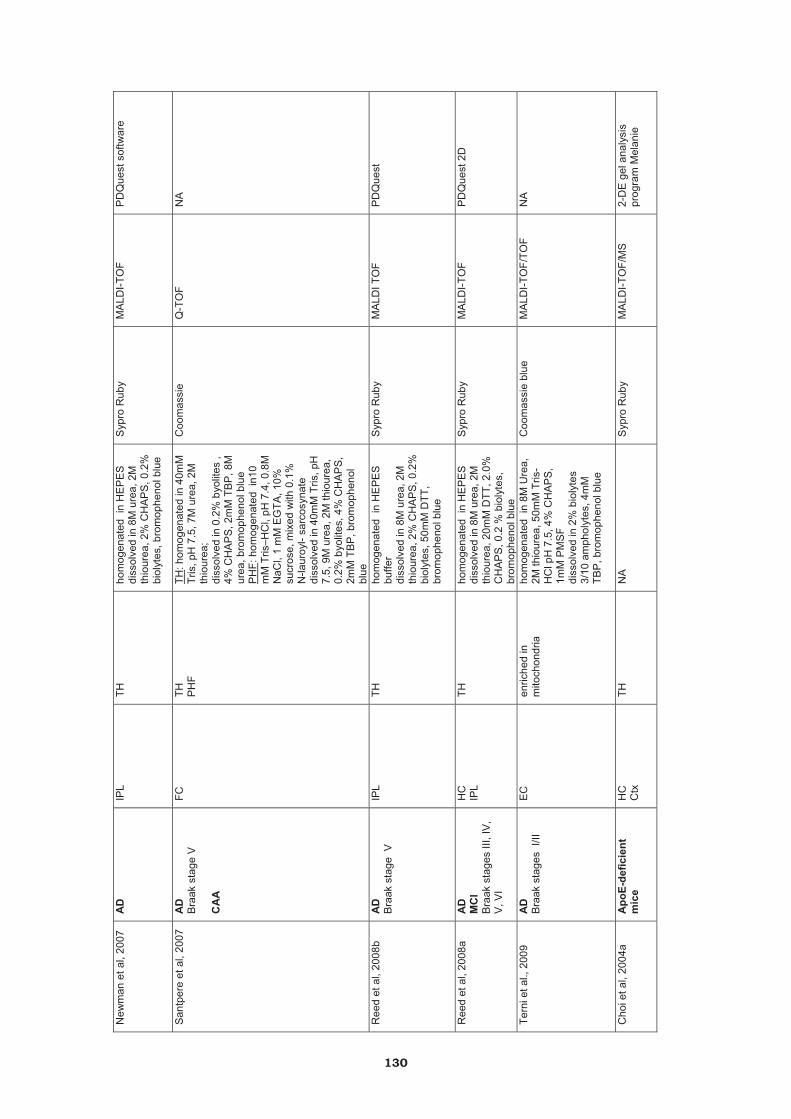
New
man
et a
l, 20
07
AD
IP
L TH
ho
mog
enat
ed i
n H
EP
ES
di
ssol
ved
in 8
M u
rea,
2M
th
iour
ea, 2
% C
HA
PS
, 0.2
%
biol
ytes
, bro
mop
heno
l blu
e
Syp
ro R
uby
MA
LDI-T
OF
PD
Que
st s
oftw
are
San
tper
e et
al,
2007
A
D
Bra
ak s
tage
V
CA
A
FC
TH
PH
FTH
: hom
ogen
ated
in 4
0mM
Tr
is, p
H 7
.5, 7
M u
rea,
2M
th
iour
ea;
diss
olve
d in
0.2
% b
yolit
es ,
4% C
HA
PS
, 2m
M T
BP
, 8M
ur
ea, b
rom
ophe
nol b
lue
PH
F : h
omog
enat
ed i
n10
mM
Tris
–HC
l, pH
7.4
, 0.8
M
NaC
l, 1
mM
EG
TA, 1
0%
sucr
ose,
mix
ed w
ith 0
.1%
N
-laur
oyl-
sarc
osyn
ate
diss
olve
d in
40m
M T
ris, p
H
7.5,
9M
ure
a, 2
M th
iour
ea,
0.2%
byo
lites
, 4%
CH
AP
S,
2mM
TB
P, b
rom
ophe
nol
blue
Coo
mas
sie
Q-T
OF
NA
Ree
d et
al,
2008
b A
D
Bra
ak s
tage
V
IPL
TH
hom
ogen
ated
in
HE
PE
S
buffe
rdi
ssol
ved
in 8
M u
rea,
2M
th
iour
ea, 2
% C
HA
PS
, 0.2
%
biol
ytes
, 50m
M D
TT,
brom
ophe
nol b
lue
Syp
ro R
uby
MA
LDI T
OF
PD
Que
st
Ree
d et
al,
2008
a A
D
MC
I B
raak
sta
ges
III, I
V,
V, V
I
HC
IP
LTH
ho
mog
enat
ed i
n H
EP
ES
di
ssol
ved
in 8
M u
rea,
2M
th
iour
ea, 2
0mM
DTT
, 2.0
%
CH
AP
S, 0
.2 %
bio
lyte
s,
brom
ophe
nol b
lue
Syp
ro R
uby
MA
LDI-T
OF
PD
Que
st 2
D
Tern
i et a
l., 2
009
AD
B
raak
sta
ges
I/II
EC
en
riche
d in
m
itoch
ondr
iaho
mog
enat
ed i
n 8M
Ure
a,
2M th
iour
ea, 5
0mM
Tris
-H
Cl p
H 7
.5, 4
% C
HA
PS
, 1m
M P
MS
F di
ssol
ved
in 2
% b
ioly
tes
3/10
am
phol
ytes
, 4m
M
TBP
, bro
mop
heno
l blu
e
Coo
mas
sie
blue
M
ALD
I-TO
F/TO
F N
A
Cho
i et a
l, 20
04a
Apo
E-de
ficie
nt
mic
eH
C
Ctx
TH
NA
S
ypro
Rub
y M
ALD
I-TO
F/M
S
2-D
E g
el a
naly
sis
pr
ogra
m M
elan
ie
130
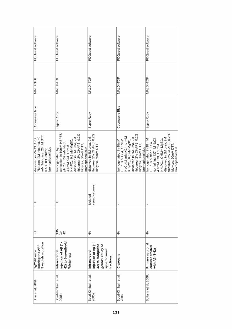
Shi
n et
al,
2004
Tg
2576
mic
e be
arin
g th
e A
PP
Swed
ish
mut
atio
n
FC
TH
diss
olve
d in
2%
CH
AP
S,
7M u
rea,
2M
thio
urea
, 40
mM
Tris
-bas
e, 2
0mM
DTT
, 0.
5 %
IPG
buf
fer,
brom
ophe
nol b
lue
Coo
mas
sie
blue
M
ALD
I/TO
F P
DQ
uest
sof
twar
e
Boy
d-K
imba
ll e
t al,
2005
b In
trac
ereb
ral
inje
ctio
n of
A�
(1-
42) t
o 3-
mon
th-o
ld
Wis
tar r
ats
NB
MC
txH
C
TH
hom
ogen
ated
by
soni
catio
n in
10m
M H
EP
ES
pH
7.4
, 137
mM
NaC
l, 4.
6mM
KC
l, 1.
1mM
K
H2P
O4,
0.6m
M M
gSO
4di
ssol
ved
in 8
M u
rea,
2M
th
iour
ea, 2
% C
HA
PS
, 0.2
%
biol
ytes
, 50m
M D
TT,
brom
ophe
nol b
lue
Syp
ro R
uby
MA
LDI-T
OF
PD
Que
st s
oftw
are
Boy
d-K
imba
ll e
t al,
2005
a In
trac
ereb
ral
inje
ctio
n of
A�
(1-
42) t
o M
ongo
lian
gerb
ils. S
tudy
of
syna
ptos
omal
fr
actio
ns
NA
is
olat
ed
syna
ptos
omes
di
ssol
ved
in 8
M u
rea,
2M
th
iour
ea, 2
% C
HA
PS
, 0.2
%
biol
ytes
, 50m
M D
TT
Syp
ro R
uby
MA
LDI-T
OF
PD
Que
st s
oftw
are
Boy
d-K
imba
ll e
t al,
2006
C
.ele
gans
N
A
- ho
mog
enat
ed i
n 10
mM
H
EP
ES
pH
7.4
, 137
mM
N
aCl,4
.6 m
M K
Cl,
1.1m
M
KH
2PO
4, 0.
6mM
MgS
O4
diss
olve
d in
8M
ure
a, 2
M
thio
urea
, 2%
CH
AP
S, 0
.2%
bi
olyt
es, 5
0mM
DTT
, br
omop
heno
l blu
e
Coo
mas
sie
Blu
e M
ALD
I-TO
F P
DQ
uest
sof
twar
e
Sul
tana
et a
l, 20
06c
Prim
ary
neur
onal
cu
lture
s tr
eate
d w
ith A
� (1
-42)
NA
-
hom
ogen
ated
in
10 m
M
HE
PE
S b
uffe
r pH
7.4
co
ntai
ning
137
mM
NaC
l, 4.
6mM
KC
l, 1.
1 m
M
KH
2PO
4, O
.6M
m M
gSO
4di
ssol
ved
in 8
M u
rea,
2M
th
iour
ea, 2
% C
HA
PS
, 0.2
%
biol
ytes
, 50m
M D
TT,
brom
ophe
nol b
lue
Syp
ro R
uby
MA
LDI-T
OF
PD
Que
st s
oftw
are
131

Tabl
e SI
II. S
ynuc
leop
athi
es
R
efer
ence
D
isea
se/s
tage
R
egio
n Fr
actio
nB
uffe
r G
el s
tain
ing
MS
Type
of a
naly
sis
Cho
i et a
l, 20
04a
PD
FC
TH
hom
ogen
ated
in 5
0 m
M T
ris-H
Cl
SYP
RO
Rub
y
MA
LDI-T
OF/
MS
H
PLC
-ES
I/MS
/MS
an
alys
es
prog
ram
PD
Que
st
Chu
ng e
t al.,
200
4 PD
N
A
NA
N
A
NA
N
A
NA
Yao
et a
l., 2
004
PD
TC
TH
hom
ogen
ated
in 2
50 m
M H
epes
, pH
7.5
,1 m
M E
DTA
,0.1
mM
ne
ocup
roin
e 0.
4% 3
-[(3-
chol
amid
opro
pyl)d
imet
hyla
mm
onio
]- 1-
prop
anes
ulfo
nate
or 1
% T
riton
X-
100
NA
N
A
NA
Cho
i et a
l, 20
05
PD
FC
TH
hom
ogen
ated
in 5
0 m
M T
ris-H
Cl
SYP
RO
Rub
y M
ALD
I-TO
F-(E
SI)
tand
em M
S/M
S
PD
Que
st
Dal
fó e
t al,
2005
iP
D
FC
TH
hom
ogen
ated
in 4
0 m
M T
ris p
H 7
.4
cont
aini
ng 7
M u
rea,
2 M
th
iour
ea,4
% C
HA
PS
, 1 m
M E
DTA
di
ssol
ved
in 0
.2%
byo
lites
, 2 m
M
TBP
, bro
mop
heno
l blu
e
Silv
er s
tain
ing
MA
LDI-T
OF/
TOF
or E
SI-M
S-M
S
Inve
stig
ator
HT
anal
yzer
Cho
i et a
l, 20
06
PD
FC
TH
acco
rdin
g to
Cho
i et a
l., 2
004a
S
YPR
O R
uby
M
ALD
I/TO
F M
ALD
I/TO
F/TO
F P
DQ
uest
Kee
ney
et a
l., 2
006
PD
FC
Mito
chon
dria
l en
richm
ent
hom
ogen
ated
in
0.1
5 M
KC
l, 20
m
M K
pho
spha
te, 1
mM
ED
TA, p
H
7.6
diss
olve
d in
RIP
A
SYP
RO
Rub
y LC
-MS
mas
s sp
ectro
met
ry
N
A
Dal
fó a
nd F
erre
r, 20
08
iPD
, PD
, DLB
FC
SN
TH
hom
ogen
ated
in P
BS
, 1m
M E
DTA
N
A
NA
N
A
Góm
ez, F
erre
r, 20
09
iPD
, PD
, DLB
FC
M
itoch
ondr
ial
enric
hmen
t ho
mog
enat
ed in
20
mM
HE
PE
S-
KO
H, 2
50 m
M s
ucro
se, 1
0 m
M K
Cl,
1.5
mM
MgC
l 2, 1
mM
ED
TA, 1
mM
E
GTA
, 1 m
M D
TT
diss
olve
d in
40
mM
Tris
pH
7.5
co
ntai
ning
7 M
ure
a, 2
M th
iour
ea,
0.2%
byo
lites
, 4%
CH
AP
S, 2
mM
TB
P, 0
.1%
bro
mop
heno
l blu
e
Silv
er s
tain
ing
Q-T
OF
NA
Poo
n et
al,
2005
a A
30P
�-sy
nucl
ein
tran
sgen
ic m
ice
LH
TH
hom
ogen
ated
in T
BS
, pH
7.4
, 1 m
M
ED
TA, 5
mM
sod
ium
py
roph
osph
ate,
30
mM
gly
cero
l 2-
phos
phat
e, 3
0 m
M s
odiu
m fl
uorid
e,
1 m
M E
DTA
Syp
ro R
uby
M
ALD
I-TO
F P
D Q
uest
132

VI. DISCUSSIÓ


VI.DISCUSSIÓ
1. Sensibilitat del RNA i de les proteïnes del teixit cerebral humà als factors post mortem
L’estudi de les malalties neurològiques té un alt grau de complexitat per la
dificultat de desenvolupar un model animal que reprodueixi la patologia o els
símptomes i els marcadors histopatològics que es troben en els humans. La utilització
de material post mortem dels bancs de teixit és una molt bona opció per tal de
realitzar els experiments directament en el teixit en el que s’ha donat el desordre. Ara
bé, la utilització d’aquesta mostra presenta una sèrie de variables que s’han de tenir
en compte. El tipus i el temps de fixació de la mostra, la inclusió en parafina, la
congelació, el temps de retardament post mortem (RPM) i la temperatura, són una
sèrie d’elements post mortem que necessiten ser caracteritzats per saber fins a quin
punt poden o no modificar les diferents molècules. D’aquests factors, el temps de RPM
i la temperatura són les variables més fàcils de poder controlar necessitant, per tant,
estudis per determinar la seva importància pel que fa a la preservació i a la degradació
del DNA, del RNA i de les proteïnes.
En estudis multicèntrics de l’European Brain Bank Network es va veure que el
DNA era relativament resistent a la degradació post mortem i que resultava ser útil per
fer estudis genètics, encara que les mostres estiguessin guardades a -80ºC durant
diversos anys. En solucions líquides, però, el DNA resultava vulnerable recomanant
que la mostra es fixi durant un temps curt (un mes) en formol al 4% per preservar-la.
Pel que fa a al protocol d’extracció de DNA el mètode de QIAmp Micro Qiagen va ser el
sistema que donava resultats més reproduïbles (Ferrer i col. 2007). El 2005 es va
publicar que les modificacions epigenètiques com la metilació de les cues d’histones i
l’estabilitat dels nucleosomes es mantenen estables en el teixit després de la mort del
pacient, sense veure’s afectades pel RPM (Stadler i col. 2005). Barrachina i col. 2009
també van detmostrar que la metilació de les illes CpG dels gens promotors es troben
conservades amb el RPM.
A nivell del RNA es troben resultats antagònics: diferents estudis han suggerit
que és estable en mostres humanes de cervell post mortem (Cummings i col. 2001,
Yasojima i col. 2001) i, per altra banda, altres treballs han demostrat que la integritat
del RNA és en gran part depenent de diferents factors tant pre mortem com poden ser
l’estat agònic, coma, l’hospitalització, la hipòxia,…com post mortem tals com el temps
de RPM, la temperatura,… (Durrenberger i col. 2010, Harrison i col. 1995, Preece i col.
2003). Tenint en compte aquests últims resultats és important determinar que per
millorar la preservació del RNA es recomana fer una ràpida immersió de la mostra en
Trizol després d’extreure-la i que l’anàlisi de la seva qualitat es faci utilitzant el
135
VI.DISCUSSIÓ
Bioanalyzer que resulta ser una eina molt més eficaç i sensible que no pas els gels
d’agarosa. En aquest context de possibles alteracions del RNA, es van plantejar dos
estudis per fer en el nostre treball: el primer es va centrar en determinar les
modificacions del RNA segons el temps de RPM i la temperatura per guardar el teixit.
Es van utilizar microarrays de RNA de mostres humanes de cervell congelades a les 2h
després de la mort del pacient, guardades a 4ºC o bé a temperatura ambient durant
diferents hores i més tard congelades a -80ºC. Hi havia una disminució de l’expressió
de diferents RNAs depenent del temps de RPM i de la temperatura d’emmagatzematge
de la mostra i l’expressió de petits RNAs augmentava quan també ho feia el temps de
RPM. Aquests resultats indiquen una vulnerabilitat del RNA pel RPM corroborada
també pel grup de Buesa i col. 2004 on van fer estudis de RPM en mostres humanes
finalment congelades en nitrogen líquid.
L’objectiu del segon estudi va ser el d’identificar canvis en la qualitat del RNA en
diferents regions del cervell d’un mateix pacient. Es van utilitzar els valors del RIN
(RNA Integrity Number) a través del Bioanalyzer com a indicador de la preservació del
RNA. El valor del RIN va mostrar que no era depenent del temps de RPM i que aquest
valor variava entre les diferents zones. Aquesta diferència individual no se sap a què
pot ser deguda. Una explicació podria ser que les estructures internes del cervell
triguen més a refredar-se a 4ºC i que, per altra banda, l’escorça cerebral es refreda
més ràpidament gràcies a la seva localització superficial podent mantenir més
preservat el RNA.
Pel que a les proteïnes, s’han realitzat estudis en models animals (rata, ratolí)
per veure còm afecta el RPM en el teixit on es va demostrar una sensibilitat de les
proteïnes a la degradació, comportant-se diferent unes que d’altres (Fountoulakis i col.
2001, Hilbig i col. 2004). En el cas del cervell humà post mortem s’ha pogut concloure
que la temperatura en la que s’han guardat les mostres afecta a la preservació de les
proteïnes. En un treball de la degradació proteïca, emmarcat dins del BrainNet Europe,
Ferrer i col. 2007a van utilitzar mostres humanes de cervell per realitzar un estudi del
temps de RPM i de la temperatura (congelació directa a -80ºC, a temperatura ambient,
a 4ºC, a 1ºC en diferents temps i finalment congelació a -80ºC). Es va determinar que
a 1ºC les mostres eren més estables i que hi havia una diferència de vulnerabilitat en
les proteïnes sent unes més resistents que altres a la degradació. Altre cop va quedar
patent la importància d’aquests dos factors pel que fa a la preservació de les proteïnes
on s’observava una disminució de l’expressió de determinades proteïnes però també
un augment del nivell d’altres (Crecelius i col. 2008). El fet que hi hagi una disminució
de l’expressió de certes proteïnes és esperable en estudis de degradació proteïca, ara
136
VI.DISCUSSIÓ
bé un augment del seu nivell és més difícil d’explicar: una disminució del recanvi de la
proteïna o de l’eliminació normal d’aquestes proteïnes, un augment de la seva síntesi
donada per la situació cel·lular en que es troba, la formació de proteïnes
truncades,…podrien ser possibles explicacions d’aquest increment.
Les modificacions post translacionals de les proteïnes també poden estar subjectes a
possibles alteracions degudes al RPM i a la temperatura. Aquestes són importants en
les malalties neurodegeneratives ja que les proteïnes que formen les inclusions
presenten aquest tipus d’alteracions. En el cas de les taupaties, per exemple, la
proteïna tau es desfosforila ràpidament en condicions normals però la tau
hiperfosforilada és més resistent a la degradació. En la MA el triplet de 60, 64 i 68
kDa, i les bandes de baix pes molecular, presenten una bona preservació durant les
primeres 24h i una disminució de la seva intensitat cap a les 48h a 4ºC (Santpere i
col. 2006). En aquest context, ens vam proposar de valorar les modificacions
d’oxidació i de nitració que podien patir les proteïnes exposant les mostres d’escorça
frontal, diagnosticades per la MA, a 2h, 5h, 8h i 20h a temperatura ambient per ser
després congelades a -80ºC. Els marcadors utilitzats de lipoxidació (MDA-L, CML), de
glicoxidació (CEL, CML, AGE) i de nitració (N-Tyr) reconeixien una sèrie de bandes on
cadascun d’ells tenia un patró propi. En general, però, s’observava que durant les
primeres 8h la densitat i la distribució de les bandes es mantenia bastant uniforme i
que a partir de les 20h hi havia variacions en el marcatge. Com en el cas de la
degradació proteïca, en algun marcador es podia veure la disminució de la intensitat
de les bandes (CEL, CML), en d’altres un augment (MDA-L, CEL) i també l’aparició de
noves (MDA-L, N-Tyr) podent ser les proteïnes possibles dianes d’oxidació durant el
RPM. En un treball publicat al 2009 van fer estudis en mostres de cervells humans
(controls que no presentaven desordres neurològics) per valorar també l’efecte del
temps de RPM (4-18h) en la nitració proteïca (Chandana i col. 2009): van utilitzar slot
blot amb l’anticòs de la 3-nitrotirosina com a marcador de nitració proteïca. Es va
trobar un increment significatiu en els nivells de nitració en la substància negra, un
augment més suau en el cerebel però sense canvis en l’escorça frontal. Pel que fa a
l’anàlisi dels grups carbolins de les proteïnes, el temps de RPM va influir només en la
substància negra amb valors més elevats. Aquests dades concorden, en part, amb els
nostres resultats de les mostres d’escorça frontal: el rang de temps de RPM que van
utilizar per l’estudi va de les 4 a les 18h, sense observar canvis durant aquest temps, i
en el nostre cas va ser de les 2 a les 20h començant a trobar alteracions importants a
partir de les 20h. Ara bé, per poder estar segurs d’aquests resultats es necessitarien
fer altres estudis on les hores de RPM fossin més continues doncs en el nostre
experiment va haver-hi un salt de les 8h a les 20h.
137
VI.DISCUSSIÓ
El fet que trobessin modificacions en la substància negra podria suportar la idea que
les regions profundes del cervell poden tenir més tendència a patir modificacions
oxidatives i alteracions enzimàtiques per la diferència de refrigeració, podent alterar la
integritat d’algunes proteïnes.
Així doncs és important caracteritzar els diferents factors post mortem per saber còm
poden modificar a les diferents molècules (DNA, RNA, proteïnes) i buscar les vies per
minimitzar aquestes variacions. Aquests estudis prenen especial rellevància si es
consideren una via per estandaritzar protocols entre els diferents hospitals i bancs de
teixit, ajudant a l’homogeneïtat de les mostres entre diversos centres i permetent una
millor reproductibilitat de resultats entre grups.
2. Efecte de l’estrès oxidatiu en les proteïnes a la malaltia de PSP
Havent caracteritzat el patró de certs marcadors d’oxidació en funció del RPM a
temperatura ambient, es va passar a estudiar l’efecte d’aquests en les proteïnes de
determinades malalties neurodegeneratives. Per a dur a terme el nostre objectiu es va
fer ús d’eines proteòmiques per tal de poder seleccionar i identificar quina era la
proteïna diana de l’oxidació.
La primera patologia que es va estudiar va ser la PSP, del grup de les taupaties, on es
va realitzar un estudi dels diferents marcadors d’oxidació en mostres d’escorça frontal
de patològics i dels respectius controls. Es van trobar diferències significatives amb el
marcatge de lipoxidació, anticòs anti-HNE, en les mostres patològiques. Odetti i col.
2000 van descriure un augment d’aquest marcador en la zona del mesencèfal de teixit
de PSP. D’altres estudis també havien observat un augment de lesió lipoxidativa en
mostres de pacients al nucli sutbtalàmic (Albers i col. 1999) i a l’escorça frontal
superior (Albers i col. 2000), utilitzant ambdós estudis tècniques d’HPLC i el MDA com
a marcador de dany lipoxidatiu. El fet que en els nostres resultats no s’hagués
observat un augment de MDA en les mostres patològiques pot ser degut a la separació
de les diferents regions de l’escorça frontal, en el nostre estudi no es va fer i potser es
van emmascarar resultats o bé per possibles diferències individuals.
En el nostre cas es va prosseguir l’estudi passant a localitzar i determinar les
proteïnes modificades per l’HNE. Es van identificar dos enzims implicats en la ruta
metabòlica de la glicòlisi: la fructosa 1,6-difosfat aldolasa A (aldolasa A) i la
fosfoglicerat cinasa-1 (FGC-1).
138

VI.DISCUSSIÓ
No hi ha estudis previs de proteïnes que representin possibles dianes afectades pels
radicals lliures i que estiguin implicades en el metabolisme glicolític a PSP. Ara bé
Albers i col. 2000 van observar una disminució en l’activitat de l’�-cetoglutarat
deshidrogenasa demostrant una alteració mitocondrial en mostres d’escorça frontal
quedant patent una disfuncionalitat del metabolisme energètic. Pel que fa a estudis in
vitro de cèl·lules cíbrides, introducció de mitocondris de teixit de pacients de PSP en
línies cel·lulars que no tenen DNA mitocondrial, es va descriure una reducció de
l’activitat del complex I i del III del mitocondri (Chirichigno i col. 2002).
L’aldolasa A catalitza la conversió reversible de la fructosa 1,6-difosfat a gliceraldehid
3-fosfat i a dihidroxiacetona fosfat. Se n’han descrit tres d’isoformes: la isoforma A es
produeix durant el desenvolupament embrionari i està molt expressada en el múscul
de l’adult (podent representar el 5% de la proteïna cel·lular total), la B es troba
majoritàriament en el fetge, en els ronyons i l’intestí. Pel que fa al cervell i al sistema
nerviós l’aldolasa A i la C estan expressades per igual. En mostres de pacients amb
MA es van mesurar els grups carbonils. Es van trobar diferències en determinades
proteïnes: en el cas de l’aldolasa A, i de la isoforma C, es van observar canvis en el
contingut de carbonils però no arribaven a presentar diferències estadísticament
significatives degut als canvis en la quantitat de proteïna (Korolainen i col. 2006).
Després d’haver publicat el nostre treball, Reed i col. 2008b van trobar l’aldolasa A
modificada per nitració en el lòbul parietal inferior de cervells afectats per la MA.
Pel que fa a la FGC-1, és un dels enzims més importants de la via glicolítica
participant en la catalització de la reacció de l’1,3-difosfoglicerat a 3-fosfoglicerat amb
la formació d’una molècula d’ATP. Es van detectar una sèrie de proteïnes modificades
per l’HNE en mostres d’Alzheimer (hipocamp), sent la FGC-1 una d’elles (Reed i col.
2008a).
Diferents enzims relacionats amb la glicòlisi i la glicogènesi han estat identificats per
estar modificats oxidativament en diferents sistemes com per exemple en
sinaptosomes tractats amb A� (1-42) (Boyd-Kimball i col. 2005), en cultius primaris de
rates amb inducció d’estrès oxidatiu per l’amiloide � 1-42 (Sultana i col. 2006a), en
ratolins envellits (Poon i col. 2006), en models de ratolins que mimetitzen la malaltia
d’Huntington (Perluigi i col. 2005), en models murins d’envelliment accelerat (SAMP-8)
de A� (Poon i col. 2004).
Sobre els anys 80 es va descriure en un estudi, amb Escherichia coli, el fet que certs
enzims metabòlics es podien inactivar per efectes de sistemes d’oxidació. Es van trobar
la FGC, l’alcohol deshidrogenasa, l’enolasa, la piruvat cinasa entre d’altres,
139
VI.DISCUSSIÓ
emfatitzant que molts d’aquests enzims presenten una histidina o un altre residu
fàcilment oxidable en el seu centre catalític (Fucci i col. 1983). Anys després s’ha anat
demostrant que diversos enzims són suceptibles a ser inactivats per l’acció d’adductes
d’HNE com per exemple la glucosa-6-fosfat deshidrogenasa, la gliceraldehid-3-fosfat
deshidrogenasa, la glutatió S-transferasa, la glutatió reductasa, l’aldosa reductasa
(Uchida K. 2003a).
En el cas de l’aldolasa A, una reducció de l’activitat enzimática pot provocar un
augment dels nivells de fructosa 1,6-difosfat induïnt una inhibició de la glicòlisi i una
disminució d’ATP. Pel que fa a la FGC-1, la seva disfuncionalitat podria comportar una
davallada en la producció d’energia.
L’any 1992 Karbe i col. 1992 van determinar l’índex metabòlic de la glucosa en
cervells de pacients de PSP, a través de tècniques de neuroimatge com la FDG (18F-2-
Fluoro-2-deoxi-D-Glucosa) i la PET (tomografia d’emissió de positrons). Van descriure
en els individus patològics un hipometabolisme molt sever en les estructures
subcorticals i una afectació menor, però igualment significativa, en l’escorça frontal
corroborant d’aquesta manera estudis anteriors d’altres grups. Resultats publicats
recentment, demostren in vivo una disminució de metabòlits rics energèticament (ATP,
ADP, creatina fosforilada, creatina no fosforilada, fosfat inorgànic, lactat) en l’escorça
frontal de cervells patològics (Stamelou i col. 2009) aportant més informació pel que fa
a l’alteració del metabolisme energètic d’individus amb PSP.
En el cas de la MA també s’ha descrit una marcada disfuncionalitat del metabolisme
de la glucosa cerebral (Pietrini i col. 2000) que pot ser donada per l’alteració de les
activitats enzimàtiques de certs enzims claus en la glicòlisi (Bigl i col. 1999) o per una
reducció en la densitat de les diferents classes de transportadors de glucosa corticals
GLUT1 i GLUT3 (Mooradian i col. 1997).
Fa 30 anys es van fer estudis en sistemes biològics utilitzant models d’envelliment
cel·lular, per tal de determinar els nivells de proteïnes modificades oxidativament. Per
una banda, es van fer servir eritròcits d’individus normals de diferents edats per
determinar els valors de proteïnes oxidades i, com a segon experiment, cultius
cel·lulars de fibroblasts de pacients amb el síndrome de Werner (o progènia, desordre
associat a l’envelliment accelerat). Els resultats van mostrar que els nivells de
proteïnes modificades oxidativament augmentaven amb l’edat en els eritròcits i que
aquests canvis estaven correlacionats amb una pèrdua de marcadors d’activitat
enzimática. Pel que als estudis dels cultius de fibroblasts es va trobar una pujada dels
140
VI.DISCUSSIÓ
valors de carbonils a partir dels 60 anys d’edat, obtenint valors molt més alts i
significatius en els patològics (Oliver i col. 1987). Al llarg de tots aquests anys s’han
anat realitzant altres estudis on s’ha corroborat l’augment d’oxidació en funció de
l’envelliment, com per exemple en el cas de l’enzim aldolasa trobant-se modificat
oxidativament en el bulb olfactori de ratolins envellits (Vaishnav i col. 2007). Un altre
treball per destacar fa referència a models murins d’envelliment on es va trobar una
disminució de les activitats enzimàtiques mitocondrials (òxid nítric sintasa
mitocondrial, NADH deshidrogenasa, citocrom oxidasa) directament relacionada amb
el contingut de productes d’oxidació en els animals vells (Navarro A. 2004). En aquest
context s’enquadren els nostres resultats on es va observar la presència de l’aldolasa A
i de la FGC-1 oxidades en 3 dels 8 casos controls podent-se relacionar amb
l’envelliment propi d’aquests individus.
3. Efecte de l’estrès oxidatiu en les proteïnes de les malalties englobades dins de la
degeneració frontotemporal lobar (DFTL)
Seguint en la línia d’estudiar l’efecte de l’estrès oxidatiu sobre les proteïnes a
l’escorça frontal de diferents malalties, es van caracteritzar tres grups de malalties
englobades dins del marc de la DFTL: DFTL-tau (amb inclusions de tau), DFTL-U
(sense inclusions de tau però immunoreactives per la ubiqüitina) i DFTL-ELA (sense
inclusions de tau però positives per la ubiqüitina amb afectació de les motoneurones).
Com a resultat de l’estudi de diferents marcadors d’estrès oxidatiu, es va observar la
presència de lesió lipoxidativa (augment de la immunoreactivitat de l’anticòs anti-HNE)
en l’escorça frontal de mostres patològiques però mostrant una gran diferència entre
les diferents classes de malalties.
Pocs treballs s’han centrat en el paper de l’estrès oxidatiu en aquestes malalties. En
ratolins transgènics amb mutació de la tau P301L es van medir els nivells de ROS
(radical anió superòxid i peròxid d’hidrogen) de manera indirecta, a través de la DHE -
DiHidroEtidi- que s’oxida a catió etidi fluorescent per l’anió superòxid i la H2DCF -
DiClorFluoresceïna- pels peròxids citosòlics, trobant-se augmentats ambdós
marcadors en els ratolins transgènics (David i col. 2005). A través d’estudis funcionals
i proteòmics també van observar defectes en el mitocondri amb una reducció de
l’activitat del complex I i del complex V, corroborant l’alteració de l’activitat d’aquest
últim en cervells humans diagnosticats amb DFTL-tau (mutació P301L) (David i col.
2005).
141
VI.DISCUSSIÓ
Pel que fa al grup de DFTL-U no s’han realitzat treballs previs semblants, en el
subgrup de DFTL-ELA Ilieva i col 2007 van demostrar la presència de lesió oxidativa,
anormalitats del mitocondri i estrès del reticle endoplasmàtic en la medul·la espinal de
pacients.
Al fer la identificació dels diferents punts es va trobar que corresponien a la GFAP
(glial fibrillary acidic protein), confirmant-ho per la col·localització de l’anticòs anti-
HNE amb la GFAP en els astròcits gràcies al doble marcatge d’immunofluorescència i
a la microscopia confocal, per gel d’electroforesis i western blot.
La GFAP és un filament intermedi que, juntament amb els microtúbuls i els
microfilaments, formen part del citoesquelet. Representa la proteïna més important
dels astròcits, havent-hi expressades d’altres com la nestina i la vimentina però en
menor grau. Els astròcits són molt més abundants que les neurones, amb un ràtio
d’astròcit-neurona de 1:10, en la majoria de regions del cervell.
Aquest tipus cel·lular és el principal sistema homeostàtic del cervell exercint diferents
papers en el sistema nerviós: control del moviment dels fluids entre l’espai
intracel·lular i l’extracel·lular, capacitat d’acceptar el glutamat i reduir l’excitoxicitat,
els astròcits estan interconnectats via gap junctions permetent la redistribució d’ions i
de metabòlits perjudicials a llarga distància, participant en la regulació de la barrera
hematoencefàlica, defensa del sistema nerviós central, alliberament de substàncies
vasoactives que controlen el flux sanguini, coordinar també l’activitat neuronal
(Anderson i col. 2003, Pekny i col. 2004, Pekny i col. 2005).
Els filaments intermedis de GFAP tenen importància en el manteniment mecànic o
estructural del sistema nerviós quan aquest està sotmés a estressors mecànics greus.
Pekny i col. 2004 van trobar en experiments amb ratolins knock out de filaments
intermedis, on se’ls hi havia aplicat un estrès mecànic sever, que les retines dels
controls estaven intactes però que les dels ratolins GFAP -/- Vim -/- es trobaven
alterades. Els astròcits també tenen un paper molt important en el sistema de defensa
antioxidant del cervell amb concentracions altes d’antioxidants comparant amb
d’altres tipus cel·lulars del cervell (Dringen i col. 2000). Aquesta idea es basa en
estudis de cultius de neurones que resulten més vulnerables al dany produït per
H2O2, peroxinitrit o 6-hidroxidopamina que els cultius de cèl·lules astroglials (Bolanos
i col. 1995). A més a més, els astròcits contribueixen als sistemes de defensa contra
l’estrès oxidatiu de cèl·lules endotelials: en un model in vitro de la barrera
hematoencefàlica els astròcits tenien augmentades les activitats de la SOD, la catalasa
i la GPx amb una disminució de la quantitat de radicals produïts per peroxidació
142

VI.DISCUSSIÓ
lipídica (Schroeter i col. 1999). L’expressió de GPX i de catalasa estan augmentades en
cultius cel·lulars d’astròcits després de l’exposició a H2O2 (Rohrdanz i col. 2001), la
inhibició de catalasa redueix el ràtio d’eliminació del H2O2 pels astròcits.
Els astròcits estan implicats en una gran varietat de malalties del sistema nerviós
central com per exemple en un trauma, isquèmia o bé en malalties neurodegeneratives
(mutacions en el gen de la GFAP donen lloc a una patologia anomenada Alexander
(Liem i col. 2009)). En aquestes situacions les cèl·lules de la glia s’activen: la microglia
produeix resposta inflamatòria i els astròcits canvien la seva morfologia (hi ha una
hipertrofia dels seus processos cel·lulars i un augment de la producció de filaments
intermedis, principalment de la GFAP encara que també de la nestina i de la
vimentina). Aquest procés s’anomena gliosi reactiva i quan es parla només de
l’activació dels astròcits es fa referència a l’astrocitosi (astrogliosi). És una reacció de
defensa del cervell per tal d’aïllar la zona afectada de la resta del teixit del sistema
nerviós central, reconstruir la barrera hematoencefàlica i ajudar a modificar els
circuits del cervell en regions al voltant de la zona lesionada (Rodriguez i col. 2009).
S’han desrit cinc isoformes de la GFAP: �, �, �, � i � (Nielsen i col. 2002) on l’� és la
més abundant en situacions d’astrocitosi, encara que en el nostre treball no se sap
quina de les cinc isoformes es va identificar.
Tot i que que està descrit que hi ha un augment de l’expressió de GFAP amb
l’envelliment (Major i col. 1997), s’ha vist un increment molt més marcat en diferents
patologies. Schweitzer i col. 2006 van detectar, en cervells diagnosticats de DFTL-tau,
un augment dels nivells de GFAP (multiplicada 5.4 vegades la seva expressió) respecte
dels controls. Els nostres resultats d’immunohistoquímica mostren una pèrdua
neuronal consistent amb una forta presència d’astròcits podent indicar un canvi de
població cel·lular. Aquesta astrocitosi està molt accentuada (resultats també
corroborats per l’anàlisi de western blot) en les mostres de DFTL-tau i també en els
casos de DFTL-U i de DFTL-ELA observant una gran variabilitat en aquestes últimes
patologies. Aquest augment dels nivell d’expressió de la GFAP correlaciona amb la
intensitat de la lesió oxidativa (lipoxidativa). Es va intentar fer un anàlisi quantitatiu
de l’expressió de la GFAP, i també de l’HNE, però no va ser possible: l’astrocitosi era
molt severa en uns casos i en d’altres no tant i, a més a més, els diferents casos
expressaven més una banda que unes altres presentant problemes de quantificació.
143
VI.DISCUSSIÓ
La GFAP ha estat descrita oxidada en d’altres malalties neurodegeneratives, en
algunes taupaties com en la MA (Pamplona i col. 2005), la malaltia de Pick (Muntané i
col. 2006) i en la PSP (Santpere i col. 2009). Ara bé, també s’ha trobat la GFAP com a
diana d’oxidació en d’altres tipus de patologies no associades a inclusions de tau com,
per exemple, en la malaltia de Huntington on els nivells dels grups carbonils es van
trobar doblats en la zona de l’estriat dels patològics comparant amb els controls
(Sorolla i col. 2008). En mostres d’escorça de pacients amb ceruloplasminèmia (un
desordre autosòmic recessiu on està afectat el metabolisme del ferro per mutacions en
el gen de la ceruloplasmina) es va trobar la GFAP com una de les proteïnes més
importants carbonilades (Kaneko i col. 2002). S’han descrit d’altres modificacions, a
part de l’oxidació, com són l’O-glicosilació, la N-glicosilació o la fosforilació: es va
observar un augment de la GFAP N-glicosilada i fosforilada en mostres de MA
(Korolainen i col. 2005). Ara bé, aquestes modificacions oxidatives que pateix la GFAP
encara que no se sap quin tipus d’efecte produeix en la seva funcionalitat, indiquen
que els astròcits són dianes d’estrès oxidatiu.
Seguint aquesta línia, es va analitzar la composició d’àcids grassos de la membrana
trobant un canvi en el seu perfil comparant les mostres de DFTL-tau amb els controls:
es va determinar un contingut menor d’àcids grassos saturats i de PUFA
(majoritàriament de PUFA n-3) amb una disminució en els índexs de dobles enllaços i
de peroxidabilitat. Aquests resultats donen suport als valors de MDA analitzats per
GC/MS (gas chromatography/mass spectometry) on es van trobar significativament
elevats en els casos de DFTL-tau, ja que és un aldehid que es forma a partir de la
peroxidació lipídica dels �-3 i també dels �-6. Segurament per la diferència de
sensiblitat de les tècniques, aquestes resultats no es van trobar per western blot. Pel
que fa als casos de DFTL-ELA, només van resultar significatius els baixos continguts
de PUFA n-3 i l’augment de PUFA n-6. Els marcadors indicadors de dobles enllaços i
de peroxidació lipídica van disminuir respecte als controls però no significativament.
No se sap l’origen dels canvis en la composició dels àcids grassos de les membranes
però podrien ser deguts per una combinació de diferents factors: deplaçament d’una
població cel·lular per una altra on cada una conté patrons específics d’àcids grassos
(pèrdua neuronal i astrocitosi), canvis produïts perque la membrana cel·lular s’adapti
a l’estat patològic, alteracions del metabolisme i del transport dels àcids grassos
essencials, acció directa de lesió oxidativa en la membrana cel·lular, una disfunció
mitocondrial podria promoure la fugida de ROS podent atacar a les membranes amb el
conseqüent augment de peroxidació lipídica i producció d’adductes (HNE, MDA-L).
144

VI.DISCUSSIÓ
En el nostre treball es va trobar un augment de l’expressió, per western blot, de
l’enzim catalasa en la majoria de casos de DFTL que correlacionava amb els resultats
obtinguts amb l’HNE. Aquest increment indica una resposta de les cèl·lules per tal de
compensar la situació de desequilibri provocat per l’augment de radicals lliures
reactius. Pel que fa als enzims antioxidants SOD, la SOD-1 col·localitzava amb la
GFAP en mostres de DFTL-tau i de DFTL-U i la SOD-2 es trobava tant en els astròcits
(col·localització amb GFAP) com en les neurones (reconegudes per la morfologia del
citoplasma) de DFTL-tau i de DFTL-U.
4. Proteïnes modificades nitro/oxidativament en les malalties neurodegeneratives
En el marc dels estudis presentats, junt amb d’altres treballs del grup i veient
la gran quantitat d’informació publicada, ens vam plantejar de fer una revisió dels
diferents treballs de proteòmica redox en les malalties neurodegeneratives.
La tècnica dels gels bidimensionals va ser desenvolupada sobre els anys 70 però la
seva aplicació útil va arribar uns anys després amb l’ús d’eines microanalítiques que
poguessin identificar les proteïnes. A mitjans dels anys 90 la introducció de
determinades tècniques, com la separació de proteïnes a través de gels 2D-PAGE amb
gradients de pH immovilitzats (IPGs) i l’espectometria de masses per analitzar
proteïnes i pèptids, van permetre el desenvolupament del camp de la proteòmica. El
terme de proteoma es va utilizar per primer cop l’any 1995 per definir el “conjunt de
PROTEïnes d’un genOMA, una cèl·lula o un teixit”, donant lloc a la seva corresponent
disciplina: la proteòmica, on el seu objectiu és l’ ”estudi a gran escala dels productes
gènics d’un genoma a través de mètodes bioquímics, on la idea final és obtenir una
visió global i integrada dels processos cel·lulars”.
S’han publicat interessants treballs (Butterfield i col.. 2006a, Dalle-Donne I., Scaloni
A., Butterfield DA. 2006, Sultana i col. 2009) que han llistat les proteïnes modificades
nitro/oxidativament de diferents patologies, principalment de la MA, utilitzant gels
bidimensionals.
En el nostre cas es va voler emfatitzar:
• actualitzant les dades fins ara obtingudes i ampliant la revisió a d’altres taupaties,
sinucleopaties, malaltia d’Huntington i a l’ELA. També pel que fa als models animals i
cel·lulars que mimetitzen les malalties.
145

VI.DISCUSSIÓ
• identificant vies metabòliques i proteïnes vulnerables a la nitració/oxidació, comuns
en les diferents patologies.
• examinant els resultats descrits en estadiatges primerencs de les malalties.
• analitzant si les dianes proteïques identificades estan vinculades a estudis
d’activitat enzimàtica.
• revisant la metodologia utilitzada en els diferents estudis.
• discutint les limitacions de la proteòmica redox i suggerint millores per futurs
estudis.
Les majoria de treballs que han identificat proteïnes que són dianes de
nitració/oxidació, s’han centrat en la MA. Per tal de fer més entenedor el gran volum
d’informació que hi ha al respecte, les proteïnes modificades es van classificar en
diferents grups: a) metabolisme de la glicòlisi i energètic, b) cadena de transport
d’electrons, fosforilació oxidativa i elements mitocondrials, c) proteïnes estructurals, d)
xaperones, proteïnes d’estrès i de resposta a l’estrès, e) sistema ubiqüitina-
proteasoma, f) altres proteïnes aïllades dins de vies metabòliques concretes.
En les altres malalties no va ser necessari agrupar les proteïnes ja que hi ha pocs
estudis al respecte.
En la nostra revisió es va observar la identificació de la �-actina (Aksenov i col. 2001,
Castegna i col. 2003, Korolainen i col. 2005, Pamplona i col. 2005, Reed i col. 2008a),
la �-tubulina (Aksenov i col. 2001, Pamplona i col. 2005), la GFAP (Korolainen i col.
2005, Pamplona i col. 2005), l’�-enolasa (Butterfield i col. 2006b, Castegna i col. 2003,
Korolainen i col. 2005, Newman i col. 2007, Pamplona i col. 2005, Reed i col. 2008a,
Reed i col. 2008b, Sultana i col. 2006b, Sultana i col. 2009), la �-enolasa (Butterfield i
col. 2006c, Castegna i col. 2003, Pamplona i col. 2005), l’aldolasa A (Korolainen i col.
2006, Reed i col. 2008b), la glutamat deshidrogenasa (Korolainen i col. 2006,
Pamplona i col. 2005), la glutamina sintetasa (Butterfield i col. 2006b, Pamplona i col.
2005), l’ATP sintasa (Pamplona i col. 2005, Reed i col. 2008a, Sultana i col. 2006b,
Terni i col. 2009), la piruvat cinasa (Butterfield i col. 2006b, Reed i col. 2008a),
l’UCHL-1 (Butterfield i col. 2006c, Sultana i col. 2009), la CK BB (Aksenov i col. 2001,
Korolainen i col. 2005, Pamplona i col. 2005), la Pin 1 (Butterfield i col. 2006b,
Sultana i col. 2006c, Sultana i col. 2009) en, com a mínim, dos estudis diferents en
pacients de la MA demostrant la repoductibiliat dels resultats i també que
determinades proteïnes tenen tendència a modificar-se.
També es va poder veure l’elevada presència de les mateixes proteïnes modificades
(aldolasa A, enolasa, GAPDH,…) relacionades amb la glicòlisi i el metabolisme
146
VI.DISCUSSIÓ
energètic, en diferents malalties (en la MA, en la MP, a Huntington, a PSP, a la
malaltia de Pick). Els nostres resultats de la malaltia de PSP queden emmarcats dins
d’aquesta categoria d’enzims identificats i relacionats amb la glicòlisi. Així doncs
s’observen dianes d’oxidació comuns entre patologies i per altra banda determinades
proteïnes, com per exemple del mitocondri on el complex V (ATP sintasa) que estava
modificat en la MA mentres que la subunitat del complex I ho estava en la MP,
específiques de patologia.
Petrak i col. 2008 van dissenyar un interessant estudi: un meta-anàlisi proteòmic in
silico per valorar si la presència de les mateixes proteïnes en diferents experiments,
teixits i espècies era una resposta comú de la cèl·lula o bé un reflex de certes
limitacions tècniques dels gels bidimensionals. Van recollir i llistar les proteïnes
diferencialment expressades, detectades per gels bidimensionals, dels treballs
publicats en tres volums de la revista Proteomics. Dins de les “TOP 15” es trobaven
enzims glicolítics (enolasa 1, piruvat cinasa 1), proteïnes d’estrès (HSP 27,
peroxiredoxin) i proteïnes relacionades amb el citoesquelet (vimentina). Van passar a
fer també un anàlisi computacional dels resultats dels respectius RNAm que hi havia
publicats. Trobant valors dels RNAm alterats de les proteïnes majoritàries, van
suggerir una relació entre la diferent expressió a nivell de proteïna i la patologia
(Petrak i col. 2008).
El fet que s’hagin utilitzat models animals murins (ratolins i rates) i cultius cel·lulars
primaris, obtenint resultats semblants als de les mostres humanes, indica que
aquesta modificació proteïca està relacionada principalment amb el procés
degeneratiu i no amb possibles factors pre i/o post mortem.
No se sap el perquè de la vulnerablilitat a l’oxidació de determinades proteïnes però
alguns dels següents factors podrien donar una possible explicació: la presència de
llocs d’unió als metalls, la pròpia conformació molecular proteïca que augmenti la
predisposició, l’índex de proteòlisi, l’abundància de determinats aminoàcids
susceptibles a l’oxidació catalitzada per metalls i la quantitat de la proteïna.
Aquesta última variable és important de comentar una mica més ja que les proteïnes
abundants són més fácils d’identificar que les minoritàries. El fet de carregar més
quantitat de proteïna a la tira (per córrer la primera dimensió) no comporta que les
proteïnes (minoritàries) esdevindran en punts més visibles. Això es deu a que, per una
banda, les tires tenen un llindar a partir del qual la concentració de proteïna total no
interfereix amb la capacitat de la solució tampó i, per altra banda, pot haver-hi una
possible fusió de punts i comigració que poden portar a identificacions ambigües
147
VI.DISCUSSIÓ
(Corthals i col. 2000). Així doncs, potser proteïnes importants però menys presents
podrien estar emmascarades i no poder-se detectar.
La majoria d’experiments s’han fet en homogenats totals de cervell (taula d’informació
adicional de l’article) de la regió seleccionada, una via alternativa podria ser la de fer
enriquiment o fraccionament cel·lular (Gomez i col. 2009, Santpere i col. 2007, Terni i
col. 2009) per poder augmentar la quantitat total de les possibles proteïnes
minoritàries (Rabilloud T. 2002). Possibles mètodes que es poden aplicar podrien ser
la separació per gradients de densitats, immuno-aïllament, citometria de flux,
extracció seqüencial de cèl·lules i de teixit utilitzant solucions tampons amb diferents
detergents (Corthals i col. 2000). Aquesta seria una possible estratègia per poder
ajudar a entendre la vulnerabilitat selectiva vs de la no selectiva.
En la mateixa línia, Bland i col. 2006 van fer estudis comparatius de diferents
solucions tampons per homogenitzar i enfocar la mostra (teixit cardíac) en gels
bidimensionals. Van concloure que homogenitzant el teixit en el tampó A (5 M urea, 2
M tiourea, 2% CHAPS, 2% amfolits 3-10, 0.2% biolits, 1% DTT) i enfocant en el tampó
B (7 M urea, 2 M tiourea, 2% CHAPS, 1% ASB 14, 0.2% biolits) s’augmentava el
número de punts. Per altra banda també van determinar que tots els gels tenien la
majoria de proteïnes en el rang de pI de 5-7. Així doncs si s’optimitzessin les
condicions es podria augmentar el nombre de proteïnes per identificar i, per altra
banda, combinant les diferents solucions tampons es milloraria la reproductiblitat
dels extrems àcids o bàsics augmentant la resolució. Fer ús d’aquests diferents
protocols seria molt útil i interessant en el cas del nostre treball d’oxidació en la DLFT.
La identificació que es va fer de la GFAP com a diana oxidativa proporciona una bona
informació descriptiva, lligada amb la pèrdua neuronal i la modificació del perfil dels
àcids grassos de membrana. Ara bé, és una proteïna molt expressada en les malalties
neurodegeneratives i aquests nivells alts podrien enmascarar d’altres proteïnes també
modificades oxidativament.
Per altra banda, es fa patent la importància de detectar els residus específics de les
modificacions oxidatives de les proteïnes identificades gràcies a la combinació de
MALDI-TOF/MS i d’anàlisis d’HPLC-ESI/MS/MS (Choi i col. 2004). Aquesta
informació podria ajudar a relacionar l’oxidació de determinats residus amb la seva
importància en la funcionalitat de la proteïna.
Els enzims poden patir modificacions nitro/oxidatives que modifiquin la seva activitat,
ara bé s’haurien de fer estudis d’activitat per poder verificar la seva disfuncionalitat.
148

VI.DISCUSSIÓ
Aquesta disminució de l’activitat enzimática podria ser deguda a una reducció del
nombre de cèl·lules específiques, a una distribució tissular, a una reducció de la
quantitat d’enzim,…En la revisió feta la majoria d’estudis enfocats a identificar
l’oxidació de proteïnes no anaven acompanyats d’estudis funcionals. En els treballs
que sí la feien, un augment de la lesió oxidativa en certes proteïnes correlacionava
amb una disminució de l’activitat enzimática com en el cas de la CK BB (Aksenov i col.
2000), l’enolasa 1 (Butterfield i col. 2006b, Newman i col. 2007, Sultana i col. 2006b),
la glutamina sintetasa (Butterfield i col. 2006b), la Pin-1 (Butterfield i col. 2006b,
Sultana i col. 2006c), la CA II (Sultana i col. 2006b), l’UCHL-1 (Sultana i col. 2006b),
la GAPDH (Newman i col. 2007), la GDH (Reed i col. 2008b), la LDH (Reed i col.
2008a), l’ATP sintasa (Reed i col. 2008a), la piruvat cinasa (Butterfield i col. 2006b,
Reed i col. 2008a, Terni i col. 2009) en la MA. Ara bé, cal tenir en compte els nivells de
la proteïna total: una disminució de l’activitat amb una reducció del nivell total de
proteïna no permetria concloure que aquest descens ve donat per l’oxidació que pateix
l’enzim. Seria interessant a partir de la identificació de la proteïna modificada
oxidativament, determinar el seu nivell d’expressió i la seva activitat enzimàtica. En el
cas del nostre estudi de la PSP, l’aldolasa A i la FGC-1 van presentar iguals nivells
proteïcs en els controls que en els patològics (aquests resultats no estan a l’article). No
es van realitzar, però, estudis funcionals de les activitats. Aquests dada seria molt útil
per tal de poder relacionar més fermement que la lesió lipoxidativa produïa disminució
de l’activitat dels enzims glicolítics junt amb l’hipometabolisme descrit en aquests
pacients. La lesió oxidativa proteïca relacionada amb la disfuncionalitat del
metabolisme també ha estat descrita en casos de la MA i de la MP, així que una
correlació d’estudis neurològics, de neuroimatge i bioquímics seria clau per reconèixer
l’important paper de l’estrès oxidatiu amb la conseqüent clínica. Anant un pas més
enllà, permetria tenir identificades les dianes per futures aplicacions terapèutiques.
149


VII. CONCLUSIONS


VII.CONCLUSIONS
• La integritat del RNA en el cervell post mortem varia en les diferents regions del
cervell d’un mateix individu sent una informació molt valuosa a l’hora de fer estudis
comparatius entre zones. És necessari l’estudi individual de cada una de les mostres
d’estudi per tal de valorar la conservacio del RNA amb paràmetres del valor del RIN.
• Les modificacions d’oxidació i de nitració de les proteïnes són sensibles al temps de
RPM podent ser les proteïnes dianes d’alteracions en el post mortem.
• Les mostres d’escorça frontal de paràlisi supranuclear progressiva (PSP) presenten
un augment de la lesió oxidativa, concretament de lipoxidació, comparant amb els
controls.
• L’aldolasa A i la fosfoglicerat cinasa-1 (FGC-1) són dianes de lipoxidació en els
patològics, suggerint una possible disfunció de la via glicolítica.
• Aquests enzims es poden trobar modificats lipoxidativament amb l’edat, indicant
una alteració associada amb l’envelliment.
• L’escorça frontal de pacients amb degeneració frontotemporal lobar (DFTL) tenen
nivells més alts de lesió lipoxidativa comparant amb els controls.
• La GFAP és una diana de lipoxidació en les diferents patologies estudiades de DFTL
(DFTL amb parkinsonisme lligada al cromosoma 17 associada a mutacions de la tau -
DFTL-tau-, DFTL amb inclusions positives d'ubiqüitina -DFTL-U-, DFTL associada a
patologia de motoneurona i esclerosi lateral amiotròfica -DLFT-ELA-).
• La lesió lipoxidativa correlaciona amb l’astrocitosi en cada grup de patologies.
• Les mostres de DFTL-tau i de DFTL-ELA tenen alteracions en la composició dels
àcids grassos de membrana.
• S’observa la presència de respostes antioxidants com la sobrexpressió de catalasa en
els patològics correlacionant amb els casos amb més lesió oxidativa.
• Pel que fa a les respostes de les superòxids dismutases (SODs), la SOD-1 col·localitza
amb la GFAP en mostres de DFTL-tau i de DFTL-U i en el cas de la SOD-2 es troba
tant en astròcits com en neurones.
153

VII.CONCLUSIONS
• Determinades proteïnes són dianes d’oxidació comuns en diferents malalties
neurodegeneratives, per contra n’hi ha d’altres que són selectives de patologia.
• Les proteïnes relacionades amb les vies del metabolisme energètic són les
majoritàries dianes vulnerables a l’estrès oxidatiu.
• La utilització de models animals i de cultius cel·lulars demostren que la modificació
proteïca no és deguda a factors pre o post mortem sinó que està relacionada amb el
procés de degeneració.
• Realitzar protocols d’enriquiment o fraccionament cel·lular, la utilització de diferents
solucions tampons, l’augment de la resolució dels extrems àcids i bàsics, poden
permetre una millora en la detecció de proteïnes minoritàries.
• Els anàlisis funcionals de les proteïnes oxidades, i la quantitat de proteïna, permeten
relacionar la modificació proteïca amb la disfuncionalitat de l’enzim.
154

VIII.BIBLIOGRAFIA

VIII.BIBLIOGRAFIA
Acker T, Acker H. Cellular oxygen sensing need in CNS function: Physiological and
pathological implications. J Exp Biol 2004; 207: 3171-88. Adibhatla RM, Hatcher JF. Lipid oxidation and peroxidation in CNS health and
disease: From molecular mechanisms to therapeutic opportunities. Antioxid Redox Signal 2009.
Ahmed KA, Muniandy S, Ismail IS. N(epsilon)-(carboxymethyl)lysine and coronary
atherosclerosis-associated low density lipoprotein abnormalities in type 2 diabetes: Current status. J Clin Biochem Nutr 2009; 44: 14-27.
Aksenov M, Aksenova M, Butterfield DA, Markesbery WR. Oxidative modification of
creatine kinase BB in alzheimer's disease brain. J Neurochem 2000; 74: 2520-7. Aksenov MY, Aksenova MV, Butterfield DA, Geddes JW, Markesbery WR. Protein
oxidation in the brain in alzheimer's disease. Neuroscience 2001; 103: 373-83. Alberts B, Bray D, Lewis J. Biología molecular de la célula. Editorial Omega, 1996. Albers DS, Augood SJ, Martin DM, Standaert DG, Vonsattel JP, Beal MF. Evidence for
oxidative stress in the subthalamic nucleus in progressive supranuclear palsy. J Neurochem 1999; 73: 881-4.
Albers DS, Augood SJ, Park LC, Browne SE, Martin DM, Adamson J, Hutton M,
Standaert DG, Vonsattel JPG, Gibson GE, Flint Beal M. Frontal lobe dysfunction in progressive supranuclear palsy: Evidence for oxidative stress and mitochondrial impairment. J Neurochem 2000; 74: 878-81.
Albers DS, Augood SJ. New insights into progressive supranuclear palsy. Trends
Neurosci 2001; 24: 347-53. Aldini G, Dalle-Donne I, Facino RM, Milzani A, Carini M. Intervention strategies to
inhibit protein carbonylation by lipoxidation-derived reactive carbonyls. Med Res Rev 2007; 27: 817-68.
Alvarez-Garcia O, Vega-Naredo I, Sierra V, Caballero B, Tomas-Zapico C, Camins A,
García JJ, Pallàs M, Coto-Montes A. Elevated oxidative stress in the brain of senescence-accelerated mice at 5 months of age. Biogerontology 2006; 7: 43-52.
Ames BN, Cathcart R, Schwiers E, Hochstein P. Uric acid provides an antioxidant
defense in humans against oxidant- and radical-caused aging and cancer: A hypothesis. Proc Natl Acad Sci U S A 1981; 78: 6858-62.
Andersen O, Markham K. Flavonoids: Chemistry, biochemistry and applications. CRC
Publication, 2005. Anderson MF, Blomstrand F, Blomstrand C, Eriksson PS, Nilsson M. Astrocytes and
stroke: Networking for survival? Neurochem Res 2003; 28: 293-305. Aoyama K, Matsubara K, Kobayashi S. Aging and oxidative stress in progressive
supranuclear palsy. Eur J Neurol 2006; 13: 89-92. Armstrong RA, Lantos PL, Cairns NJ. Overlap between neurodegenerative disorders.
Neuropathology 2005; 25: 111-24.
157
VIII.BIBLIOGRAFIA
Arosio P, Levi S. Ferritin, iron homeostasis, and oxidative damage. Free Radic Biol
Med 2002; 33: 457-63. Arvanitakis Z. Update on frontotemporal dementia. Neurologist. 2010 Jan;16(1):16-22. Baker M, Litvan I, Houlden H, Adamson J, Dickson D, Perez-Tur J, Hardy J, Lynch T,
Bigio E, Hutton E. Association of an extended haplotype in the tau gene with progressive supranuclear palsy. Hum Mol Genet 1999; 8: 711-5.
Baker M, Mackenzie IR, Pickering-Brown SM, Gass J, Rademakers R, Lindholm C i
col. Mutations in progranulin cause tau-negative frontotemporal dementia linked to chromosome 17. Nature 2006; 442: 916-9.
Ballatore C, Lee VM, Trojanowski JQ. Tau-mediated neurodegeneration in alzheimer's
disease and related disorders. Nat Rev Neurosci 2007; 8: 663-72. Bamburg JR, Bloom GS. Cytoskeletal pathologies of alzheimer disease. Cell Motil
Cytoskeleton 2009; 66: 635-49. Barber SC, Shaw PJ. Oxidative stress in ALS: Key role in motor neuron injury and
therapeutic target. Free Radic Biol Med. 2009 Dec 4. [Epub ahead of print]. Barghorn S, Zheng-Fischhofer Q, Ackmann M, Biernat J, von Bergen M, Mandelkow
EM, Mandelkow E. Structure, microtubule interactions, and paired helical filament aggregation by tau mutants of frontotemporal dementias. Biochemistry 2000; 39: 11714-21.
Barrachina M, Ferrer I. DNA methylation of Alzheimer disease and tauopathy-related
genes in postmortem brain. J Neuropathol Exp Neurol. 2009 Aug;68(8):880-91. Basta G, Castagnini M, Del Turco S, Epistolato MC, Righini P, Sangiorgi GM, De
Caterina R, Tanganelli T. High plasma levels of the soluble receptor for advanced glycation endproducts in patients with symptomatic carotid atherosclerosis. Eur J Clin Invest 2009; 39: 1065-72.
Beal MF, Ferrante RJ, Browne SE, Matthews RT, Kowall NW, Brown RH Jr. Increased
3-nitrotyrosine in both sporadic and familial amyotrophic lateral sclerosis. Ann Neurol. 1997 Oct;42(4):644-54.
Beckman KB, Ames BN. The free radical theory of aging matures. Physiol Rev 1998;
78: 547-81. Benistant C, Dehouck MP, Fruchart JC, Cecchelli R, Lagarde M. Fatty acid
composition of brain capillary endothelial cells: Effect of the coculture with astrocytes. J Lipid Res 1995; 36: 2311-9.
Berlett BS, Stadtman ER. Protein oxidation in aging, disease, and oxidative stress. J
Biol Chem 1997; 272: 20313-6. Bernoud N, Fenart L, Benistant C, Pageaux JF, Dehouck MP, Moliere P, Lagarde M,
Cecchelli R, Lecerf J. Astrocytes are mainly responsible for the polyunsaturated fatty acid enrichment in blood-brain barrier endothelial cells in vitro. J Lipid Res 1998; 39: 1816-24.
158
VIII.BIBLIOGRAFIA
Bigl M, Bruckner MK, Arendt T, Bigl V, Eschrich K. Activities of key glycolytic enzymes
in the brains of patients with alzheimer's disease. J Neural Transm 1999; 106: 499-511.
Billingsley ML, Kincaid RL. Regulated phosphorylation and dephosphorylation of tau
protein: Effects on microtubule interaction, intracellular trafficking and neurodegeneration. Biochem J 1997; 323 ( Pt 3): 577-91.
Bland AM, D'Eugenio LR, Dugan MA, Janech MG, Almeida JS, Zile MR, Arthur JM.
Comparison of variability associated with sample preparation in two-dimensional gel electrophoresis of cardiac tissue. J Biomol Tech 2006; 17: 195-9.
Bohm B. Introduction to flavonoids. Harwood Academic Publishers, 1998. Bolanos JP, Heales SJ, Land JM, Clark JB. Effect of peroxynitrite on the
mitochondrial respiratory chain: Differential susceptibility of neurones and astrocytes in primary culture. J Neurochem 1995; 64: 1965-72.
Bourre JM. Effects of nutrients (in food) on the structure and function of the nervous
system: Update on dietary requirements for brain. part 2 : Macronutrients. J Nutr Health Aging 2006; 10: 386-99.
Boveris A. La evolución del concepto de radicales libres en biología y medicina. Ars
Pharm 2005; 46: 85-95. Boyd-Kimball D, Castegna A, Sultana R, Poon HF, Petroze R, Lynn BC, Klein JB,
Butterfield DA. Proteomic identification of proteins oxidized by abeta(1-42) in synaptosomes: Implications for alzheimer's disease. Brain Res 2005; 1044: 206-15.
Brenna JT, Diau GY. The influence of dietary docosahexaenoic acid and arachidonic
acid on central nervous system polyunsaturated fatty acid composition. Prostaglandins Leukot Essent Fatty Acids 2007; 77: 247-50.
Brun A, Englund B, Gustafson L, Passant U, Mann DMA, Snowden JS, Neary D.
Clinical and neuropathological criteria for frontotemporal dementia. The Lund and Manchester Groups. J Neurol Neurosurg Psychiatry. 1994 Apr;57(4):416-8.
Bruun-Jensen L, Skovgaard IM, Skibsted LH, Bertelsen G. Antioxidant synergism
between tocopherols and ascorbyl palmitate in cooked, minced turkey. Z Lebensm Unters Forsch 1994; 199: 210-3.
Buesa C, Maes T, Subirada F, Barrachina M, Ferrer I. DNA chip technology in brain
banks: Confronting a degrading world. J Neuropathol Exp Neurol 2004; 63: 1003-14.
Butterfield DA, Abdul HM, Newman S, Reed T. Redox proteomics in some age-related
neurodegenerative disorders or models thereof. NeuroRx 2006a; 3: 344-57. Butterfield DA, Poon HF, St Clair D, Keller JN, Pierce WM, Klein JB, Markesbery WR.
Redox proteomics identification of oxidatively modified hippocampal proteins in mild cognitive impairment: Insights into the development of alzheimer's disease. Neurobiol Dis 2006b; 22: 223-32.
Butterfield DA, Gnjec A, Poon HF, Castegna A, Pierce WM, Klein JB, Martins RN.
Redox proteomics identification of oxidatively modified brain proteins in inherited alzheimer's disease: An initial assessment. J Alzheimers Dis 2006c; 10: 391-7.
159
VIII.BIBLIOGRAFIA
Cairns NJ, Bigio EH, Mackenzie IR, Neumann M, Lee VM, Hatanpaa KJ i col.
Neuropathologic diagnostic and nosologic criteria for frontotemporal lobar degeneration: Consensus of the consortium for frontotemporal lobar degeneration. Acta Neuropathol 2007a; 114: 5-22.
Cairns NJ, Neumann M, Bigio EH, Holm IE, Troost D, Hatanpaa KJ, Foong C, White
CL 3rd, Schneider JA, Kretzschmar HA i col. TDP-43 in familial and sporadic frontotemporal lobar degeneration with ubiquitin inclusions. Am J Pathol. 2007b Jul;171(1):227-40.
Calabrese V, Scapagnini G, Ravagna A, Giuffrida Stella AM, Butterfield DA. Molecular
chaperones and their roles in neural cell differentiation. Dev Neurosci 2002; 24: 1-13.
Candeias LP, Patel KB, Stratford MR, Wardman P. Free hydroxyl radicals are formed
on reaction between the neutrophil-derived species superoxide anion and hypochlorous acid. FEBS Lett 1993; 333: 151-3.
Cantuti-Castelvetri I, Keller-McGandy CE, Albers DS, Beal MF, Vonsattel JP,
Standaert DG, Augood SJ. Expression and activity of antioxidants in the brain in progressive supranuclear palsy. Brain Res 2002; 930: 170-81.
Castegna A, Thongboonkerd V, Klein JB, Lynn B, Markesbery WR, Butterfield DA.
Proteomic identification of nitrated proteins in alzheimer's disease brain. J Neurochem 2003; 85: 1394-401.
Chambers CB, Lee JM, Troncoso JC, Reich S, Muma NA. Overexpression of four-
repeat tau mRNA isoforms in progressive supranuclear palsy but not in alzheimer's disease. Ann Neurol 1999; 46: 325-32.
Chandana R, Mythri RB, Mahadevan A, Shankar SK, Srinivas Bharath MM.
Biochemical analysis of protein stability in human brain collected at different post-mortem intervals. Indian J Med Res 2009; 129: 189-99.
Chinta SJ, Andersen JK. Redox imbalance in parkinson's disease. Biochim Biophys
Acta 2008; 1780: 1362-7. Chirichigno JW, Manfredi G, Beal MF, Albers DS. Stress-induced mitochondrial
depolarization and oxidative damage in PSP cybrids. Brain Res 2002; 951: 31-5. Choi J, Levey AI, Weintraub ST, Rees HD, Gearing M, Chin LS, Li L. Oxidative
modifications and down-regulation of ubiquitin carboxyl-terminal hydrolase L1 associated with idiopathic Parkinson's and Alzheimer's diseases. J Biol Chem. 2004 Mar 26;279(13):13256-64. Epub 2004 Jan 13.
Cleveland DW. From charcot to SOD1: Mechanisms of selective motor neuron death in
ALS. Neuron 1999; 24: 515-20. Corthals GL, Wasinger VC, Hochstrasser DF, Sanchez JC. The dynamic range of
protein expression: A challenge for proteomic research. Electrophoresis 2000; 21: 1104-15.
Crecelius A, Gotz A, Arzberger T, Frohlich T, Arnold GJ, Ferrer I, Kretzschmar HA.
Assessing quantitative post-mortem changes in the gray matter of the human frontal cortex proteome by 2-D DIGE. Proteomics 2008; 8: 1276-91.
160

VIII.BIBLIOGRAFIA
Culotta VC, Yang M, O'Halloran TV. Activation of superoxide dismutases: Putting the
metal to the pedal. Biochim Biophys Acta 2006; 1763: 747-58. Cummings TJ, Strum JC, Yoon LW, Szymanski MH, Hulette CM. Recovery and
expression of messenger RNA from postmortem human brain tissue. Mod Pathol 2001; 14: 1157-61.
Dalle-Donne I, Scaloni A, Giustarini D, Cavarra E, Tell G, Lungarella G, Colombo R,
Rossi R, Mizani A. Proteins as biomarkers of oxidative/nitrosative stress in diseases: The contribution of redox proteomics. Mass Spectrom Rev 2005; 24: 55-99.
Dalle-Donne I, Scaloni A, Butterfield DA. Redox proteomics: From protein
modifications to cellular dysfunction and diseases. Hoboken, NJ: John Wiley and Sons; 2006.
Danielson SR, Andersen JK. Oxidative and nitrative protein modifications in
parkinson's disease. Free Radic Biol Med 2008; 44: 1787-94. David DC, Hauptmann S, Scherping I, Schuessel K, Keil U, Rizzu P, Ravid R, Dröse S,
Brandt U, Müller WE, Eckert A, Götz J. Proteomic and functional analyses reveal a mitochondrial dysfunction in P301L tau transgenic mice. J Biol Chem 2005; 280: 23802-14.
Davies KJ, Sevanian A, Muakkassah-Kelly SF, Hochstein P. Uric acid-iron ion
complexes. A new aspect of the antioxidant functions of uric acid. Biochem J 1986; 235: 747-54.
Dayanandan R, Van Slegtenhorst M, Mack TG, Ko L, Yen SH, Leroy K, Brion JP,
Anderton BH, Hutton M, Lovestone S. Mutations in tau reduce its microtubule binding properties in intact cells and affect its phosphorylation. FEBS Lett 1999; 446: 228-32.
Dean RT, Fu S, Stocker R, Davies MJ. Biochemistry and pathology of radical-mediated
protein oxidation. Biochem J 1997; 324 ( Pt 1): 1-18. Dib M, Garrel C, Favier A, Robin V, Desnuelle C. Can malondialdehyde be used as a
biological marker of progression in neurodegenerative disease? J Neurol 2002; 249: 367-74.
Dickson DW. Neuropathologic differentiation of progressive supranuclear palsy and
corticobasal degeneration. J Neurol 1999; 246 Suppl 2: II6-15. Dobson CM. Protein folding and misfolding. Nature 2003; 426: 884-90. Dohm CP, Kermer P, Bahr M. Aggregopathy in neurodegenerative diseases:
Mechanisms and therapeutic implication. Neurodegener Dis 2008; 5: 321-38. Doorn JA, Petersen DR. Covalent modification of amino acid nucleophiles by the lipid
peroxidation products 4-hydroxy-2-nonenal and 4-oxo-2-nonenal. Chem Res Toxicol 2002; 15: 1445-50.
Drewes G, Trinczek B, Illenberger S, Biernat J, Schmitt-Ulms G, Meyer HE, Mandelkov
EA, Mandelkov E. Microtubule-associated protein/microtubule affinity-regulating kinase (p110mark). A novel protein kinase that regulates tau-microtubule
161
VIII.BIBLIOGRAFIA
interactions and dynamic instability by phosphorylation at the alzheimer-specific site serine 262. J Biol Chem 1995; 270: 7679-88.
Dringen R. Metabolism and functions of glutathione in brain. Prog Neurobiol 2000a;
62: 649-71. Dringen R, Gutterer JM, Hirrlinger J. Glutathione metabolism in brain metabolic
interaction between astrocytes and neurons in the defense against reactive oxygen species. Eur J Biochem 2000b; 267: 4912-6.
Durrenberger PF, Fernando S, Kashefi SN, Ferrer I, Hauw JJ, Seilhean D, Smith C,
Walker R, Al-Sarraj S, Troakes C, Palkovits M, Kasztner M, Huitinga I, Arzberger T, Dexter DT, Kretzschmar H, Reynolds R. Effects of antemortem and postmortem variables on human brain mRNA quality: A BrainNet europe study. J Neuropathol Exp Neurol 2010; 69: 70-81.
Ervin JF, Heinzen EL, Cronin KD, Goldstein D, Szymanski MH, Burke JR, Welsh-
Bohmer KA, Hulette CM. Postmortem delay has minimal effect on brain RNA integrity. J Neuropathol Exp Neurol 2007; 66: 1093-9.
Farooqui AA, Horrocks LA, Farooqui T. Glycerophospholipids in brain: Their
metabolism, incorporation into membranes, functions, and involvement in neurological disorders. Chem Phys Lipids 2000; 106: 1-29.
Farooqui T, Farooqui AA. Aging: An important factor for the pathogenesis of
neurodegenerative diseases. Mech Ageing Dev 2009; 130: 203-15. Fattman CL, Chu CT, Kulich SM, Enghild JJ, Oury TD. Altered expression of
extracellular superoxide dismutase in mouse lung after bleomycin treatment. Free Radic Biol Med 2001; 31: 1198-207.
Ferrante RJ, Browne SE, Shinobu LA, Bowling AC, Baik MJ, MacGarvey U i col.
Evidence of increased oxidative damage in both sporadic and familial amyotrophic lateral sclerosis. J Neurochem. 1997 Nov;69(5):2064-74.
Ferrer I, Santpere G, Arzberger T, Bell J, Blanco R, Boluda S, Budka H, Carmona M,
Giaccone G, Krebs B, Limido L, Parchi P, Puig B, Strammiello R, Ströbel T, Kretzschmar H. Brain protein preservation largely depends on the postmortem storage temperature: Implications for study of proteins in human neurologic diseases and management of brain banks: A BrainNet europe study. J Neuropathol Exp Neurol 2007a; 66: 35-46.
Ferrer I, Armstrong J, Capellari S, Parchi P, Arzberger T, Bell J, Budka H, Ströbel T,
Giaccone G, Rossi G, Bogdanovic N, Fakai P, Schmitt A, Riederers P, Al-Sarraj S, Ravid R, Kretzschmar H. Effects of formalin fixation, paraffin embedding, and time of storage on DNA preservation in brain tissue: A BrainNet europe study. Brain Pathol 2007b; 17: 297-303.
Ferrer I, Martinez A, Boluda S, Parchi P, Barrachina M. Brain banks: Benefits,
limitations and cautions concerning the use of post-mortem brain tissue for molecular studies. Cell Tissue Bank 2008; 9: 181-94.
Floyd RA. Antioxidants, oxidative stress, and degenerative neurological disorders. Proc
Soc Exp Biol Med 1999; 222: 236-45.
162
VIII.BIBLIOGRAFIA
Forman HJ, Fukuto JM, Miller T, Zhang H, Rinna A, Levy S. The chemistry of cell
signaling by reactive oxygen and nitrogen species and 4-hydroxynonenal. Arch Biochem Biophys 2008; 477: 183-95.
Forman MS, Trojanowski JQ, Lee VM. Neurodegenerative diseases: A decade of
discoveries paves the way for therapeutic breakthroughs. Nat Med 2004; 10: 1055-63.
Fountoulakis M, Hardmeier R, Hoger H, Lubec G. Postmortem changes in the level of
brain proteins. Exp Neurol 2001; 167: 86-94. Fratiglioni L, Qiu C. Prevention of common neurodegenerative disorders in the elderly.
Exp Gerontol 2009; 44: 46-50. Fu MX, Requena JR, Jenkins AJ, Lyons TJ, Baynes JW, Thorpe SR. The advanced
glycation end product, nepsilon-(carboxymethyl)lysine, is a product of both lipid peroxidation and glycoxidation reactions. J Biol Chem 1996; 271: 9982-6.
Fucci L, Oliver CN, Coon MJ, Stadtman ER. Inactivation of key metabolic enzymes by
mixed-function oxidation reactions: Possible implication in protein turnover and ageing. Proc Natl Acad Sci U S A 1983; 80: 1521-5.
Gao F, Kinnula VL, Myllarniemi M, Oury TD. Extracellular superoxide dismutase in
pulmonary fibrosis. Antioxid Redox Signal 2008; 10: 343-54. Gass J, Cannon A, Mackenzie IR, Boeve B, Baker M, Adamson J, Crook R i col.
Mutations in progranulin are a major cause of ubiquitin-positive frontotemporal lobar degeneration. Hum Mol Genet. 2006 Oct 15;15(20):2988-3001. Epub 2006 Sep 1.
Gendron TF, Petrucelli L. The role of tau in neurodegeneration. Mol Neurodegener
2009; 4: 13. Gerst JL, Siedlak SL, Nunomura A, Castellani R, Perry G, Smith MA. Role of oxidative
stress in frontotemporal dementia. Dement Geriatr Cogn Disord 1999; 10 Suppl 1: 85-7.
Ghetti B, Hutton ML, Wszolek ZK. Frontotemporal dementia and parkinsonism linked
to chromosome 17 associated with tau gene mutations (FTDP-17). In: Dickson D, editor. Neurodegeneration: the molecular pathology of dementia and movement disorders. Basel: ISN Neuropath Press, 2003 p. 86-102.
Glomset JA. Role of docosahexaenoic. Sci STKE 2006; 7: 1-4. Godbolt AK, Josephs KA, Revesz T, Warrington EK, Lantos P, King A i col. Sporadic
and familial dementia with ubiquitin-positive tau-negative inclusions: clinical features of one histopathological abnormality underlying frontotemporal lobar degeneration. Arch Neurol. 2005 Jul;62(7):1097-101.
Goedert M, Jakes R. Expression of separate isoforms of human tau protein:
Correlation with the tau pattern in brain and effects on tubulin polymerization. EMBO J 1990; 9: 4225-30.
Goldstein IM, Kaplan HB, Edelson HS, Weissmann G. Ceruloplasmin. A scavenger of
superoxide anion radicals. J Biol Chem 1979; 254: 4040-5.
163
VIII.BIBLIOGRAFIA
Gomez A, Ferrer I. Increased oxidation of certain glycolysis and energy metabolism
enzymes in the frontal cortex in lewy body diseases. J Neurosci Res 2009; 87: 1002-13.
Halliwell B. Biochemistry of oxidative stress. Biochem Soc Trans 2007; 35: 1147-50. Harrison PJ, Heath PR, Eastwood SL, Burnet PW, McDonald B, Pearson RC. The
relative importance of premortem acidosis and postmortem interval for human brain gene expression studies: Selective mRNA vulnerability and comparison with their encoded proteins. Neurosci Lett 1995; 200: 151-4.
Hartzler AW, Zhu X, Siedlak SL, Castellani RJ, Avilá J, Perry G, Smith MA. The p38
pathway is activated in Pick disease and progressive supranuclear palsy: a mechanistic link between mitogenic pathways, oxidative stress, and tau. Neurobiol Aging. 2002 Sep-Oct;23(5):855-9.
Hauw JJ. Progressive supranuclear palsy (PSP) or steele-richardson-olszewski disease.
In: Dickson D, editor. Neurodegeneration: the molecular pathology of dementia and movement disorders. Basel: ISN Neuropath Press, 2003 p.103-114.
Henderson JP, Byun J, Williams MV, McCormick ML, Parks WC, Ridnour LA,
Heinecke JW. Bromination of deoxycytidine by eosinophil peroxidase: A mechanism for mutagenesis by oxidative damage of nucleotide precursors. Proc Natl Acad Sci U S A 2001; 98: 1631-6.
Hilbig H, Bidmon HJ, Oppermann OT, Remmerbach T. Influence of post-mortem delay
and storage temperature on the immunohistochemical detection of antigens in the CNS of mice. Exp Toxicol Pathol 2004; 56: 159-71.
Hogg N. The biochemistry and physiology of S-nitrosothiols. Annu Rev Pharmacol
Toxicol 2002; 42: 585-600. Horton AA, Fairhurst S. Lipid peroxidation and mechanisms of toxicity. Crit Rev
Toxicol 1987; 18: 27-79. Hsu PC, Guo YL. Antioxidant nutrients and lead toxicity. Toxicology 2002; 180: 33-44. Ilieva EV, Ayala V, Jove M, Dalfo E, Cacabelos D, Povedano M, Bellmunt MJ, Ferrer I,
Pamplona R, Portero-Otín M. Oxidative and endoplasmic reticulum stress interplay in sporadic amyotrophic lateral sclerosis. Brain 2007; 130: 3111-23.
Jackson M, Lennox G, Lowe J. Motor neurone disease-inclusion dementia.
Neurodegeneration 1996; 5: 339-50. Jellinger KA. Recent advances in our understanding of neurodegeneration. J Neural
Transm 2009; 116: 1111-62. Josephs KA, Holton JL, Rossor MN, Godbolt AK, Ozawa T, Strand K, Khan N, Al-Sarraj
S, Revesz T. Frontotemporal lobar degeneration and ubiquitin immunohistochemistry. Neuropathol Appl Neurobiol 2004; 30: 369-73.
Kaneko K, Nakamura A, Yoshida K, Kametani F, Higuchi K, Ikeda S. Glial fibrillary
acidic protein is greatly modified by oxidative stress in aceruloplasminemia brain. Free Radic Res 2002; 36: 303-6.
164

VIII.BIBLIOGRAFIA
Karbe H, Grond M, Huber M, Herholz K, Kessler J, Heiss WD. Subcortical damage and
cortical dysfunction in progressive supranuclear palsy demonstrated by positron emission tomography. J Neurol 1992; 239: 98-102.
Kato S, Shaw PJ, Wood-Allum C, Leigh PN, Shaw C. Motor neuron disorders. In:
Dickson D, editor. Neurodegeneration: the molecular pathology of dementia and movement disorders. Basel: ISN Neuropath Press, 2003 p.349-377.
Kauffman GB. Moses gomberg (1866–1947), father of organic free radical chemistry: A
retrospective view on the 60th anniversary of his death. Chem. Educator 2008;13: 28-33.
Kawashima T, Kikuchi H, Takita M, Doh-ura K, Ogomori K, Oda M, Iwaki T. Skein-like
inclusions in the neostriatum from a case of amyotrophic lateral sclerosis with dementia. Acta Neuropathol 1998; 96: 541-5.
Kondo M, Oya-Ito T, Kumagai T, Osawa T, Uchida K. Cyclopentenone prostaglandins
as potential inducers of intracellular oxidative stress. J Biol Chem 2001; 276: 12076-83.
Korolainen MA, Auriola S, Nyman TA, Alafuzoff I, Pirttila T. Proteomic analysis of glial
fibrillary acidic protein in alzheimer's disease and aging brain. Neurobiol Dis 2005; 20: 858-70.
Korolainen MA, Goldsteins G, Nyman TA, Alafuzoff I, Koistinaho J, Pirttila T. Oxidative
modification of proteins in the frontal cortex of alzheimer's disease brain. Neurobiol Aging 2006; 27: 42-53.
Kumar-Singh S, Van Broeckhoven C. Frontotemporal lobar degeneration: Current
concepts in the light of recent advances. Brain Pathol 2007; 17: 104-14. Lee VM, Goedert M, Trojanowski JQ. Neurodegenerative tauopathies. Annu Rev
Neurosci 2001; 24: 1121-59. Liem RK, Messing A. Dysfunctions of neuronal and glial intermediate filaments in
disease. J Clin Invest 2009; 119: 1814-24. Limon-Pacheco J, Gonsebatt ME. The role of antioxidants and antioxidant-related
enzymes in protective responses to environmentally induced oxidative stress. Mutat Res 2009; 674: 137-47.
Litvan I, Mega MS, Cummings JL, Fairbanks L. Neuropsychiatric aspects of
progressive supranuclear palsy. Neurology 1996; 47: 1184-9. Liu F, Grundke-Iqbal I, Iqbal K, Gong CX. Contributions of protein phosphatases PP1,
PP2A, PP2B and PP5 to the regulation of tau phosphorylation. Eur J Neurosci 2005; 22: 1942-50.
Lomen-Hoerth C, Murphy J, Langmore S, Kramer JH, Olney RK, Miller B. Are
amyotrophic lateral sclerosis patients cognitively normal? Neurology. 2003 Apr 8;60(7):1094-7.
Lovell MA, Ehmann WD, Butler SM, Markesbery WR. Elevated thiobarbituric acid-
reactive substances and antioxidant enzyme activity in the brain in alzheimer's disease. Neurology 1995; 45: 1594-601.
165
VIII.BIBLIOGRAFIA
Lowe J RM. Frontotemporal lobar degeneration. In: Dickson D, editor.
Neurodegeneration: the molecular pathology of dementia and movement disorders. Basel: ISN Neuropath Press, 2003 p.342-347.
Mackenzie IR. The neuropathology of FTD associated With ALS. Alzheimer Dis Assoc
Disord. 2007a Oct-Dec;21(4):S44-9. Mackenzie IR, Rademakers R. The molecular genetics and neuropathology of
frontotemporal lobar degeneration: recent developments. Neurogenetics. 2007b Nov;8(4):237-48. Epub 2007 Sep 6.
Mackenzie IR, Bigio EH, Ince PG, Geser F, Neumann M, Cairns NJ, Kwong LK, Forman
MS, Ravits J, Stewart H i col. Pathological TDP-43 distinguishes sporadic amyotrophic lateral sclerosis from amyotrophic lateral sclerosis with SOD1 mutations. Ann Neurol. 2007c May;61(5):427-34.
Mackenzie IR, Neumann M, Bigio EH, Cairns NJ, Alafuzoff I, Kril J, Kovacs GG, Ghetti
B, Halliday G, Holm IE i col. Nomenclature for neuropathologic subtypes of frontotemporal lobar degeneration: consensus recommendations. Acta Neuropathol. 2009 Jan;117(1):15-8. Epub 2008 Nov 18.
Mackenzie IR, Neumann M, Bigio EH, Cairns NJ, Alafuzoff I, Kril J, Kovacs GG, Ghetti
B, Halliday G, Holm IE i col. Nomenclature and nosology for neuropathologic subtypes of frontotemporal lobar degeneration: an update. Acta Neuropathol. 2010 Jan;119(1):1-4. Epub 2009 Nov 19.
Maines MD. Heme oxygenase: Function, multiplicity, regulatory mechanisms, and
clinical applications. FASEB J 1988; 2: 2557-68. Major DE, Kesslak JP, Cotman CW, Finch CE, Day JR. Life-long dietary restriction
attenuates age-related increases in hippocampal glial fibrillary acidic protein mRNA. Neurobiol Aging 1997; 18: 523-6.
Markesbery WR. The role of oxidative stress in alzheimer disease. Arch Neurol 1999;
56: 1449-52. Markesbery WR, Lovell MA. Damage to lipids, proteins, DNA, and RNA in mild
cognitive impairment. Arch Neurol 2007; 64: 954-6. Marklund SL. Extracellular superoxide dismutase and other superoxide dismutase
isoenzymes in tissues from nine mammalian species. Biochem J 1984; 222: 649-55.
Marlatt MW, Lucassen PJ, Perry G, Smith MA, Zhu X. Alzheimer's disease:
Cerebrovascular dysfunction, oxidative stress, and advanced clinical therapies. J Alzheimers Dis 2008; 15: 199-210.
Mattson MP, Magnus T. Ageing and neuronal vulnerability. Nat Rev Neurosci 2006; 7:
278-94. Mazanetz MP, Fischer PM. Untangling tau hyperphosphorylation in drug design for
neurodegenerative diseases. Nat Rev Drug Discov 2007; 6: 464-79. McCord JM, Fridovich I. Superoxide dismutase. an enzymic function for
erythrocuprein (hemocuprein). J Biol Chem 1969; 244: 6049-55.
166
VIII.BIBLIOGRAFIA
McCord JM, Edeas MA. SOD, oxidative stress and human pathologies: A brief history
and a future vision. Biomed Pharmacother 2005; 59: 139-42. McKhann GM, Albert MS, Grossman M, Miller B, Dickson D, Trojanowski JQ. Clinical
and pathological diagnosis of frontotemporal dementia: Report of the work group on frontotemporal dementia and pick's disease. Arch Neurol 2001; 58: 1803-9.
Mikkelsen RB, Wardman P. Biological chemistry of reactive oxygen and nitrogen and
radiation-induced signal transduction mechanisms. Oncogene 2003; 22: 5734-54. Mirra SS, Murrell JR, Gearing M, Spillantini MG, Goedert M, Crowther RA i col. Tau
pathology in a family with dementia and a P301L mutation in tau. J Neuropathol Exp Neurol 1999; 58: 335-45.
Miyata T, van Ypersele de Strihou C, Kurokawa K, Baynes JW. Alterations in
nonenzymatic biochemistry in uremia: Origin and significance of "carbonyl stress" in long-term uremic complications. Kidney Int 1999; 55: 389-99.
Monnier VM. Intervention against the maillard reaction in vivo. Arch Biochem Biophys
2003; 419: 1-15. Mooradian AD, Chung HC, Shah GN. GLUT-1 expression in the cerebra of patients
with alzheimer's disease. Neurobiol Aging 1997; 18: 469-74. Morris HR, Gibb G, Katzenschlager R, Wood NW, Hanger DP, Strand C, Lashley T,
Daniel SE, Lees AJ, Anderton BH, Revesz T. Pathological, clinical and genetic heterogeneity in progressive supranuclear palsy. Brain 2002; 125: 969-75.
Muchowski PJ, Wacker JL. Modulation of neurodegeneration by molecular
chaperones. Nat Rev Neurosci 2005; 6: 11-22. Muntane G, Dalfo E, Martinez A, Rey MJ, Avila J, Perez M, Portero M, Pamplona R,
Ayala V, Ferrer I. Glial fibrillary acidic protein is a major target of glycoxidative and lipoxidative damage in pick's disease. J Neurochem 2006; 99: 177-85.
Mustacich D, Powis G. Thioredoxin reductase. Biochem J 2000; 346 Pt 1: 1-8. Nabeshi H, Oikawa S, Inoue S, Nishino K, Kawanishi S. Proteomic analysis for protein
carbonyl as an indicator of oxidative damage in senescence-accelerated mice. Free Radic Res 2006; 40: 1173-81.
Nasreddine ZS, Loginov M, Clark LN, Lamarche J, Miller BL, Lamontagne A,
Zhukareva V, Lee M-Y V, Wilhelmsen KC, Geschwind D-H. From genotype to phenotype: A clinical pathological, and biochemical investigation of frontotemporal dementia and parkinsonism (FTDP-17) caused by the P301L tau mutation. Ann Neurol 1999; 45: 704-15.
Navarro A. Mitochondrial enzyme activities as biochemical markers of aging. Mol
Aspects Med 2004; 25: 37-48. Neary D, Snowden JS, Gustafson L, Passant U, Stuss D, Black S i col. Frontotemporal
lobar degeneration: A consensus on clinical diagnostic criteria. Neurology 1998; 51: 1546-54.
Neary D, Snowden J, Mann D. Frontotemporal dementia. Lancet Neurol 2005; 4: 771-
80.
167
VIII.BIBLIOGRAFIA
Negre-Salvayre A, Coatrieux C, Ingueneau C, Salvayre R. Advanced lipid peroxidation
end products in oxidative damage to proteins. potential role in diseases and therapeutic prospects for the inhibitors. Br J Pharmacol 2008; 153: 6-20.
Neumann M, Sampathu DM, Kwong LK, Truax AC, Micsenyi MC, Chou TT i col.
Ubiquitinated TDP-43 in frontotemporal lobar degeneration and amyotrophic lateral sclerosis. Science 2006; 314: 130-3.
Neumann M. Molecular Neuropathology of TDP-43 Proteinopathies. Int J Mol Sci.
2009 Jan;10(1):232-46. Epub 2009 Jan 9. Newman SF, Sultana R, Perluigi M, Coccia R, Cai J, Pierce WM, Klein JB, Turner DM,
Butterfield DA. An increase in S-glutathionylated proteins in the alzheimer's disease inferior parietal lobule, a proteomics approach. J Neurosci Res 2007; 85: 1506-14.
Nielsen AL, Holm IE, Johansen M, Bonven B, Jorgensen P, Jorgensen AL. A new splice
variant of glial fibrillary acidic protein, GFAP epsilon, interacts with the presenilin proteins. J Biol Chem 2002; 277: 29983-91.
Niki E. Action of ascorbic acid as a scavenger of active and stable oxygen radicals. Am
J Clin Nutr 1991; 54: 1119S-24S. Odetti P, Garibaldi S, Norese R, Angelini G, Marinelli L, Valentini S, Menini S, Traverso
N, Zaccheo D, Siedlak S, Perry G, Smith MA, Tabaton M. Lipoperoxidation is selectively involved in progressive supranuclear palsy. J Neuropathol Exp Neurol 2000; 59: 393-7.
Oliver CN, Ahn BW, Moerman EJ, Goldstein S, Stadtman ER. Age-related changes in
oxidized proteins. J Biol Chem 1987; 262: 5488-91. Orino K, Lehman L, Tsuji Y, Ayaki H, Torti SV, Torti FM. Ferritin and the response to
oxidative stress. Biochem J 2001; 357: 241-7. Oshino N, Oshino R, Chance B. The characteristics of the "peroxidatic" reaction of
catalase in ethanol oxidation. Biochem J 1973; 131: 555-63. Pamplona R, Dalfo E, Ayala V, Bellmunt MJ, Prat J, Ferrer I, Portero-Otín M. Proteins
in human brain cortex are modified by oxidation, glycoxidation, and lipoxidation. effects of alzheimer disease and identification of lipoxidation targets. J Biol Chem 2005; 280: 21522-30.
Pamplona R. Membrane phospholipids, lipoxidative damage and molecular integrity: A
causal role in aging and longevity. Biochim Biophys Acta 2008; 1777: 1249-62. Papp E, Nardai G, Soti C, Csermely P. Molecular chaperones, stress proteins and
redox homeostasis. Biofactors 2003; 17: 249-57. Pappolla MA, Chyan YJ, Poeggeler B, Frangione B, Wilson G, Ghiso J, Reiter RJ. An
assessment of the antioxidant and the antiamyloidogenic properties of melatonin: Implications for alzheimer's disease. J Neural Transm 2000; 107: 203-31.
Pekny M, Pekna M. Astrocyte intermediate filaments in CNS pathologies and
regeneration. J Pathol 2004; 204: 428-37.
168

VIII.BIBLIOGRAFIA
Pekny M, Nilsson M. Astrocyte activation and reactive gliosis. Glia 2005; 50: 427-34. Perluigi M, Poon HF, Maragos W, Pierce WM, Klein JB, Calabrese V, Cini C, De Marco
C, Butterfield DA. Proteomic analysis of protein expression and oxidative modification in r6/2 transgenic mice: A model of huntington disease. Mol Cell Proteomics 2005; 4: 1849-61.
Petersen DR, Doorn JA. Reactions of 4-hydroxynonenal with proteins and cellular
targets. Free Radic Biol Med 2004; 37: 937-45. Petrak J, Ivanek R, Toman O, Cmejla R, Cmejlova J, Vyoral D, Zivny J, Vulpe CD. Deja
vu in proteomics. A hit parade of repeatedly identified differentially expressed proteins. Proteomics 2008; 8: 1744-9.
Pietrini P, Alexander GE, Furey ML, Hampel H, Guazzelli M. The neurometabolic
landscape of cognitive decline: In vivo studies with positron emission tomography in alzheimer's disease. Int J Psychophysiol 2000; 37: 87-98.
Polidori MC, Mecocci P, Browne SE, Senin U, Beal MF. Oxidative damage to
mitochondrial DNA in huntington's disease parietal cortex. Neurosci Lett 1999; 272: 53-6.
Poon HF, Castegna A, Farr SA, Thongboonkerd V, Lynn BC, Banks WA, Morley JE,
Klein JB, Butterfield DA. Quantitative proteomics analysis of specific protein expression and oxidative modification in aged senescence-accelerated-prone 8 mice brain. Neuroscience 2004; 126: 915-26.
Poon HF, Vaishnav RA, Getchell TV, Getchell ML, Butterfield DA. Quantitative
proteomics analysis of differential protein expression and oxidative modification of specific proteins in the brains of old mice. Neurobiol Aging 2006; 27: 1010-9.
Preece P, Cairns NJ. Quantifying mRNA in postmortem human brain: Influence of
gender, age at death, postmortem interval, brain pH, agonal state and inter-lobe mRNA variance. Brain Res Mol Brain Res 2003; 118: 60-71.
Rabilloud T. Two-dimensional gel electrophoresis in proteomics: Old, old fashioned,
but it still climbs up the mountains. Proteomics 2002; 2: 3-10. Radi R, Turrens JF, Chang LY, Bush KM, Crapo JD, Freeman BA. Detection of
catalase in rat heart mitochondria. J Biol Chem 1991; 266: 22028-34. Rampello L, Butta V, Raffaele R, Vecchio I, Battaglia G, Cormaci G, Alvano A.
Progressive supranuclear palsy: A systematic review. Neurobiol Dis 2005; 20: 179-86.
Reed LA, Wszolek ZK, Hutton M. Phenotypic correlations in FTDP-17. Neurobiol Aging
2001; 22: 89-107. Reed T, Perluigi M, Sultana R, Pierce WM, Klein JB, Turner DM, Coccia R, Markesbery
WR, Butterfield DA. Redox proteomic identification of 4-hydroxy-2-nonenal-modified brain proteins in amnestic mild cognitive impairment: Insight into the role of lipid peroxidation in the progression and pathogenesis of alzheimer's disease. Neurobiol Dis 2008a; 30: 107-20.
169

VIII.BIBLIOGRAFIA
Reed TT, Pierce WM,Jr, Turner DM, Markesbery WR, Butterfield DA. Proteomic
identification of nitrated brain proteins in early alzheimer's disease inferior parietal lobule. J Cell Mol Med 2008b.
Requena JR, Chao CC, Levine RL, Stadtman ER. Glutamic and aminoadipic
semialdehydes are the main carbonyl products of metal-catalyzed oxidation of proteins. Proc Natl Acad Sci U S A 2001; 98: 69-74.
Rice ME. Ascorbate regulation and its neuroprotective role in the brain. Trends
Neurosci 2000; 23: 209-16. Riederer P, Sofic E, Rausch WD, Schmidt B, Reynolds GP, Jellinger K, Youdim MB.
Transition metals, ferritin, glutathione, and ascorbic acid in parkinsonian brains. J Neurochem 1989; 52: 515-20.
Rikans LE, Hornbrook KR. Lipid peroxidation, antioxidant protection and aging.
Biochim Biophys Acta 1997; 1362: 116-27. Rodriguez JJ, Olabarria M, Chvatal A, Verkhratsky A. Astroglia in dementia and
alzheimer's disease. Cell Death Differ 2009; 16: 378-85. Rohrdanz E, Schmuck G, Ohler S, Tran-Thi QH, Kahl R. Changes in antioxidant
enzyme expression in response to hydrogen peroxide in rat astroglial cells. Arch Toxicol 2001; 75: 150-8.
Ross CA, Poirier MA. Protein aggregation and neurodegenerative disease. Nat Med.
2004 Jul;10 Suppl:S10-7. Ross CA, Poirier MA. Opinion: What is the role of protein aggregation in
neurodegeneration? Nat Rev Mol Cell Biol 2005; 6: 891-8. Salvi M, Battaglia V, Brunati AM, La Rocca N, Tibaldi E, Pietrangeli P, Marcocci L,
Mondovì B, Rossi CA, Toninello A. Catalase takes part in rat liver mitochondria oxidative stress defense. J Biol Chem 2007; 282: 24407-15.
Sampathu DM, Neumann M, Kwong LK, Chou TT, Micsenyi M, Truax A, Bruce J,
Grossman M, Trojanowski JQ, Lee VM. Pathological heterogeneity of frontotemporal lobar degeneration with ubiquitin-positive inclusions delineated by ubiquitin immunohistochemistry and novel monoclonal antibodies. Am J Pathol 2006; 169: 1343-52.
Santacruz P, Uttl B, Litvan I, Grafman J. Progressive supranuclear palsy: A survey of
the disease course. Neurology 1998; 50: 1637-47. Santpere G, Puig B, Ferrer I. Low molecular weight species of tau in alzheimer's
disease are dependent on tau phosphorylation sites but not on delayed post-mortem delay in tissue processing. Neurosci Lett 2006; 399: 106-10.
Santpere G, Puig B, Ferrer I. Oxidative damage of 14-3-3 zeta and gamma isoforms in
alzheimer's disease and cerebral amyloid angiopathy. Neuroscience 2007; 146: 1640-51.
Santpere G, Ferrer I. Delineation of early changes in cases with progressive
supranuclear palsy-like pathology. astrocytes in striatum are primary targets of tau phosphorylation and GFAP oxidation. Brain Pathol 2009; 19: 177-87.
170
VIII.BIBLIOGRAFIA
Schlesinger I, Schlesinger N. Uric acid in parkinson's disease. Mov Disord 2008; 23:
1653-7. Schrag A, Ben-Shlomo Y, Quinn NP. Prevalence of progressive supranuclear palsy and
multiple system atrophy: A cross-sectional study. Lancet 1999; 354: 1771-5. Schroeter ML, Mertsch K, Giese H, Muller S, Sporbert A, Hickel B, et al. Astrocytes
enhance radical defence in capillary endothelial cells constituting the blood-brain barrier. FEBS Lett 1999; 449: 241-4.
Schweitzer K, Decker E, Zhu L, Miller RE, Mirra SS, Spina S, Ghetti B, Wang M,
Murrell J. Aberrantly regulated proteins in frontotemporal dementia. Biochem Biophys Res Commun 2006; 348: 465-72.
Shelton SB, Johnson GV. Cyclin-dependent kinase-5 in neurodegeneration. J
Neurochem 2004; 88: 1313-26. Shibata N, Nagai R, Uchida K, Horiuchi S, Yamada S, Hirano A, Kawaguchi M,
Yamamoto T i col. Morphological evidence for lipid peroxidation and protein glycoxidation in spinal cords from sporadic amyotrophic lateral sclerosis patients. Brain Res. 2001 Oct 26;917(1):97-104.
Sies H. Strategies of antioxidant defense. Eur J Biochem 1993; 215: 213-9. Sies H, Stahl W. Vitamins E and C, beta-carotene, and other carotenoids as
antioxidants. Am J Clin Nutr 1995; 62: 1315S-21S. Siew LK, Love S, Dawbarn D, Wilcock GK, Allen SJ. Measurement of pre- and post-
synaptic proteins in cerebral cortex: Effects of post-mortem delay. J Neurosci Methods 2004; 139: 153-9.
Skoglund L, Brundin R, Olofsson T, Kalimo H, Ingvast S, Blom ES, Giedraitis V,
Ingelsson M, Lannfelt L, Basun H, Glaser A. Frontotemporal dementia in a large swedish family is caused by a progranulin null mutation. Neurogenetics 2009; 10: 27-34.
Skovronsky DM, Lee VM, Trojanowski JQ. Neurodegenerative diseases: New concepts
of pathogenesis and their therapeutic implications. Annu Rev Pathol 2006; 1: 151-70.
Skulachev VP. Why are mitochondria involved in apoptosis? permeability transition
pores and apoptosis as selective mechanisms to eliminate superoxide-producing mitochondria and cell. FEBS Lett 1996; 397: 7-10.
Smith RG, Henry YK, Mattson MP, Appel SH. Presence of 4-hydroxynonenal in
cerebrospinal fluid of patients with sporadic amyotrophic lateral sclerosis. Ann Neurol. 1998 Oct;44(4):696-9.
Sorolla MA, Reverter-Branchat G, Tamarit J, Ferrer I, Ros J, Cabiscol E. Proteomic
and oxidative stress analysis in human brain samples of huntington disease. Free Radic Biol Med 2008; 45: 667-78.
Spillantini MG, Goedert M. Tau protein pathology in neurodegenerative diseases.
Trends Neurosci 1998; 21: 428-33.
171
VIII.BIBLIOGRAFIA
Spillantini MG, Van Swieten JC, Goedert M. Tau gene mutations in frontotemporal
dementia and parkinsonism linked to chromosome 17 (FTDP-17). Neurogenetics 2000; 2(4), 193-205.
Spiteller G. Peroxyl radicals: Inductors of neurodegenerative and other inflammatory
diseases. their origin and how they transform cholesterol, phospholipids, plasmalogens, polyunsaturated fatty acids, sugars, and proteins into deleterious products. Free Radic Biol Med 2006; 41: 362-87.
Spittaels K, Van den Haute C, Van Dorpe J, Geerts H, Mercken M, Bruynseels K,
Lasrado R i col. Glycogen synthase kinase-3beta phosphorylates protein tau and rescues the axonopathy in the central nervous system of human four-repeat tau transgenic mice. J Biol Chem 2000; 275: 41340-9.
Stadler F, Kolb G, Rubusch L, Baker SP, Jones EG, Akbarian S. Histone methylation
at gene promoters is associated with developmental regulation and region-specific expression of ionotropic and metabotropic glutamate receptors in human brain. J Neurochem. 2005 Jul;94(2):324-36.
Stadtman ER. Protein oxidation and aging. Free Radic Res 2006; 40: 1250-8. Stadtman ER, Levine RL. Free radical-mediated oxidation of free amino acids and
amino acid residues in proteins. Amino Acids 2003; 25: 207-18. Stamelou M, Pilatus U, Reuss A, Magerkurth J, Eggert KM, Knake S, Ruberg M,
Schade-Brittinger C, Oertel WH, Höglinger GU. In vivo evidence for cerebral depletion in high-energy phosphates in progressive supranuclear palsy. J Cereb Blood Flow Metab 2009; 29: 861-70.
Stuehr DJ. Mammalian nitric oxide synthases. Biochim Biophys Acta 1999; 1411: 217-30. Sultana R, Newman SF, Abdul HM, Cai J, Pierce WM, Klein JB, Merchant M,
Butterfield DA. Protective effect of D609 against amyloid-beta1-42-induced oxidative modification of neuronal proteins: Redox proteomics study. J Neurosci Res 2006a; 84: 409-17.
Sultana R, Boyd-Kimball D, Poon HF, Cai J, Pierce WM, Klein JB, Merchant M,
Markesbery WR, Butterfield DA. Redox proteomics identification of oxidized proteins in alzheimer's disease hippocampus and cerebellum: An approach to understand pathological and biochemical alterations in AD. Neurobiol Aging 2006b; 27: 1564-76.
Sultana R, Boyd-Kimball D, Poon HF, Cai J, Pierce WM, Klein JB, Markesbery WR,
Zhou XZ, Lu KP, Butterfield DA. Oxidative modification and down-regulation of Pin1 in alzheimer's disease hippocampus: A redox proteomics analysis. Neurobiol Aging 2006c; 27: 918-25.
Sultana R, Perluigi M, Butterfield DA. Oxidatively modified proteins in alzheimer's
disease (AD), mild cognitive impairment and animal models of AD: Role of abeta in pathogenesis. Acta Neuropathol 2009; 118: 131-50.
Taylor JP, Hardy J, Fischbeck KH. Toxic proteins in neurodegenerative disease.
Science 2002; 296: 1991-5.
172
VIII.BIBLIOGRAFIA
Terni B, Boada J, Portero-Otin M, Pamplona R, Ferrer I. Mitochondrial ATP-synthase
in the entorhinal cortex is a target of oxidative stress at stages I/II of alzheimer's disease pathology. Brain Pathol 2009.
Thomas EL, Bozeman PM, Jefferson MM, King CC. Oxidation of bromide by the
human leukocyte enzymes myeloperoxidase and eosinophil peroxidase. formation of bromamines. J Biol Chem 1995; 270: 2906-13.
Thorpe SR, Baynes JW. Maillard reaction products in tissue proteins: New products
and new perspectives. Amino Acids 2003; 25: 275-81. Togo T, Dickson DW. Tau accumulation in astrocytes in progressive supranuclear
palsy is a degenerative rather than a reactive process. Acta Neuropathol 2002; 104: 398-402.
Tokoyuni S, Uchida K, Okamoto K, Hattori-Nakakuki Y, Hiai H, Stadtman ER.
Formation of 4-hydroxy-2-nonenal-modified proteins in the renal proximal tubes of rats treated with a renal carcinogen, ferric nitrilotriacetate. Proc.Natl.Acad.Sci.U.S.A
1994; 91:7:2126-2620. Tolnay M, Probst A. The neuropathological spectrum of neurodegenerative
tauopathies. IUBMB Life 2003; 55: 299-305. Traber MG, Sies H. Vitamin E in humans: Demand and delivery. Annu Rev Nutr 1996;
16: 321-47. Tsuboi Y. Neuropathology of familial tauopathy. Neuropathology 2006; 26: 471-4. Uchida K, Shiraishi M, Naito Y, Torii Y, Nakamura Y, Osawa T. Activation of stress
signaling pathways by the end product of lipid peroxidation. 4-hydroxy-2-nonenal is a potential inducer of intracellular peroxide production. J Biol Chem 1999; 274: 2234-42.
Uchida K. Histidine and lysine as targets of oxidative modification. Amino Acids
2003a; 25: 249-57. Uchida K. 4-hydroxy-2-nonenal: A product and mediator of oxidative stress. Prog Lipid
Res 2003b; 42: 318-43. Vaishnav RA, Getchell ML, Poon HF, Barnett KR, Hunter SA, Pierce WM, Klein JB,
Butterfield DA, Getchell TV. Oxidative stress in the aging murine olfactory bulb: Redox proteomics and cellular localization. J Neurosci Res 2007; 85: 373-85.
Van Meer G, Voelker DR, Feigenson GW. Membrane lipids: Where they are and how
they behave. Nat Rev Mol Cell Biol 2008; 9: 112-24. Vassiliev V, Harris ZL, Zatta P. Ceruloplasmin in neurodegenerative diseases. Brain
Res Brain Res Rev 2005; 49: 633-40. Vlassara H, Palace MR. Glycoxidation: The menace of diabetes and aging. Mt Sinai J
Med 2003; 70: 232-41. Wainwright PE. Dietary essential fatty acids and brain function: A developmental
perspective on mechanisms. Proc Nutr Soc 2002; 61: 61-9.
173

VIII.BIBLIOGRAFIA
Wallis JG, Watts JL, Browse J. Polyunsaturated fatty acid synthesis: What will they
think of next? Trends Biochem Sci 2002; 27: 467. Wanders RJ, Waterham HR. Biochemistry of mammalian peroxisomes revisited. Annu
Rev Biochem 2006; 75: 295-332. Wang IF, Wu LS, Chang HY, Shen CK. TDP-43, the signature protein of FTLD-U, is a
neuronal activity-responsive factor. J Neurochem. 2008 May;105(3):797-806. Epub 2007 Dec 15.
Wentrup C. Radical chemistry. from reactive intermediates to stable compounds.
Science 2002; 295: 1846-7. Woulfe J. Nuclear bodies in neurodegenerative disease. Biochim Biophys Acta 2008;
1783: 2195-206. Wszolek ZK, Tsuboi Y, Ghetti B, Pickering-Brown S, Baba Y, Cheshire WP.
Frontotemporal dementia and parkinsonism linked to chromosome 17 (FTDP-17). Orphanet J Rare Dis 2006; 1: 30.
Wu KH, Penfold PL, Billson FA. Effects of post-mortem delay and storage duration on
the expression of GFAP in normal human adult retinae. Clin Experiment Ophthalmol 2002; 30: 200-7.
Yasojima K, McGeer EG, McGeer PL. High stability of mRNAs postmortem and
protocols for their assessment by RT-PCR. Brain Res Brain Res Protoc 2001; 8: 212-8.
Youdim KA, Martin A, Joseph JA. Essential fatty acids and the brain: Possible health
implications. Int J Dev Neurosci 2000; 18: 383-99. Zarkovic K. 4-hydroxynonenal and neurodegenerative diseases. Mol Aspects Med
2003; 24: 293-303.
174
Top Related