


Idiomas
Páginas
Jurídico

1
Efficient maternal to neonatal transfer of antibodies against SARS-CoV-2 and BNT162b2
mRNA COVID-19 vaccine
Ofer Beharier, PhD, MD 1, 2*, Romina Plitman Mayo, PhD2*, Tal Raz, DVM, PhD, Dipl. ACT3,
Kira Nahum Sacks MD4, Letizia Schreiber MD5, Yael Suissa-Cohen, MD 11, Rony Chen, MD6,
Rachel Gomez-Tolub, MD6, Eran Hadar MD6, Rinat Gabbay-Benziv, MD7, Yuval Jaffe
Moshkovich, M.P.H., C.N.M7. Tal Biron-Shental MD8, Gil Shechter-Maor MD8, Sivan
Farladansky-Gershnabel MD8, Hen Yitzhak Sela, MD9, Hedi Benyamini Raischer, MD10, Nitzan
Dana Sela, MD10, Debra Goldman-Wohl, PhD 1, Ziv Shulman PhD11, Ariel Many, MD12, Haim
Barr, PhD13, Simcha Yagel, MD1**, Michal Neeman, PhD2** and Michal Kovo, PhD, MD4**
* OB and RPM contributed equally to this study
** SY, MN, and MK Equal corresponding authors
1 Department of Obstetrics and Gynecology, Hadassah-Hebrew University Medical Center,
Jerusalem, Israel
2 Department of Biological Regulation, Weizmann Institute of Science, Rehovot 7610001 Israel
3 Koret School of Veterinary Medicine, The Robert H. Smith Faculty of Agriculture, Food &
Environment, The Hebrew University of Jerusalem P.O. Box 12, Rehovot 76100, Israel
4 Department Obstetrics and Gynecology, Wolfson Medical Center, Holon; affiliated to Sackler
Faculty of Medicine, Tel Aviv University, Tel Aviv, Israel
5 Department of Pathology, Wolfson Medical Center, Holon; affiliated to Sackler Faculty of
Medicine, Tel Aviv University, Tel Aviv, Israel
6 Helen Schneider Hospital for Women, Rabin Medical Center, Petach Tikva; affiliated to
Sackler Faculty of Medicine, Tel Aviv University, Israel.

2
7 The Hillel Yaffe Medical Center, Hadera, Israel, the Bruce Rappaport Faculty of Medicine,
Technion - Israel Institute of Technology, Haifa, Israel.
8 Department of Obstetrics and Gynecology, Meir Medical Center, Kfar Saba,
Israel, affiliated to Sackler School of Medicine, Tel Aviv University, Tel Aviv, Israel.
9 Department of Obstetrics and Gynecology, Shaare Zedek Medical Center and Faculty of
Medicine, Hebrew University of Jerusalem, Israel
10 Department of Obstetrics and Gynecology, Emek Medical Center, Afula, Israel affiliated with
Rappaport Faculty of Medicine, Technion, Haifa, Israel.
11 Department of Immunology, Weizmann Institute of Science, Rehovot 7610001 Israel
12 Lis Hospital for Women, Tel Aviv Sourasky Medical Center, Tel Aviv, Israel; affiliated to
Sackler Faculty of Medicine, Tel Aviv University, Tel Aviv, Israel
13 The Nancy and Stephen Grand Israel National Center for Personalized Medicine (G-
INCPM), Weizmann Institute of Science, Rehovot 7610001 Israel
Contact information the corresponding authors
Prof. Simcha Yagel
Department of Obstetrics and Gynecology,
Hadassah-Hebrew University Medical Center, Jerusalem, Israel
Email: [email protected]
Tel: +972-50-7874478
Prof Michal Neeman
Department of Biological Regulation
3
The Weizmann Institute of Science, Rehovot 76100 Israel
Email: [email protected]
Tel: +972-8-9342487
Prof Michal Kovo
Department Obstetrics and Gynecology,
Wolfson Medical Center, Holon; affiliated to Sackler Faculty of Medicine, Tel Aviv
University, Tel Aviv, Israel
Email: [email protected]
Tel: +972-50-8533119
4
ABSTRACT
Background: The significant risks posed to mothers and fetuses by COVID-19 in pregnancy have
sparked a worldwide debate surrounding the pros and cons of antenatal SARS-CoV-2 inoculation,
as we lack sufficient evidence regarding vaccine effectiveness in pregnant women and their
offspring. We aimed to provide substantial evidence for the effect of BNT162b2 mRNA vaccine
versus native infection on maternal humoral, as well as transplacentally acquired fetal immune
response, potentially providing newborn protection.
Methods: A multicenter study where parturients presenting for delivery were recruited at 8 medical
centers across Israel and assigned to three study groups: vaccinated (n=86); PCR-confirmed
SARS-CoV-2 infected during pregnancy (n=65), and unvaccinated non-infected controls (n=62).
Maternal and fetal blood samples were collected from parturients prior to delivery and from the
umbilical cord following delivery, respectively. Sera IgG and IgM titers were measured using
Milliplex MAP SARS-CoV-2 Antigen Panel (for S1, S2, RBD and N).
Results: BNT162b2 mRNA vaccine elicits strong maternal humoral IgG response (Anti-S and
RBD) that crosses the placenta barrier and approaches maternal titers in the fetus within 15 days
following the first dose. Maternal to neonatal anti-COVID-19 antibodies ratio did not differ when
comparing sensitization (vaccine vs. infection). IgG transfer ratio at birth was significantly lower
for third-trimester as compared to second-trimester infection. Lastly, fetal IgM response was
detected in 5 neonates, all in the infected group.
Conclusions: Antenatal BNT162b2 mRNA vaccination induces a robust maternal humoral
response that effectively transfers to the fetus, supporting the role of vaccination during pregnancy.
Funding: Israel Science Foundation KillCorona grant 3777/19 (to MN, MK, SY, AM). Research
grant from the Weizmann Institute Fondazione Henry Krenter (to MN).

5
INTRODUCTION
The worldwide pandemic of coronavirus disease 2019 (COVID-19) continues to spread, with
substantial morbidity and mortality. To date, over 80,000 pregnant women have been infected in
the U.S. alone, and the estimated global number of pregnant women infected with COVID-19 is
likely to reach millions this year. Recent data demonstrated that pregnant women with COVID-19
infection are at increased risk for intensive care unit (ICU) admission, mechanical ventilation, and
death, compared with properly matched nonpregnant women (1-9). Furthermore, COVID-19
illness increases the risk for pregnancy complications, such as preterm birth, pregnancy-induced
hypertensive diseases, and thromboembolic diseases (10). Although accumulating data suggest
that the risk for severe morbidity and mortality among infected pregnant women is low, the Center
for Disease Control and Prevention (CDC (11)) included pregnancy as a risk factor for severe
COVID-19 illness; a statement that was also acknowledged by the American College of
Obstetricians and Gynecologists (ACOG), the Society for Maternal-Fetal Medicine (SMFM), and
other women’s health organizations (10, 12, 13).
An important aspect of the maternal response to COVID-19 infection is the rapid resolution of
infection by neutralizing immune response and transfer of immunity to the newborn. The maternal
immune system plays a unique role in pregnancy, since the newborn depends on the active transfer
of maternal immunoglobulin G (IgG) across the placenta for its protection against pathogens (14-
16). Recent data revealed decreased placental transfer of COVID-19 specific antibodies (17),
secondary to altered glycosylation profile (17, 18). Proper transfer of neutralizing antibodies may
be critical during pregnancy, as a greater proportion of neonates and infants have severe or critical
illness upon COVID-19 infection than older pediatric counterparts (19, 20).

6
In an attempt to stop the COVID-19 pandemic spread, mass vaccination campaigns commenced
worldwide. Randomized clinical trials reported efficacy of 94% (21) to 95% (22) for mRNA-based
vaccines; however, these studies excluded pregnant women. Nevertheless, following extensive
discussion regarding the risk of COVID-19 during pregnancy, potential vaccine benefits and safety
concerns, the CDC, the Israel Ministry of Health, and other health organizations advised that the
COVID-19 vaccine should be offered to pregnant women. Accordingly, a vaccination campaign
among pregnant women in Israel began in December 2020. Herein, our objective was to evaluate
the maternal production and placental transfer of antibodies following vaccination with the
BNT162b2 mRNA vaccine and natural SARS-CoV-2 infection during pregnancy. We analyzed
BNT162b2 mRNA vaccine-induced IgG and IgM antibody concentrations in maternal and cord
blood samples from 105 deliveries at eight medical centers in Israel, between January and March
2021. Furthermore, we compared these results to IgG and IgM antibody concentrations in maternal
and cord blood samples from 74 deliveries of women with PCR confirmed SARS-CoV-2 infection,
contracted during various stages of pregnancy, as well as to 62 non-infected unvaccinated matched
pregnant controls collected between April 2020 and March 2021.
RESULTS
Participant characteristics
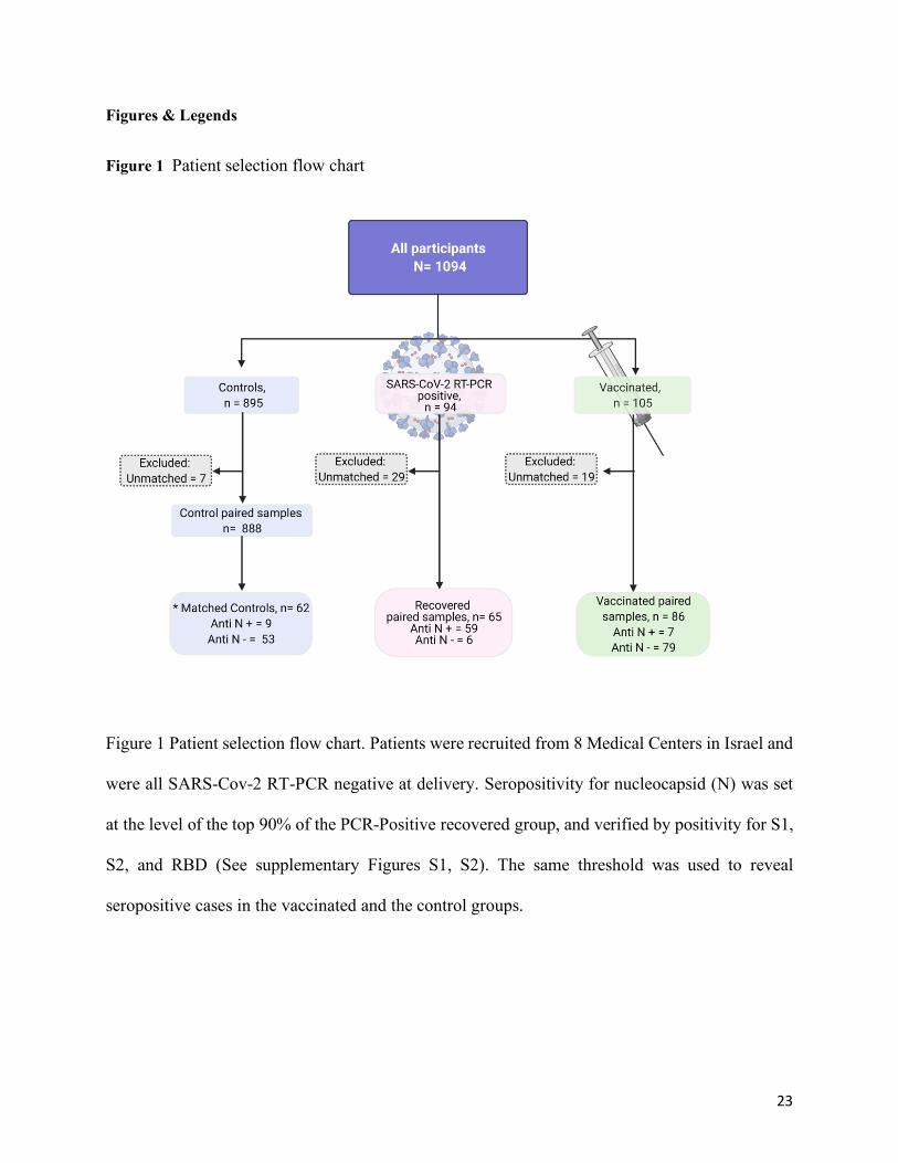
The cohort consisted of 1094 participants from eight hospitals across Israel. Samples were
collected between April 2020 and March 2021 following childbirth and stratified into three groups:
105 vaccine recipients (all collected between January to March 2021), 94 unvaccinated participants
with past SARS-CoV-2 positive RT-PCR results, and 895 unvaccinated without prior
documentation for infection (Table 1, Figure 1). Matched maternal cord blood serology results

7
were obtained for 213 dyads; n=65 with past RT-PCR positive results, n=86 vaccinated recipients
(3 of which were also PCR-positive during pregnancy), and n=62 noninfected unvaccinated; 55
enrolled participants did not have matched cord blood serological results for analysis and were
therefore excluded from the analyses. Among the 895 noninfected unvaccinated participants, 66
were selected (based on clinical parameters) as a matched comparison group, and four were later
excluded due to lack of matched maternal cord blood serology results. Participant demographic
and clinical characteristics and outcomes are provided by study groups in Table 1. Briefly, the
participants were pregnant women from all ethnic societal groups in Israel (~75% Jewish, ~20%
Arab, ~5% others). Diversity was enhanced by the large number of medical centers included in
this study, servicing all communities. Clinical parameters did not differ among the groups, except
for maternal age, which was significantly lower in the SARS-CoV-2 group, as compared to the
other two groups (Kruskal-Wallis One-Way ANOVA; P=0.0011).
Maternal and fetal serological response to SARS-Cov2 infection
Maternal and cord blood serological IgG response to S1, S2, RBD and N antigens of SARS-CoV2
were derived at the time of delivery from 65 patients who were PCR positive during pregnancy.
The data were analyzed by the gestational age (GA) of PCR diagnosis to align all patients on a
common gestational time axis to reveal possible changes in the humoral response or infection
biology across pregnancy.
Based on serology analyses at delivery (Figure 2), transmission rates of IgG to S1, S2, RBD, and
N antigens were significantly higher in participants who were PCR positive to SARS-CoV2 prior
to gestational week 30 (n=25), as compared to gestational week >30 (n=21) (Wilcoxon Rank
8
Sum Test. S1, P=0.0013; S2, P=0.0231; RBD, P=0.0010; N, P= 0.0003). Maternal to fetal
transfer ratio was defined as fetal divided by maternal antibody levels:
𝑇𝑅 = %&'()*+,-(/%+)/('&12()*+,-(/%+)
[Eq 1].
Where TR is the transfer ratio, and MFI is the mean fluorescence intensity.
Maternal to fetal IgG TR values were consistently below 1 for infection occurring at gestational
age >30 weeks, but were significantly elevated at delivery for infections prior to GA 30 (Pearson's
Chi-Square, P<0.0001; Figure 2C).
Serological based re-clustering of the study groups
Based on the robust serological response maintained from mid-pregnancy PCR verified SARS-
CoV2 infection until delivery, the multiplexed immune response was used for further clustering of
the participants based on the reactivity to the N antigen. Multiplexed serological IgG and IgM
response to S1, S2, RBD, and N were tested in maternal and neonate sera (Figure S1). In particular,
the response to N (anti-N, present in SARS-CoV2 but not in the BNT162b2 mRNA vaccine),
versus RBD (anti-RBD, present in both SARS-CoV2 and the BNT162b2 mRNA vaccine),
separated the main groups and identified additional potentially infected participants of the
vaccinated and control groups (Figure S2). Among 65 participants with past SARS-CoV2 RT-
PCR positive test, the top 90% of the maternal IgG response for N was defined as seropositive
(IgG N (MFI) > 1583; n=59). Within the control group, 9 participants (14%) were seropositive for
N using the above threshold, which together with IgG seropositivity to the other COVID-19
antigens (S1, S2 and RBD), corresponds to a preexisting induced immunity due to infection.
9
Similarly, 7 vaccinated participants (8%) were seropositive for N, of which 3 were also PCR-
positive.
Notably, within the PCR positive group, 4 neonates were identified with robust IgM response to
all SARS-CoV2 antigens, and an additional neonate showed partial response consistent with
compromised placenta barrier, fetal exposure to viral antigens, or with vertical viral transmission.
Clinical review of these cases showed that the mothers were diagnosed with mild SARS-CoV2
infection that spontaneously resolved weeks prior to childbirth. Three cases delivered at term, and
one case gave birth at 35 weeks following preterm premature rupture of the fetal membranes. In
all cases, both mother and newborn did not show any signs of illness after childbirth.
Maternal and fetal serological response to BNT162b2 vaccine
The temporal dependence of the acute maternal response to SARS-CoV2 infection (days 1-45;
Figure 3A) was compared to the response to the two-dose regime of BNT162b2; where the 1st
vaccine is administered on day 1 and the 2nd dose on day 21 (Figure 3B). A gradual rise in IgG
humoral response (Anti- S1, S2, RBD and N) was detected during the first 45 days after infection
(Figure 3A). In the same period, vaccinated participants receiving the first BNT162b2 dose showed
a rapid IgG response to S1, S2, RBD but not N, resulting in high titer values by day 15 after the
first dose. A further rise in IgG was observed following the second dose (Figure 3B). The temporal
dependence of fetal IgG for S1, S2, and RBD after vaccination trailed after the maternal IgG
showing a significant response already by day 15. As expected, a further increase was observed
following the second vaccination dose (Figure 3C). As illustrated in Supplementary Figure S3, at
the time of delivery, maternal IgG for S1 and RBD were significantly higher in vaccinated women

10
(P=0.0009, P=0.0045, respectively), while IgG for S2 and N were significantly higher in PCR-
Positive women (P=0.0016, P<0.0001, respectively). Fetal IgG for S2 and N were significantly
lower in cord blood samples of vaccinated women (P<0.0001, P<0.0001, respectively), while fetal
IgG for S1 and RBD did not differ from those of PCR-Positive women (P=0.7017, P=0.6887,
respectively).
Paired maternal-neonate serological data were grouped for statistical analysis to control,
unvaccinated mothers; as well as to mothers who presented at delivery within the first 3 weeks
after the 1st vaccine; deliveries during the first week after the 2nd vaccine; and fully vaccinated -
deliveries more than 1 week after the second vaccine. Significant increase in maternal and fetal
IgG (P<0.0001) and maternal IgM (P<0.05) to S1, S2 and RBD but not N were observed already
after the first vaccination dose and persisted at later timepoints (Table S1). Fetal IgM response to
BNT162b2 antigens (S1, S2, RBD) was negligible, consistent with no evidence for direct exposure
of the fetus to vaccine-derived antigens (Figure 3D; Figure S1).
Correlation of maternal-fetal IgG response to SARS-Cov2 infection and vaccination
The serological response in cord blood correlated positively with the maternal humoral response
for IgG against all the analyzed antigens (Figure 4). There were no differences between the
correlation slopes of the SARS-Cov2 infected group vs. the vaccinated group for any type of
antibodies (S1, P=0.2936; S2, P=0.4212; RBD, P=0.09702; N, P=0.7616), suggesting similar
placental antibodies transfers following SARS-Cov2 infection and vaccination.
Maternal to fetal IgG transfer ratio for S1, S2, RBD, and N

11
The IgG transfer ratio was derived for the PCR-positive group and for serologically positive and
negative vaccinated groups (N+ and N-; Figure 5). Note that the TR for N in the N- group is not
presented due to the low seropositivity. Significant differences were found for S1, S2 and RBD
between the PCR-positive and vaccinated anti-N- groups (P<0.0002). The transfer ratios for all
antibodies did not differ between the vaccinated anti-N+ and all the other groups (P=0.4577).
DISCUSSION
Pregnant women and their neonates are considered vulnerable populations for COVID-19
infection, with significantly greater risks for morbidity and mortality, when compared to matched
populations (17). Recent studies reported that among patients infected during the third trimester,
the transfer of anti-SARS-CoV-2 antibodies to the fetus is significantly impaired (17, 18). Indeed,
our study confirmed the low transfer ratio for infections late in pregnancy. However, with the
availability of a large cohort of patients infected earlier in pregnancy (weeks 15-30), we were able
to show for the first time that maternal and cord blood anti-COVID-19 antibodies, generated in
response to a second trimester infection, as well as transfer ratio, were high at the time of delivery,
in participants recovering from SARS-CoV-2 infection contracted months prior to childbirth. The
low transfer ratio for infections in late gestation could thus also reflect a lag in antibody transfer
across the placenta.
Following reports of a decline in antibody titers months after infection, health organizations
recently recommended vaccination following natural SARS-CoV-2 infection for boosting
immunity (23). However, the significance and relevance of this policy during pregnancy is the
subject of some debate and has not been supported by evidence. Based on our finding of persistent
humoral immunity for infections contracted during the 2nd trimester of pregnancy, titer testing

12
may be informative prior to boosting previously infected pregnant woman, unless boosting is
warranted by emerging variants (24). Unfortunately, pregnant women were excluded from
previous clinical vaccine studies. However, the significant risks and pressing need for action led
to a worldwide debate concerning SARS-CoV-2 inoculation during pregnancy, while data were
still lacking. In the present study, we drew on the unprecedented vaccination campaign undertaken
in Israel, which included pregnant women, and report on the robust humoral immune response
following antenatal immunization with the mRNA vaccine. We found that the Pfizer-BioNTech
COVID-19 mRNA vaccine elicits a rapid rise in IgG titers and effective transfer across the
placenta, exceeding the TR observed in pregnant women with third trimester SARS-CoV-2
infection, as was previously described in nonpregnant populations, and in a small pilot study in
pregnancy (25).
Importantly, maternal IgG humoral response to vaccination in non-infected patients readily
transfers across the placenta to the fetus, leading to a significant, and potentially protective, anti-
SARS-CoV-2 titer in the neonatal bloodstream, already two weeks following the first vaccine dose.
Hence, our data delivers convincing proof for the potency of COVID-19 mRNA vaccines to induce
robust humoral maternal and neonatal immunity during pregnancy. In addition to transplacental
acquired humoral defense, other investigators have recently shown that vaccine response included
the transfer of both spike-specific IgG and IgA antibodies into the maternal breastmilk, potentially
building another line of defense for breastfed infants (25). Accordingly, antenatal immunization
will potentially provide adequate maternal and neonatal protection at highly vulnerable life stages.
Nevertheless, sound evidence regarding safety is still needed and should be addressed in future
studies.

13
Utilizing multiplexed serology, we were able to distinguish between viral and vaccination-induced
immunity and uncover clusters of asymptomatic, undiagnosed infections among the control and
vaccinated groups. We describe 7 vaccinated patients that were found to have high levels of anti-
N IgG, corresponding with previous undiagnosed infection. We detected no significant changes in
maternal or neonatal titers, when compared to the titers of vaccinated and recovered participants.
In addition, among the 65 PCR positive deliveries, we found 5 fetuses (7%) who showed IgM
reactivity to all or most viral antigens, consistent with placenta barrier defect, fetal exposure to
viral antigens, or vertical viral transmission. In contrast, among the 86 vaccinated deliveries, we
found no evidence for cases of fetal IgM response to any of the vaccine-induced antigens.
Strengths and Limitations
The present study has several strengths and limitations. Its strengths include its multicenter design
and patient accrual; our relatively large cohort size; and our diverse patient population. Its
limitations include the bias in sample collection as most of the study recruitment occurred during
the day, and therefore does not includes many of the emergency cases. However, the method of
sample collection did not differ between the study groups and Medical Centers, thus minimizing
the impact of this effect on our results. Second, since sample collection began long before the
COVID-19 immunization campaign, the duration of sample collection differed between the
groups, with an extended recruitment period for COVID-19 recovered cases. We found no
differences in background or demographic parameters among the groups. Third, a history of
COVID-19 infection during pregnancy was made by positive RT-PCR results during pregnancy
with self-reporting of the time of PCR test. Stricter and more accurate symptom monitoring and
repeated sampling during pregnancy may provide a higher resolution delineation of how the

14
immune response develops and transfers following COVID-19 infection. Fourth, patients
presenting with RT-PCR positive within one week prior to delivery were excluded from this study
due to safety concerns. Thus, future studies are needed in order to characterize the maternal
humoral response to COVID-19 within days of infection.
Conclusions
We show herein a robust maternal humoral immune response coupled to a rise in protective
antibodies in the fetal circulation as early as 15 days after the first BNT162b2 mRNA vaccination.
We further show that mid-pregnancy SARS-Cov2 infection results in prolonged maternal and fetal
humoral immunity presented at delivery time.
METHODS:
Study Design
Pregnant women admitted for delivery at eight medical centers in Israel (Hadassah Mount Scopus,
Wolfson, HaEmek, Hillel Yafe, Rabin, Shaare Zedek, Meir, and Sourasky Medical Centers) were
approached for enrollment in a biorepository study, starting in April 2020. Eligibility criteria
included an age of 18 years or older, and a willingness to participate and provide informed consent.
Pregnant women with active maternal COVID-19 disease at delivery were excluded from this
study. Eligible patients were identified by dedicated study clinicians (obstetrician, nurse midwife)
present on the Labor and Delivery units enrolled in the study. Gravidae who received the
BNT162b2 mRNA vaccine during pregnancy were assigned to the vaccinated group; parturients
15
with documented COVID-19 infection during pregnancy, confirmed by positive nasopharyngeal
swab RT-PCR test, comprised the COVID-19 positive group. Unvaccinated parturients were
matched to the vaccinated group participants based on clinical parameters (Table 1) and were
assigned to a control group.
Samples Collection and handling
Maternal and fetal blood samples were collected from the enrolled patients prior to delivery, and
from the umbilical cord following delivery. The umbilical cord was wiped clean, and blood was
drawn from the vein. Blood samples were centrifuged at 1000g for 10 minutes at room
temperature, and serum samples were aliquoted into dedicated pre-coded tubes and stored at - 80°C
until analyses at the Weizmann Institute. Fourteen placental tissue samples were microscopically
blindly examined by a single experienced pathologist. The rate of malperfusion lesions was similar
in the examined placental tissue of all groups (Supplementary Table S2; Figure S4).
Quantification of Anti-COVID-19 Antibodies
Serum samples were thawed, heat-inactivated at 56oC for 30 minutes, and transferred to bar-coded
96-well plates for analysis. Serum IgG and IgM were detected using Milliplex MAP SARS-CoV-
2 Antigen Panel 1 IgG (HC12SERG-85K) and IgM (HC19SERM1-85K). Reagents were prepared
according to manufacturer instructions and dispensed to 96-well source plates (Greiner 651201,
Sigma-Aldrich). Serum samples were diluted 1:100 in assay buffer and added to antigen-
immobilized Milliplex beads in 96-well plates using a Bravo liquid handler (Agilent). Plates were
covered, shook for two hours at room temperature, and washed three times with wash buffer, using
a manual magnet and multidrop combi dispenser (Thermo). Anti-IgG-PE or Anti-IgM-PE
16
conjugate was added, and the samples were incubated (90 minutes with shaking) and washed.
Sheath fluid was added to the samples, and net fluorescent intensity (MFI) signals were detected
on a Luminex MAGPIX reader (Supplementary data). Repeat measurements of the same sample
showed less than 5% difference for all antibodies. Positive and negative controls were included in
each analysis for accuracy and reproducibility.
Statistical Analyses
Statistical analyses were performed using Statistix 8 software (Analytical Software, Tallahassee,
FL USA) and Prism 5.01 (GraphPad Software; San-Diego, CA, USA). IgG and IgM antibody (S1,
S2, RBD, N) concentrations were log10-transformed for analyses. Correlations between fetal and
maternal Ab were analyzed by Linear Regression test. Comparisons of antibody concentrations
among groups, as well as continuous parameters (e.g., clinical data), were analyzed by Kruskal–
Wallis one-way ANOVA test, following by Dunn's all-pairwise comparisons test; or alternatively,
by Wilcoxon Rank Sum Test (if only two groups were compared). Comparisons between maternal
and fetal concentrations within each group were analyzed by Paired t-test. Pearson Chi-square
analysis was used to compare proportional data. All statistical tests were based on two-tailed
hypotheses. Differences were considered significant at P < 0.05.
Study approval
The current study followed the Strengthening the Reporting of Observational Studies in
Epidemiology (STROBE) reporting guideline. The study was approved by the institutional review
boards of all participating medical centers and by the Weizmann Institute of Science. All research
participants provided written informed consent prior to enrollment.

17
Author contributions
OB, RPM, ZS, AM, HB, SY, MN, MK design of the research studies; KNS, YSC, RC, RGT,
EH, RGB, YJM, TBS, GSM, SFG, HYS, HBR, NDS, DGW, HB conducted the experiments and
collected the samples; RPM, LS, HB acquired the data; RPM, TR, LS, ZS, HB, SY, MN, MK
analyzed the data; ZS provided reagents; OB, RPM, TR, TBS, DGW, AM, HB, SY, MN, MK
participated in writing the manuscript.
Conflict of interest statement:
The authors have declared that no conflict of interest exists.
Acknowledgements
This work was supported by ISF KillCorona grant 3777/19 (to MN, MK, SY, AM); and by a
research grant from the Weizmann Institute Fondazione Henry Krenter (to MN). We would like to
thank the patients who made this research possible. We acknowledge the contribution in patient
recruitment, sample preparation and discussions by:
Weizmann Institute: Gila Meir, Leonardo Solmesky, PhD., Nava Dekel, PhD.
Hadassah Medical Center: Adva Cahen Peretz, MD; Michal Lipschuetz, RN, MPH, MSc; Nadine
Souri; Sarah M Cohen, MPH.
Wolfson Medical Center: Yasmin Farhadian MD, Hind Odeh BSc
HaEmek Medical Center: Shalva Fux BA, RN (MW)
Hillel Yaffe Medical Center Alina Wiener, MBA , Luchilla Zorzetti, MD

18
Shaare Zedek Medical Center: Itamar Glick, MD
Meir Medical Center: Avital Diamond , Yaara Hoffman

19
References
1. DeBolt CA, Bianco A, Limaye MA, Silverstein J, Penfield CA, Roman AS, et al. Pregnant women with severe or critical coronavirus disease 2019 have increased composite morbidity compared with nonpregnant matched controls. Am J Obstet Gynecol. 2020.
2. Hantoushzadeh S, Shamshirsaz AA, Aleyasin A, Seferovic MD, Aski SK, Arian SE, et al. Maternal death due to COVID-19. Am J Obstet Gynecol. 2020;223(1):109 e1- e16.
3. Pierce-Williams RAM, Burd J, Felder L, Khoury R, Bernstein PS, Avila K, et al. Clinical course of severe and critical coronavirus disease 2019 in hospitalized pregnancies: a United States cohort study. Am J Obstet Gynecol MFM. 2020;2(3):100134.
4. Juan J, Gil MM, Rong Z, Zhang Y, Yang H, and Poon LC. Effect of coronavirus disease 2019 (COVID-19) on maternal, perinatal and neonatal outcome: systematic review. Ultrasound Obstet Gynecol. 2020;56(1):15-27.
5. Dashraath P, Wong JLJ, Lim MXK, Lim LM, Li S, Biswas A, et al. Coronavirus disease 2019 (COVID-19) pandemic and pregnancy. Am J Obstet Gynecol. 2020;222(6):521-31.
6. Knight M, Bunch K, Vousden N, Morris E, Simpson N, Gale C, et al. Characteristics and outcomes of pregnant women admitted to hospital with confirmed SARS-CoV-2 infection in UK: national population based cohort study. BMJ. 2020;369:m2107.
7. Panagiotakopoulos L, Myers TR, Gee J, Lipkind HS, Kharbanda EO, Ryan DS, et al. SARS-CoV-2 Infection Among Hospitalized Pregnant Women: Reasons for Admission and Pregnancy Characteristics - Eight U.S. Health Care Centers, March 1-May 30, 2020. MMWR Morb Mortal Wkly Rep. 2020;69(38):1355-9.
8. Delahoy MJ, Whitaker M, O'Halloran A, Chai SJ, Kirley PD, Alden N, et al. Characteristics and Maternal and Birth Outcomes of Hospitalized Pregnant Women with Laboratory-Confirmed COVID-19 - COVID-NET, 13 States, March 1-August 22, 2020. MMWR Morb Mortal Wkly Rep. 2020;69(38):1347-54.
9. Jering KS, Claggett BL, Cunningham JW, Rosenthal N, Vardeny O, Greene MF, et al. Clinical Characteristics and Outcomes of Hospitalized Women Giving Birth With and Without COVID-19. JAMA Intern Med. 2021.
10. Stafford IA, Parchem JG, and Sibai BM. The coronavirus disease 2019 vaccine in pregnancy: risks, benefits, and recommendations. Am J Obstet Gynecol. 2021.
11. CDC. CDC: Data on COVID-19 during Pregnancy: Severity of Maternal Illness. https://covid.cdc.gov/covid-data-tracker/?CDC_AA_refVal=https%3A%2F%2Fwww.cdc.gov%2Fcoronavirus%2F2019-ncov%2Fcases-updates%2Fspecial-populations%2Fpregnancy-data-on-covid-19.html#pregnant-population. Accessed 30.3.21.
12. ACOG. ACOG: Vaccinating Pregnant and Lactating Patients Against COVID-19. https://www.acog.org/clinical/clinical-guidance/practice-advisory/articles/2020/12/vaccinating-pregnant-and-lactating-patients-against-covid-19. Accessed 30.3.21.
13. CDC. Centers for Disease Control and Prevention. COVID-19 (coronavirus disease): people with certain medical conditions. https://www.cdc.gov/coronavirus/2019-ncov/need-extra-precautions/people-with-medical-conditions.html. Accessed 30.3.2021.
14. Goncalves G, Cutts FT, Hills M, Rebelo-Andrade H, Trigo FA, and Barros H. Transplacental transfer of measles and total IgG. Epidemiol Infect. 1999;122(2):273-9.
15. Munoz FM, Bond NH, Maccato M, Pinell P, Hammill HA, Swamy GK, et al. Safety and immunogenicity of tetanus diphtheria and acellular pertussis (Tdap) immunization during pregnancy in mothers and infants: a randomized clinical trial. JAMA. 2014;311(17):1760-9.

20
16. Martinez DR, Fong Y, Li SH, Yang F, Jennewein MF, Weiner JA, et al. Fc Characteristics Mediate Selective Placental Transfer of IgG in HIV-Infected Women. Cell. 2019;178(1):190-201 e11.
17. Atyeo C, Pullen KM, Bordt EA, Fischinger S, Burke J, Michell A, et al. Compromised SARS-CoV-2-specific placental antibody transfer. Cell. 2021;184(3):628-42 e10.
18. Edlow AG, Li JZ, Collier AY, Atyeo C, James KE, Boatin AA, et al. Assessment of Maternal and Neonatal SARS-CoV-2 Viral Load, Transplacental Antibody Transfer, and Placental Pathology in Pregnancies During the COVID-19 Pandemic. JAMA Netw Open. 2020;3(12):e2030455.
19. Kim L, Whitaker M, O'Halloran A, Kambhampati A, Chai SJ, Reingold A, et al. Hospitalization Rates and Characteristics of Children Aged < 18 Years Hospitalized with Laboratory-Confirmed COVID-19-COVID-NET, 14 States, March 1-July 25, 2020. Mmwr-Morbidity and Mortality Weekly Report. 2020;69(32):1081-8.
20. Dong L, Tian J, He S, Zhu C, Wang J, Liu C, et al. Possible Vertical Transmission of SARS-CoV-2 From an Infected Mother to Her Newborn. Jama-Journal of the American Medical Association. 2020;323(18):1846-8.
21. Skowronski DM, and De Serres G. Safety and Efficacy of the BNT162b2 mRNA Covid-19 Vaccine. N Engl J Med. 2021;384(11).
22. Baden LR, El Sahly HM, Essink B, Kotloff K, Frey S, Novak R, et al. Efficacy and Safety of the mRNA-1273 SARS-CoV-2 Vaccine. N Engl J Med. 2021;384(5):403-16.
23. Krammer F, Srivastava K, Alshammary H, Amoako AA, Awawda MH, Beach KF, et al. Antibody Responses in Seropositive Persons after a Single Dose of SARS-CoV-2 mRNA Vaccine. N Engl J Med. 2021.
24. Stamatatos L, Czartoski J, Wan YH, Homad LJ, Rubin V, Glantz H, et al. mRNA vaccination boosts cross-variant neutralizing antibodies elicited by SARS-CoV-2 infection. Science. 2021.
25. Gray KJ, Bordt EA, Atyeo C, Deriso E, Akinwunmi B, Young N, et al. COVID-19 vaccine response in pregnant and lactating women: a cohort study. Am J Obstet Gynecol. 2021.

21
Tables
Table 1. Clinical parameters of women included in the study
Parameter* Control group
n=66
Past SARS-CoV-2 group
n=74
Vaccinated group n=92
Maternal age, mean±SD, y 31.6±5.8 28.8±5.8* 31.7±5.8
Gestational age, mean±SD, wks 39.2±1.4 39±1.6 39.3±1.3
Preterm delivery (<37), No. (%) 5 (7.6) 8 (10.8) 4 (4.3)
Pregravid BMI (kg/m2), mean±SD 25.7±6.5 26.4±9.2 24.2±5.2
Gravidity, median (IQR) 3 (2.5) 3 (2) 3 (2)
Parity, median (IQR) 2 (2) 1 (3) 1 (2)
Maternal comorbidities, No. (%)
Hypertensive disorders 1(1.5) 1 (1.4) 1(1.1)
Diabetes or gestational diabetes 9 (13.6) 4 (5.4) 8 (8.7)
Asthma 1 (1.5) 2 (2.7) 2 (2.2)
Thyroid disease 4 (6.1) 1 (1.3) 8 (8.6)
Smoker 4 (6.6) 1 (1.4) 6 (6.5)

22
Infant sex, No. (%)
Male 31 (47.7) 39 (52) 45 (49.5)
Female 34 (52.3) 36 (48) 46 (50.5)
Birthweight, mean±SD, gr 3239.4±395.7 3324.1±536.2 3281.8±420.2
NICU, No. (%) 1 (1.6) 2 (2.7) 4 (4.3)
PCR-Positivity GA, mean±SD, wks ----- 28.1±8.3 ------
1st Vaccine Dose GA, mean±SD, wks ----- ------ 34.5±7.5
Continuous parameters were analyzed by Kruskal–Wallis one-way ANOVA test, following by Dunn's all-pairwise comparisons test; Pearson Chi-square analysis was used to compare proportional data. *Clinical parameters did not differ among the groups, except for maternal age, which was significantly lower in the SARS-CoV-2 group, as compared to the other two groups (Kruskal-Wallis One-Way ANOVA; P=0.0011)
23
Figures & Legends
Figure 1 Patient selection flow chart
Figure 1 Patient selection flow chart. Patients were recruited from 8 Medical Centers in Israel and
were all SARS-Cov-2 RT-PCR negative at delivery. Seropositivity for nucleocapsid (N) was set
at the level of the top 90% of the PCR-Positive recovered group, and verified by positivity for S1,
S2, and RBD (See supplementary Figures S1, S2). The same threshold was used to reveal
seropositive cases in the vaccinated and the control groups.

24
Figure 2
Figure 2
Robust maternal and fetal seropositivity can be detected at birth after recovery from SARS-Co2 infection.
Maternal and cord blood was derived at the time of delivery from patients (n=65) who recovered from
infection with SARS-Cov-2 verified by RT-PCR at the specified GA during pregnancy. Maternal and
fetal seropositivity (A,B) and transfer ratio (TR; C) was analyzed for four SARS-Cov-2 antigens (S1, S2,
RBD and N). The data is plotted as a function of the gestational age (GA) at which positive RT-PCR was
diagnosed. (A, B) High levels of maternal (A) and fetal (B) IgG levels for S1, S2, RBD and N are found
at delivery for infections occurring prior to GA 30, and appear to be lower for late 3rd trimester infections.
Shaded areas show the 95% confidence interval. (C) Transfer ratio (TR) values at delivery are low for 3rd
trimester infections, and significantly higher for earlier, 2nd trimester infections (Wilcoxon Rank Sum
Test, P<001). S1, red; S2, green, RBD, blue; N, black. Black line marks TR = 1.

25
Figure 3
Figure 3
Dependence of immune response to SARS-Cov2 infection and to vaccination on the duration from
exposure to delivery. (A) Analysis of the level of maternal IgG at delivery for acute (<50 days) infection.
Time was derived from the GA of RT-PCR positivity and the GA of delivery (see the immune response
across pregnancy in Figure 1). (B) Analysis of maternal IgG response to BNT162b2 vaccination derived
from the GA of the first vaccine and the GA of delivery. (C) Analysis of the level of fetal IgG at delivery,

26
following BNT162b2 vaccination. Time was derived from the GA of the first vaccine and the GA of
delivery. B, C) A second dose was administered on Day 21; shaded areas and lines in (A), (B) and (C)
show the mean and 95% confidence interval. (D) Serological data of maternal-fetal pairs were derived
from control, unvaccinated serologically negative (N-) mothers; as well as vaccinated mothers grouped
for deliveries in the first 3 weeks after the 1st vaccine; deliveries during the first week after the 2nd
vaccine; and fully vaccinated who delivered more than a week after the second vaccine. Left columns,
IgG; right columns, IgM; from top to bottom, serological response to S1, S2, RBD, and N. Statistical
significance: A,B,C above the blue bars indicate significant differences among the groups in maternal
antibodies, while a,b,c above the orange bars indicate significant differences among the groups in fetal
antibodies (Kruskall–Wallis one-way ANOVA test, following by Dunn's All-Pairwise Comparisons Test);
*indicate a significant difference between maternal and fetal antibodies within the same group (Paired t-
test). Box and whiskers: Middle line= Median; Box= the 25% and 75% (25th, and 75th percentiles);
whiskers= min & max values (Table S2).

27
Figure 4
Figure 4
Maternal-fetal serological correlation of IgG for S1, S2, RBD and N. A subgroup of the Control group was
identified as serologically positive for N as well as for S1, S2, and RBD (n=9 of 62). Similarly, a subgroup
of the vaccinated was serologically identified as positive for N (n=7 of 86; marked in black). A significant
maternal-fetal correlation was observed for all groups and all antigens. Correlations between fetal and

28
maternal Ab (N, S1, S2, RBD) were analyzed by Linear Regression test (Supplementary Figure S3). Each
dot represents data from a single patient; the linear regression line is marked in black, with its 95% CI
(dotted lines). (A) R2=0.9443; Adjusted R2= 0.9438; P<0.0001. (B) R2= 0.9353; Adjusted R2= 0.9348;
P<0.0001. (C) R2= 0.9200; Adjusted R2= 0.9194; P<0.0001. (D) R2= 0.9366; Adjusted R2= 0.9361;
P<0.0001. Red, Control; Green, PCR positive; Blue, Vaccinated N-; Black, Vaccinated N+.

29
Figure 5
Figure 5
Maternal to fetal transfer ratio (TR) for the PCR positive recovered group and for serologically positive
and negative vaccinated groups. Note that the TR for N in the N- group is not presented due to the low
seropositivity. Differences among the groups were analyzed by using Kruskall–Wallis one-way ANOVA
test, following by Dunn's All-Pairwise Comparisons Test. For S1, S2 and RBD, there was a significant
difference between the PCR-positive vaccinated Anti N- groups (P<0.0002). The transfer ratios did not
differ between the vaccinated Anti N+ and all the other groups for all antigens (P=0.4577).
Top Related