


Idiomas
Páginas
Jurídico


Pr Francesco GIAMMARILE
CHLS Lyon
Faculté de Lyon Sud
« Aut tace aut loquere meliora silentio »
CURSO REGIONAL DE CAPACITACIÓN EN GANGLIO
CENTINELA Y CIRUGÍA RADIOGUIADA
El ganglio centinela en cáncer de mama
F.Giammarile
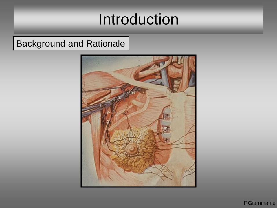
Introduction
Background and Rationale

Important staging procedure in breast cancer surgery
Trend toward early diagnosis: many negative ALND
Complications: pain, paresthesia, infection, lymphedema, mobility
Axillary lymph node dissection (ALND)
Applied in breast cancer about 20 y. ago and became a routine technique
Accurate staging (identification rates of SLN of more than 95% in
experienced multidisciplinary team: learning curve)
May also detect non-axillary sentinel nodes
Minimize the number of unnecessary ALND
morbidity, cosmetic results, shorter hospital stay
Sentinel lymph node biobsy (SLNB)
F.Giammarile
Breast cancer
Breast cancer is the most frequent cancer in women worldwide
Accurate lymph node staging is essential for prognosis and treatment
No imaging modality is accurate enough to detect lymph node metastases
Rationale

SNL in breast cancer
Techniques
and results
F.Giammarile
Author Year Patient
nb
Identified
SN
False
negative
Blue dye only
Giuliano 1994 174 66% 11%
Guenther 1997 145 71% 10%
Flett 1997 68 82% 17%
Probe only
Crossin 1998 50 84% 13%
Krag 1998 443 93% 11%
Probe and dye
Chatterjee 1998 60 97% 5%
Cox 1998 466 94% 1%
v.d. Ent 1998 70 100% 4%
Doting 1999 136 93% 5%
Tanis 2002 501 96% <5%
Technical aspects
Injection modalities
Volume
Activity
Particle size
Time between
injection and
imaging / surgery

The breast: multiple drainage patterns
Axillary / Internal Mammary Chain (IMC)
SNL in breast cancer
F.Giammarile
EANM Oncology Committee:
F.Giammarile
• Francesco Giammarile
• Jolanta Kunikowska
• Wim J.G. Oyen
ESSO • Riccardo Audisio
• Marjut Leidenius
SNM • Naomi Alazraki
Reviewers • Renato Valdes Olmos
• Sergi Vidal Sicart
Indication
EANM procedure guidelines
for sentinel node in breast cancer
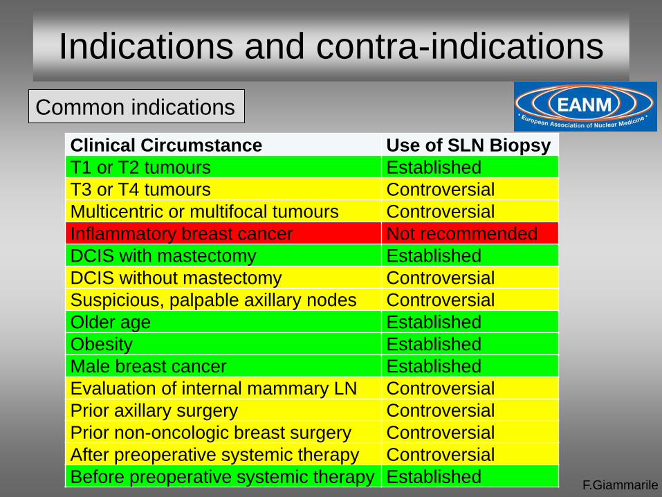
Common indications
F.Giammarile
Indications and contra-indications
Clinical Circumstance Use of SLN Biopsy
T1 or T2 tumours Established
T3 or T4 tumours Controversial
Multicentric or multifocal tumours Controversial
Inflammatory breast cancer Not recommended
DCIS with mastectomy Established
DCIS without mastectomy Controversial
Suspicious, palpable axillary nodes Controversial
Older age Established
Obesity Established
Male breast cancer Established
Evaluation of internal mammary LN Controversial
Prior axillary surgery Controversial
Prior non-oncologic breast surgery Controversial
After preoperative systemic therapy Controversial
Before preoperative systemic therapy Established

Precautions
F.Giammarile
Indications and contra-indications
• Pregnancy and lactation
Pregnancy is not a contraindication for probe-guided SLN biopsy (the
dose to the foetus is negligible)
The use of blue dye is not recommended during pregnancy
In nursing mothers, lactating should be suspended for 24 h after
radiopharmaceutical administration
• Training
Studies should only be performed by a surgeon and a nuclear medicine
specialist who have undergone specific training in this technique
At this time, no definition of the required training has been validated for
the surgeon or the nuclear physician, although a minimum of 30
procedures has been proposed for the surgeon

F.Giammarile
Nuclear Medicine procedure
Injection technique, detection technique
SPECT-CT: 3D
Volume rendering
F.Giammarile
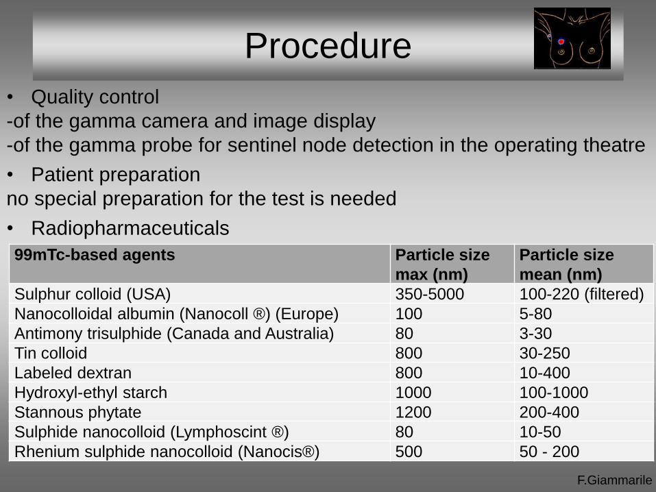
Procedure
• Quality control
-of the gamma camera and image display
-of the gamma probe for sentinel node detection in the operating theatre
• Patient preparation
no special preparation for the test is needed
• Radiopharmaceuticals
99mTc-based agents Particle size
max (nm)
Particle size
mean (nm)
Sulphur colloid (USA) 350-5000 100-220 (filtered)
Nanocolloidal albumin (Nanocoll ®) (Europe) 100 5-80
Antimony trisulphide (Canada and Australia) 80 3-30
Tin colloid 800 30-250
Labeled dextran 800 10-400
Hydroxyl-ethyl starch 1000 100-1000
Stannous phytate 1200 200-400
Sulphide nanocolloid (Lymphoscint ®) 80 10-50
Rhenium sulphide nanocolloid (Nanocis®) 500 50 - 200

Injection techniques
Volume - Concentration
F.Giammarile
Total injected volume: 0.2-0.5 mL (physiologic)
- if less: slow drainage
- if more: saturation
Size of colloids
sulfur colloid (5-80 nm) or
albumin nanocolloid (5-30 nm)

Injection techniques
F.Giammarile
Activity - Timing
- If injection the same day of surgery : 5-30 MBq
- If injection the day after the surgery: 150 MBq max
10 min 2 h
Early:
intravascular
activity;
multiple vessels
leading to
a single sentinel
node
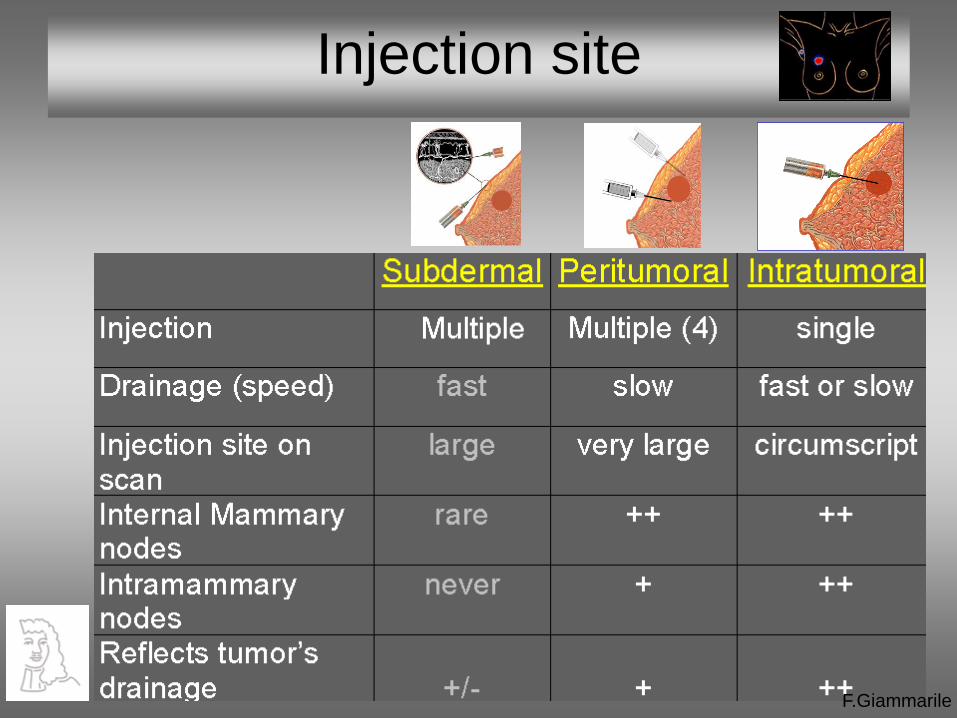
Injection site
F.Giammarile

F.Giammarile
In non-palpable breast lesion replace hooked wired localization
Intratumoral administration guided by ultrasound/stereotaxis
Followed by immediate surgical resection, guided by probe
Can be associated to SLN
ROLL technique
Radioguided Occult Lesion Localisation

Peritumoral injection site
Injection technique
3-4 injection around the tumor F.Giammarile
• Non-palpable breast carcinoma
Administration guided by
ultrasound/stereotaxis
• Palpable breast carcinoma

Visualization of IMC
ROLL and SLN detection
Internal Mammary Chain
(5-20%) Axilla (98%)
F.Giammarile
Peritumoral injection site
Characteristics

Superficial injection sites
F.Giammarile
Injection technique
Subareolar/Periareolar (Sappey’s subareolar plexus) Over the lesion
1 – 4 injection at the skin level
(subdermal/intradermal)

Subdermal injection sites
Subareolar/Periareolar Over the lesion
F.Giammarile
- Impalpable and multicentric tumours
Easy technique, rapid uptake Characteristics
- Better accuracy (elderly)

F.Giammarile
Injection site
Injection method does not significantly affect the
identification of SLN
-superficial injection is easy to perform
-deep injections may require ultrasound or sterotaxic
guidance but allow detection of extra-axillary nodes

F.Giammarile
Image acquisition
Imaging is strongly recommended before any operative procedure
(variability in breast lymphatic drainage into the axilla and extra-axillary)
• Patient position:
Supine or prone (with hanging breast position or upright)
Anterior, 45° anterior oblique and lateral imaging can be obtained
• Imaging:
Performed within 15-30 min after the injection and 2-4 hours after
Images acquired for 3-5 min, pixel size of about 2mm (256x256 or
128x128 matrix)
57Co or 99mTc flood source for delineation of patient’s body contour
SPECT/CT optional images: better contrast and spatial resolution
• Localisation:
The site of any suspected SLN can be localized on overlying skin,
preferably on the 45° anterior oblique image

DRENAJE LINFÁTICO
Ganglios axilares (Niveles I, II y III de Berg) (N1-N3) G. del parénquima mamario G. de la cadena mamaria interna (N1-N3) G. de la zona supraclavicular (N3) G. de la cara lateral del cuello (M1) G. de la axila contralateral (M1)

Detection techniques
F.Giammarile
Localization
17%
30%
46%
4%
IM: incidence = 20%
Tanis P et al. Br J Cancer 2002

Detection techniques
F.Giammarile
Localization
Tanis P et al. Br J Cancer 2002
Others: incidence = 11%
Supraclavicular 0,3%
Interpectoral 2,0 %
Intramamary lateral 5,3 %
Intramamary medial 2,0 %
Infraclavicular 1,2%

Detection techniques
F.Giammarile
Landmark
Anterior projection
Axillary projection
Lateral projection

Detection techniques
F.Giammarile
Imaging
The ideal
case!

Detection techniques
F.Giammarile
Timing
SN?
SN
SN
Role of
early/
delayed
images

Detection techniques
F.Giammarile
Imaging
Two SLN

Detection techniques
F.Giammarile
Imaging
One SLN
Detection techniques
F.Giammarile
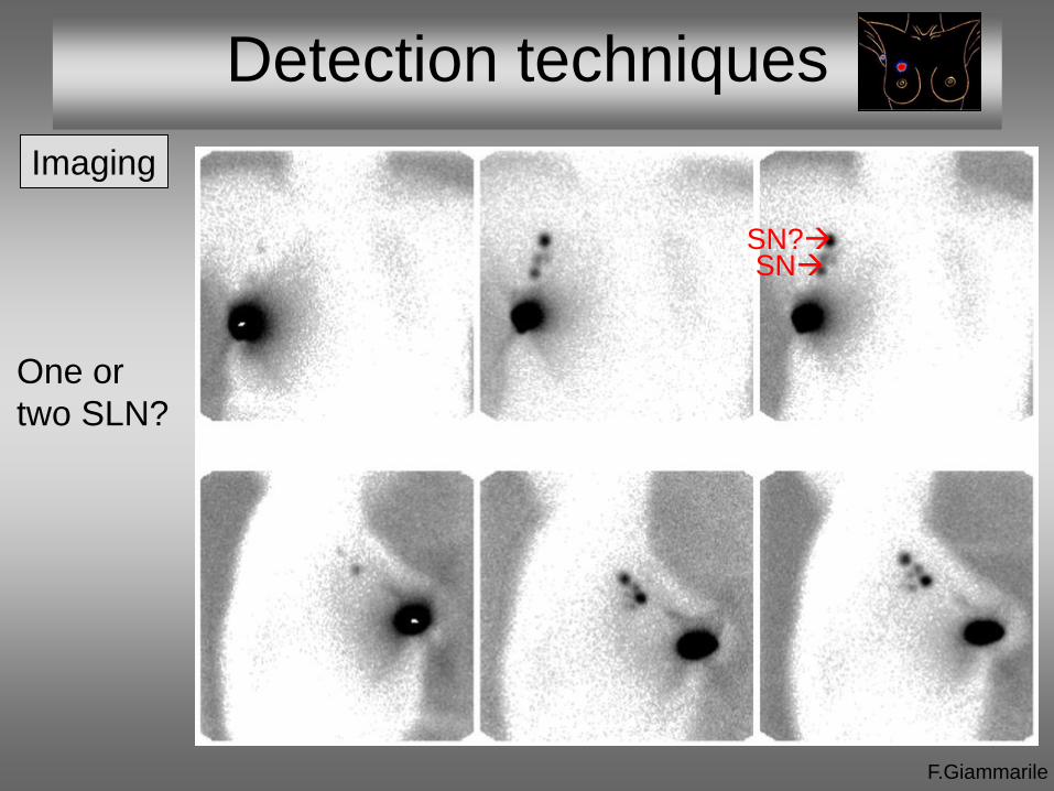
Imaging
One or
two SLN?
SN SN?

F.Giammarile
Detection techniques
Imaging
Intra-
mammary
SLN
Detection techniques
F.Giammarile
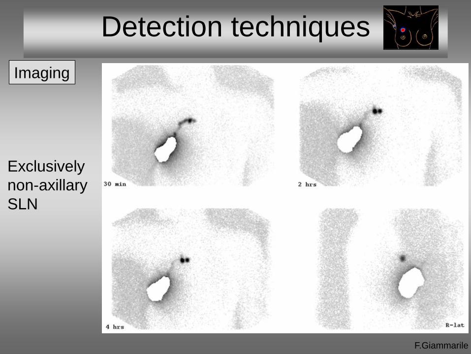
Imaging
Exclusively
non-axillary
SLN

Detection techniques
F.Giammarile
Imaging
All in one!

SLN non visualization
The majority of patients with preoperative lymphoscintigraphic SLN non
visualization will have at least one SLN detected intraoperatively
In approximately 1%-2% of patients, SLN will not be detected
intraoperatively, and the status of axillary nodes cannot be determined
Old age, obesity, tumour location other than in the upper outer
quadrant, and non-visualization of SLN on preoperative
lymphoscintigraphy may be associated with failed SLN localization
There is no definitive consensus on what to do if the SLN cannot be
visualised. However, current standard patient care recommends axillary
lymph node dissection
Detection techniques
F.Giammarile

Captación débil o ausente en el
ganglio centinela
F.Giammarile
1. Radiotrazador
a. Dosis escasa
b. Baja calidad
c. Cifra de partículas insuficiente
2. Imágenes
Intervalos demasiado cortos o largos entre la administración del
radiotrazador y la linfogammagrafía
3. Paciente
a. Edad avanzada
b. Mamas grandes / grasas
c. Cuadrante superoexterno
d. Invasión tumoral
e. Cirugía / biopsia de mama previa
Tanis P et al. Eur J Nucl Med 2002;29:993

SPECT-CT
F.Giammarile
Overweighted patients with breast cancer
Improved sentinel node
identification by SPECT/CT
Lerman et al. JNM, 2007

F.Giammarile
SPECT-CT

F.Giammarile
Advantages
SLN better
localization
Two parietal lymph
nodes clearly shown
only on SPECT-CT
SPECT-CT

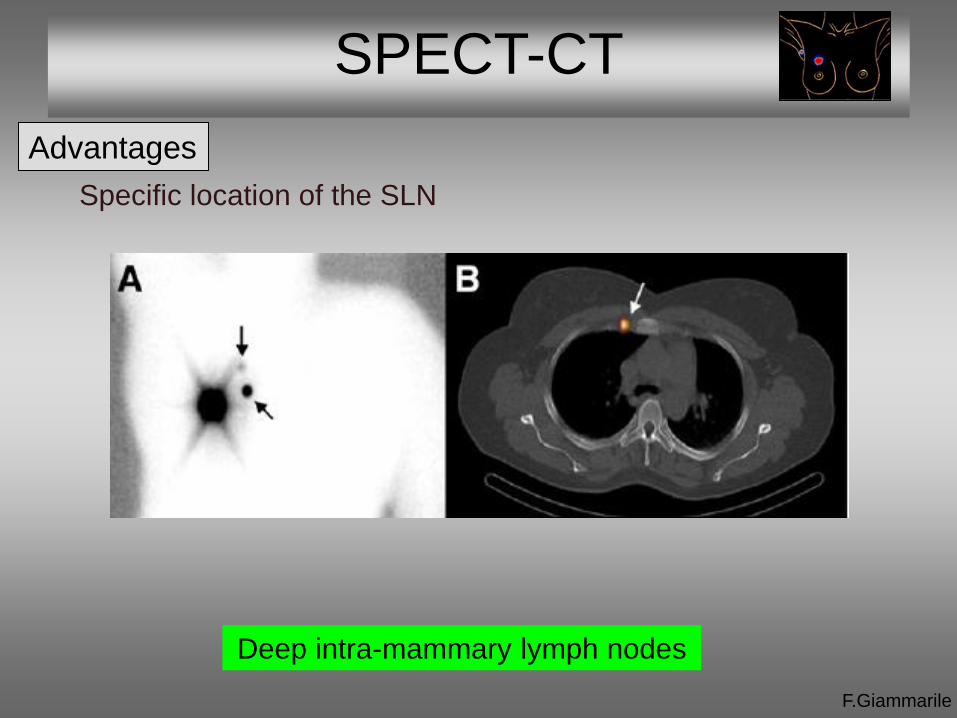
F.Giammarile
Advantages
Specific location of the SLN
Level I and Level II axillar lymph
nodes clearly shown on SPECT-CT
SPECT-CT
F.Giammarile
Advantages
Specific location of the SLN
Deep intra-mammary lymph nodes
SPECT-CT
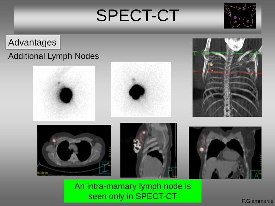
F.Giammarile
Advantages
Additional Lymph Nodes
An intra-mamary lymph node is
seen only in SPECT-CT
SPECT-CT

F.Giammarile
Advantages
Intra-mamary lymph node detection
SPECT-CT

F.Giammarile
Advantages
Intra-mamary and axillary lymph node detection
SPECT-CT
F.Giammarile
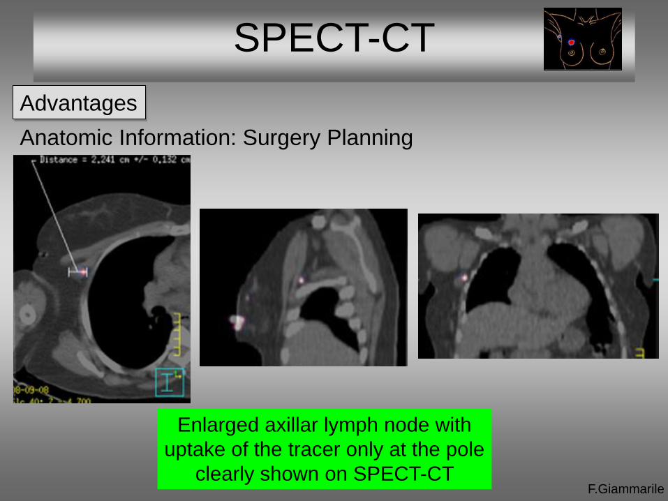
Advantages
Anatomic Information: Surgery Planning
Enlarged axillar lymph node with
uptake of the tracer only at the pole
clearly shown on SPECT-CT
SPECT-CT

F.Giammarile
Advantages
Anatomic Information: Surgical Planning
SPECT-CT

Procedures during surgery
F.Giammarile
SN exeresis and analysis
While removing too few nodes may result in missing potential
metastasis in regional lymph nodes, indiscriminate removal of axillary
nodes may cause morbidity similar to that of axillary lymphadenectomy
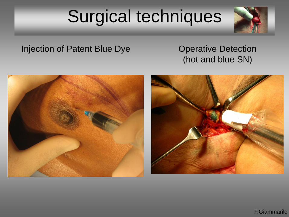
Surgical techniques
F.Giammarile
Injection of Patent Blue Dye Operative Detection
(hot and blue SN)

Blue dye localisation
F.Giammarile
patent blue V, isosulfan blue,
methylene blue
0.5-1 mL of blue dye
injected around primary tumour,
10-20 min prior surgery
Within 5-15 min the SLN is coloured.
Washout after about 45 min
In most cases, the same SLNs are
detected by the two methods

F.Giammarile
Detection probes
Able to detect the SLN from the skin
surface and within the exposed
surgical cavity:
- must be well collimated to
discriminate activity
- can be used to select the
optimum location for incision
- must be used to check the wound
site for remaining activity
- must be used to re-check
removed SLN before sending for
histology
Deep SLN are difficult to detect (tissue attenuation): to avoid false
negative results, the open axilla should be palpated and suspicious
lymph nodes harvested, even if these are neither hot nor blue

Resultados
BIOPSIA DEL GANGLIO CENTINELA
INDICACION CLINICA
VISUALIZACION 95%
LOCALIZACION QUIRURGICA
98%
T1 <2cm
T2 2-5 cm
Ausencia clínica de metástasis linfáticas
Falsos negativos = 3 % (0-10%) VPN = 98%

SNL in breast cancer
F.Giammarile
Local anesthesia: None
Tracer activity: 40 MBq in 1/4 injection (0.2 mL), for surgery the
day after
Particle type: Nanocis® or Nanocoll®
Injection site: Intradermal/Periareolar
Time between injection and imaging: early static images at 30’,
eventually late static images at 2h (if Nanocis®, normally not
necessary if Nanocoll®)
Acquisition: 5’, 256² matrix, parallel hole high resolution collimator,
Projections: anterior (supine) and lateral (prone, hanging breast)
57Co flood source: eventually (medical option)
SPECT-CT: Yes if BMI > 25, otherwise eventually (medical option)
SPECT parameters: 128² matrix, 32 images of 15”, zoom 1.23
CT parameters: 3mm slices, pitch 1.5
Landmarks: Yes
HCL protocol

T3-T4 tumours
F.Giammarile
Issues requiring further clarification
False negative rate and axillary recurrence reported similar to T1-T2
Multifocal and multicentric tumours
Prevalence of axillary metastases is higher
High false negative rates
However, the reported axillary recurrence rates are acceptable
DCIS and breast conservation
DCIS does not metastasize to regional lymph nodes. However, invasion
is missed in up to 40% of patients. Therefore, sentinel node biopsy is
recommended only in patients undergoing mastectomy
Suspicious palpable nodes.
Palpable axillary nodes may be tumour negative in 40% of cases. Thus,
SLN biopsy could be performed also in patients with palpable nodes, if
negative in the preoperative diagnosis (FNAB)

Evaluation of internal mammary and other extra-axillary nodes
F.Giammarile
Issues requiring further clarification
Prior excisional biopsy or breast surgery
The lymph drainage is probably changed. However, previous breast
surgery probably do not affect the accuracy of SLN procedure
Neoadjuvant chemotherapy
Before nCT, SLN biopsy is useful but can postpone the treatment
After nCT, SLN biopsy may lead to an underestimation of the initial
stage, with unknown clinical significance
Detection rate is affected by the depth of injection
The significance of internal mammary SLN biopsy is under debate
More evidence is necessary to support the idea that mapping of IMN
will improve the outcome of treatment and survival

F.Giammarile
New trends in SLN

Small field of view portable imaging devices
F.Giammarile
Last generation
portable
gamma camera
fitted with a pin-
hole collimator,
with improved
ergometrical
details and
adequate
support system
for intra-
operative use
Instrumentation
Gamma-Node ® (Clerad)
Sentinella S102®
(Oncovision)

F.Giammarile
Radiopharmaceuticals
Bimodal tracers
Brouwer OR,
Ann Surg
Oncol (2012)
Simultaneous radioguided and fluorescent detection
Congress 2013
Thanks for your attention
• Lyon
Top Related