SISTEMA DE IMPLANTES CORTOS. PARTE II: … · 202 Revista Facultad de Odontología Universidad de...
12
Revista Facultad de Odontología Universidad de Antioquia - Vol. 24 N.º 2 - Primer semestre, 2013 202 SISTEMA DE IMPLANTES CORTOS. PARTE II: ESTABILIDAD DE TEJIDOS BLANDOS 1 SHORT IMPLANT SYSTEMS. PART II: SOFT TISSUE STABILITY 1 MANUEL CABRALES 2 , JORGE ALBERTO ARISMENDI 3 , ALEJANDRO ZAPATA 4 , JUAN CARLOS GÓMEZ 4 RESUMEN. Introducción: el propósito de este estudio fue evaluar la variación en la posición de los tejidos blandos alrededor de implantes cortos de doble fase quirúrgica, seis meses posterior a la restauración con coronas individuales cementadas extraoralmente. Métodos: se hicieron mediciones clínicas y radiográficas sobre restauraciones individuales metal-cerámicas en 22 pacientes, soportadas por implantes cortos Bicon ® (30 implantes), al momento de instalar la restauración y a los 6 meses poscarga. Las mediciones clínicas hechas fueron: distancia margen gingival-borde incisal (MG-BI), distancia vértice de papila-punto de contacto mesial (VP-PCM), distancia vértice de papila-punto de contacto distal (VP-PCD), distancia vértice de papila-ángulo incisomesial (VP-AIM), distancia vértice de papila-ángulo incisodistal (VP-AID), distancia margen gingival-línea mucogingival (MG-LMG), y las mediciones radiográficas hechas fueron: distancia cresta ósea-punto de contacto mesial (CO-PCM), distancia cresta ósea-punto de contacto distal (CO-PCD). Resultados: no se encontraron cambios estadísticamente significativos en las variables enunciadas, a excepción de la distancia VP-PCD que registró la medida de 2,55 mm al momento de la carga y de 1,49 mm a los 6 meses poscarga, y se reportó como una diferencia estadísticamente significativa (p = 0,003). Conclusión: los resultados muestran un comportamiento estable del tejido blando y del soporte óseo alrededor de los implantes, con un cambio favorable en la posición de las papilas y leve modificación hacia apical en la posición del margen gingival. Palabras clave: estabilidad de tejidos blandos, implante corto, restauración de diente único, cementación extraoral. Cabrales M, Arismendi JA, Zapata A, Gómez JC. Sistema de implantes cortos. Parte II: estabilidad de tejidos blandos. Rev Fac Odontol Univ Antioq 2013; 24(2): 202-213. ABSTRACT. Introduction: the purpose of this study was to evaluate position change of the soft tissues surrounding double-stage surgery short implants six months after restoration with individual crowns extra-orally cemented. Methods: clinical and radiographic measurements were made on metal-ceramic single restorations in 22 patients, supported by Bicon ® short implants (30 implants in total), at the time of installing the restoration and 6 months after load. The clinical distances measured were: gingival margin-incisal border (GM–IB), papillary vertex-mesial contact point (PV-MCP), papillary vertex-distal contact point (PV–DCP), papillary vertex-incisomesial angle (PV-IMA), papillary vertex-incisodistal angle (PV-IDA), gingival margin-mucogingival line (GM-MGL), and the radiographic distances measured were: bone ridge-mesial contact point (BR-MCP), and bone ridge–distal contact point (BR-DCP). Results: no statistically significant differences were found in the aforementioned variables, except for the PV–DCP distance which measured 2.55 mm at the time of installation and 1.49 mm six months afterwards, considered as a statistically significant difference (p = 0.003). Conclusion: the results show a stable behavior of soft tissue and bone support around the implants, with a favorable change in the position of the papillae and a slight apically modification in the position of gingival margin. Key words: soft tissue stability, short implant, single-tooth restoration, extra-oral cementation. Cabrales M, Arismendi JA, Zapata A, Gómez JC. Short implant systems. Part II: soft tissue stability. Rev Fac Odontol Univ Antioq 2013; 24(2): 202-213. RECIBIDO: MAYO 24/2011-ACEPTADO: OCTUBRE 23/2012 1 Trabajo de grado presentado como requisito parcial para optar al título de especialista clínico en Odontología Integral del Adulto, énfasis en Periodoncia, Facultad de Odontología, Universidad de Antioquia. Medellín, Colombia. 2 Odontólogo, especialista en Odontología Integral del Adulto, profesor asociado, Facultad de Odontología, Universidad de Antioquia, Medellín, Colombia. [email protected]. 3 Odontólogo, especialista en Odontología Integral del Adulto, profesor titular, grupo POPCAD, Facultad de Odontología, Universidad de Antioquia, Medellín, Colombia. 4 Odontólogos, residentes Especialidad Clínica en Odontología Integral del Adulto énfasis en Periodoncia, Facultad de Odontología, Universidad de Antioquia, Medellín, Colombia. 1 Thesis presented as a partial requirement for the degree of Specialist in Comprehensive Dentistry of the Adult, with a focus on Periodontics. School of Dentistry, Universidad de Antioquia, Medellín, Colombia. 2 Dentist, Specialist in Comprehensive Dentistry of the Adult. Associate Professor, Universidad de Antioquia, School of Dentisty, Medellín, Colombia. [email protected]. 3 Dentist, Specialist in Comprehensive Dentistry of the Adult. Professor, POPCAD Group. Universidad de Antioquia, School of Dentisty, Medellín, Colombia. 4 Dentists, Comprehensive Dentistry of the Adult Interns with a focus on Periodontics. Universidad de Antioquia, School of Dentisty, Medellín, Colombia. SUBMITTED: MAY 24/2011-ACCEPTED: OCTOBER 23/2012
Transcript of SISTEMA DE IMPLANTES CORTOS. PARTE II: … · 202 Revista Facultad de Odontología Universidad de...
Revista Facultad de Odontología Universidad de Antioquia - Vol. 24 N.º 2 - Primer semestre, 2013202
SISTEMA DE IMPLANTES CORTOS. PARTE II: ESTABILIDAD DE TEJIDOS BLANDOS1
SHORT IMPLANT SYSTEMS. PART II: SOFT TISSUE STABILITY1
MANUEL CABRALES 2, JORGE ALBERTO ARISMENDI 3, ALEJANDRO ZAPATA 4, JUAN CARLOS GÓMEZ 4
RESUMEN. Introducción: el propósito de este estudio fue evaluar la variación en la posición de los tejidos blandos alrededor de implantes cortos de doble fase quirúrgica, seis meses posterior a la restauración con coronas individuales cementadas extraoralmente. Métodos: se hicieron mediciones clínicas y radiográficas sobre restauraciones individuales metal-cerámicas en 22 pacientes, soportadas por implantes cortos Bicon® (30 implantes), al momento de instalar la restauración y a los 6 meses poscarga. Las mediciones clínicas hechas fueron: distancia margen gingival-borde incisal (MG-BI), distancia vértice de papila-punto de contacto mesial (VP-PCM), distancia vértice de papila-punto de contacto distal (VP-PCD), distancia vértice de papila-ángulo incisomesial (VP-AIM), distancia vértice de papila-ángulo incisodistal (VP-AID), distancia margen gingival-línea mucogingival (MG-LMG), y las mediciones radiográficas hechas fueron: distancia cresta ósea-punto de contacto mesial (CO-PCM), distancia cresta ósea-punto de contacto distal (CO-PCD). Resultados: no se encontraron cambios estadísticamente significativos en las variables enunciadas, a excepción de la distancia VP-PCD que registró la medida de 2,55 mm al momento de la carga y de 1,49 mm a los 6 meses poscarga, y se reportó como una diferencia estadísticamente significativa (p = 0,003). Conclusión: los resultados muestran un comportamiento estable del tejido blando y del soporte óseo alrededor de los implantes, con un cambio favorable en la posición de las papilas y leve modificación hacia apical en la posición del margen gingival.
Palabras clave: estabilidad de tejidos blandos, implante corto, restauración de diente único, cementación extraoral.
Cabrales M, Arismendi JA, Zapata A, Gómez JC. Sistema de implantes cortos. Parte II: estabilidad de tejidos blandos. Rev Fac Odontol Univ Antioq 2013; 24(2): 202-213.
ABSTRACT. Introduction: the purpose of this study was to evaluate position change of the soft tissues surrounding double-stage surgery short implants six months after restoration with individual crowns extra-orally cemented. Methods: clinical and radiographic measurements were made on metal-ceramic single restorations in 22 patients, supported by Bicon® short implants (30 implants in total), at the time of installing the restoration and 6 months after load. The clinical distances measured were: gingival margin-incisal border (GM–IB), papillary vertex-mesial contact point (PV-MCP), papillary vertex-distal contact point (PV–DCP), papillary vertex-incisomesial angle (PV-IMA), papillary vertex-incisodistal angle (PV-IDA), gingival margin-mucogingival line (GM-MGL) , and the radiographic distances measured were : bone ridge-mesial contact point (BR-MCP), and bone ridge–distal contact point (BR-DCP). Results: no statistically significant differences were found in the aforementioned variables, except for the PV–DCP distance which measured 2.55 mm at the time of installation and 1.49 mm six months afterwards, considered as a statistically significant difference (p = 0.003). Conclusion: the results show a stable behavior of soft tissue and bone support around the implants, with a favorable change in the position of the papillae and a slight apically modification in the position of gingival margin.
Key words: soft tissue stability, short implant, single-tooth restoration, extra-oral cementation.
Cabrales M, Arismendi JA, Zapata A, Gómez JC. Short implant systems. Part II: soft tissue stability. Rev Fac Odontol Univ Antioq 2013; 24(2): 202-213.
RECIBIDO: MAYO 24/2011-ACEPTADO: OCTUBRE 23/2012
1 Trabajo de grado presentado como requisito parcial para optar al título de especialista clínico en Odontología Integral del Adulto, énfasis en Periodoncia, Facultad de Odontología, Universidad de Antioquia. Medellín, Colombia.
2 Odontólogo, especialista en Odontología Integral del Adulto, profesor asociado, Facultad de Odontología, Universidad de Antioquia, Medellín, Colombia. [email protected].
3 Odontólogo, especialista en Odontología Integral del Adulto, profesor titular, grupo POPCAD, Facultad de Odontología, Universidad de Antioquia, Medellín, Colombia.
4 Odontólogos, residentes Especialidad Clínica en Odontología Integral del Adulto énfasis en Periodoncia, Facultad de Odontología, Universidad de Antioquia, Medellín, Colombia.
1 Thesis presented as a partial requirement for the degree of Specialist in Comprehensive Dentistry of the Adult, with a focus on Periodontics. School of Dentistry, Universidad de Antioquia, Medellín, Colombia.
2 Dentist, Specialist in Comprehensive Dentistry of the Adult. Associate Professor, Universidad de Antioquia, School of Dentisty, Medellín, Colombia. [email protected].
3 Dentist, Specialist in Comprehensive Dentistry of the Adult. Professor, POPCAD Group. Universidad de Antioquia, School of Dentisty, Medellín, Colombia.
4 Dentists, Comprehensive Dentistry of the Adult Interns with a focus on Periodontics. Universidad de Antioquia, School of Dentisty, Medellín, Colombia.
SUBMITTED: MAY 24/2011-ACCEPTED: OCTOBER 23/2012
Revista Facultad de Odontología Universidad de Antioquia - Vol. 24 N.º 2 - Primer semestre, 2013 203
INTRODUCCIÓN
Los procedimientos restauradores actuales brindan al paciente múltiples opciones para el reemplazo de los dientes perdidos. Desde el desarrollo del concepto de oseointegración para la rehabilitación de pacientes edén-tulos totales en el arco inferior, propuesto por Brånemark, fue posible la utilización de los implantes con resultados predecibles en cuanto a la cicatrización del tejido óseo, obteniéndose la fijación del implante de titanio en íntimo contacto con la superficie ósea.1 Los implantes han mos-trado clínicamente un comportamiento predecible a largo plazo con tasas de supervivencia a 15 años de 78% en el maxilar y de 86% en la mandíbula, por lo que se presentan como alternativa atractiva de tratamiento para pacientes parcial o totalmente edéntulos.1, 2 Estos procedimientos de rehabilitación evolucionaron con mejorías notables en el diseño de los implantes y de los aditamentos protésicos. Buscando mejor respuesta del tejido blando alrededor de los implantes, algunos autores han sugerido diferentes diseños de los componentes transmucosos. Así, mientras algunos autores estudiaron la reacción de los tejidos duros y blandos alrededor de un implante tipo tornillo con paredes paralelas,3 el desarrollo de implantes ahusados propuesto posteriormente facilitaba la colocación de estos y hacía predecible la preservación de la estructura ósea alrededor del sitio de extracción, en conjunción con el uso de dispositivos especiales de cicatrización que contribuían significativamente a la preservación de los tejidos blandos supracrestales, incluyendo la papila.4 Otros autores propusieron el uso de un implante con hombro parabólico, armónico con el espacio biológico de los tejidos periimplantares.5 Se ha planteado el uso de pilares cóncavos con perfil convergente para mejorar la estabilidad vertical del tejido blando en el aspecto facial principalmente, evidenciando mayor preocupación por el manejo de los tejidos periodontales de soporte relacio-nados con el implante más allá de su oseointegración.6
La estabilidad del tejido blando alrededor del implante se convierte en uno de los principales objetivos porque permite la apariencia estética y natural al final del proceso restaurativo y, más importante aún, que se conserve a través del tiempo. Algunos estudios se han enfocado en el análisis del aspecto estético del tejido blando que rodea El implante en dientes del sector anterior, tanto en su porción marginal bucal como en cuanto al tejido papilar, por el gran impacto que este tiene en la apariencia estética de las restauraciones definitivas.7, 8
INTRODUCTION
Current restoration procedures provide patients with multiple options for replacing missing teeth. Since the emergence of the concept of osseointegration for rehabilitation of fully edentulous patients in the lower arch, as suggested by Brånemark, it became possible to use implants yielding predictable results in terms of bone tissue healing, as well as fixation of titanium implants in close contact with bone surface.1 Implants have shown a long-term clinically predictable behavior, with survival rates of 78% after 15 years in the maxilla and 86% in the jaw, so they have become an attractive treatment alternative to partially or fully edentulous patients.1, 2
These rehabilitation procedures have evolved with remarkable improvements in terms of implant design and prosthetic attachments. In search for a better response of the soft tissues surrounding the implants, some authors have suggested different transmucosal component designs. Therefore, while some authors have studied the reaction of hard and soft tissues around implants that include a screw and parallel walls,3 the subsequent development of tapered implants facilitated their insertion and made it possible to predict the preservation of bone structure around the site of extraction in conjunction with the use of special healing devices that significantly contributed to the preservation of supracrestal soft tissues including the papilla.4 Other authors proposed using implants with parabolic shoulders, in harmony with the surrounding biological tissues.5 The use of concave pillars with convergent profile has also been suggested as a way to improve soft tissue vertical stability mainly in the facial aspect, with greater concern for implant-related periodontal supporting tissue beyond its osseointegration.6
Stability of the soft tissue surrounding the implant becomes one of the main treatment objectives because it allows aesthetic and natural appearance at the end of the restorative process and, more importantly, long-term results. Some studies have focused on the aesthetic appearance of the soft tissues surrounding implants in the anterior teeth in both their oral marginal portion and their papillary tissue, due to their great impact on the aesthetic appearance of permanent restorations.7, 8
SHORT IMPLANT SYSTEMS. PART II: SOFT TISSUE STABILITY
Revista Facultad de Odontología Universidad de Antioquia - Vol. 24 N.º 2 - Primer semestre, 2013204
En el análisis de estos tejidos se han propuesto igual-mente índices como el contorno papilar interproximal, la distancia de la cresta ósea al punto de contacto y vértice de la papila, distancia del borde marginal vestibular y tope de la papila al borde incisal de la corona y de dientes adyacentes. El propósito de estos índices es permitir la valoración del comportamiento y regeneración del tejido blando luego de la rehabilitación protésica.9-11 Una vez llevado a cabo el procedimiento quirúrgico con el adecuado manejo de los tejidos blandos y posterior-mente ubicada la restauración implanto-soportada, es imperioso el mantenimiento de la arquitectura lograda del tejido blando.12 Al respecto, se pueden mencionar las mediciones efectuadas por dos estudios, encontró el primero la ganancia en altura papilar de 0,37 mm, mientras que el segundo reporta la ganancia de 0,65 mmen la papila mesial y 0,62 mm en la papila distal; se repor ta además una contracción en la posición del margen de 0,6 mm un año después de la inserción dela prótesis.8, 13 Varios autores concluyeron además que la presencia de esa papila estaba relacionada con un ade-cuado grosor del tejido blando, la apariencia estética de la restauración y no con el nivel óseo del implante, sino con el nivel óseo del diente adyacente.13-16 Se reporta un hallazgo similar en 45 coronas implanto-soportadas en el maxilar anterior, con un periodo de seguimiento promedio de 32,5 meses;17 adicionalmente se observó mayor dimensión de la mucosa periimplantar en biotipos gruesos.18
En cuanto al comportamiento de la papila en los meses siguientes a la ubicación de la restauración, se ha encon-trado la regeneración del tejido, sugiriendo incluso que la acumulación natural de placa y posterior inflamación e hiperplasia papilar, puede llegar a ser un factor a favor en el desarrollo y reorganización de la papila.10 Una eva-luación clínica y radiográfica retrospectiva de implantes únicos en la par te anterosuperior, en 26 pacientes y 27 implantes con su respectivo diente natural control, indica que la medida del contacto proximal a la cresta ósea de 5 mm o menos favorecía la presencia de la papila en casi el 100% de los casos; cuando la distancia fue mayor o igual a 6 mm la papila se presentó en el 50% de los casos.19 Lo anterior permite concluir que la cresta ósea influye en la presencia o ausencia de papila entre implantes y dientes adyacentes.20, 21
Several analysis of these tissues have also proposed some indices such as interproximal papillary contour, the distance from the bone ridge to the contact point and the papilla apex, and the distance from the vestibular marginal edge and the top of the papilla to the incisal edge of the crown and adjacent teeth. The purpose of these indices is to assess the behavior and regeneration of soft tissue after prosthetic rehabilitation.9-11 Once the surgical procedure has been performed taking good care of soft tissues and after inserting the implant-supported restoration, it is critical to successfully maintain the soft tissue architecture.12 The measurements from two studies may be mentioned here: the first one found out a papillary height gain of 0.37 mm, while the second one reports a gain of 0.65 mm in the mesial papilla and 0.62 mm in the distal papilla; a contraction in the margin position of 0.6 mm is also reported one year after prosthesis insertion.8, 13
Several authors have also concluded that the presence of papilla is associated with adequate soft tissue thickness and with the aesthetic appearance of the restoration, and also that it is not associated with the implant bone level but with adjacent teeth bone level.13-16 A similar finding was reported in 45 implant-supported crowns in the anterior maxilla, with a mean follow-up period of 32.5 months;17
also, a greater dimension of the mucosa surrounding the implant was observed in thick biotypes.18
Concerning papilla behavior during the months following placement of the restoration, tissue regeneration has been observed, even suggesting that the natural accumulation of plaque and the subsequent inflammation and papillary hyperplasia may become a favorable factor for papilla development and reorganization.10 A retrospective clinical and radiographic evaluation of single implants in the upper anterior region in 26 patients and 27 implants with their own natural tooth control indicates that a 5 mm or less proximal contact distance to the bone ridge favored papilla presence in almost 100% of the cases, but when this distance was greater than or equal to 6 mm the papilla was present in only 50% of the cases.19 This allows to conclude that the bone ridge influences the presence or absence of papilla between implants and adjacent teeth.20, 21
SISTEMA DE IMPLANTES CORTOS. PARTE II: ESTABILIDAD DE TEJIDOS BLANDOS
Revista Facultad de Odontología Universidad de Antioquia - Vol. 24 N.º 2 - Primer semestre, 2013 205
Cabe destacar el comportamiento del tejido blando en las primeras semanas luego de la conexión del pilar proté-sico, mostrando leve inflamación en la primera semana y posteriormente la clara tendencia hacia la recesión en la gran mayoría de sitios bucales dentro de los prime-ros 3 meses.7, 22 Se concluye que, como regla general, aproximadamente 1 mm de recesión puede ser anticipado desde el tiempo de la cirugía de conexión del pilar.7 La estabilidad del tejido blando dependerá en gran medida del control de factores bacterianos que pueden llevar al establecimiento de una lesión inflamatoria. El selle dado por el diseño de la conexión ahusada ha demostrado ser hermético con respecto a la invasión bacteriana in vitro.23
En la actualidad no existen medidas precisas acerca del comportamiento que tendrá este tejido en el tiempo, para tener como referencia en el desarrollo de protocolos de manejo de los tejidos blandos y establecer un pronóstico más predecible. Con este proyecto se pretende evaluar la variación en la posición de los tejidos blandos alrededor de implantes cortos de dos fases que no utilizan tornillo protésico, posterior a la restauración con coronas indivi-duales cementadas extraoralmente, con un seguimiento a seis meses después de la restauración protésica.
MÉTODOS
Diseño del estudio y muestra
El proyecto parte de la muestra obtenida del estudio “Implantes cortos. Reporte de 30 implantes. Parte I: oseointegración”,24 en el cual se instalaron 30 implantes cortos Bicon® (Boston MA) con restauraciones individuales metal-cerámicas cementadas extraoralmente; distribuidos por sexo, ubicación y zona anatómica como se muestra en la tabla 1. Estos implantes no utilizan tornillo protésico sino un sistema de ajuste por interferencia cónica en la conexión implante-pilar. El tratamiento con este tipo de implantes se hizo con la técnica quirúrgica a dos fases. En la primera fase quirúrgica se colocó el implante; en la segunda fase se expuso el implante con técnica de sacabocado, se retiró el tapón de cicatrización y seguidamente se instaló un pilar de cicatrización cuatro semanas logrando la cicatrización y maduración del tejido blando alrededor del implante. La restauración final se hizo de forma indirecta, luego de la toma de impresión con el elemento de transferencia al nivel del implante, para luego hacer la restauración, la cemen-tación extraoral y el asentamiento definitivo de la corona.
It is important to note the behavior of soft tissue during the first weeks after the prosthetic abutment connection, showing mild inflammation in the first week and then a clear trend towards recession in most oral sites within the first 3 months.7, 22 We may conclude then, as a general rule, that about 1 mm recession may be anticipated from the time of abutment connection surgery.7 Soft tissue stability will largely depend on factors of bacterial control that may lead to the production of inflammatory lesions. The seal provided by tapered connections has proven to be hermetic to bacterial invasion in vitro.23
Currently, there are no precise studies on how this tissue will behave over time, as a reference in developing protocols for handling soft tissues and establishing more predictable prognoses. The goal of the present study is to assess the change in position of the soft tissues around two-phase short implants with no prosthetic screw, following restoration of single crowns extra-orally cemented, and with a six-month follow-up after prosthetic restoration.
METHODS
Study design and sample
The starting point of this project was the sample obtained from the study “Short implants. Report of 30 implants. Part I: Osseointegration”,24 in which 30 Bicon 30® (Boston MA) short implants were installed with single metal-ceramic restorations extra-orally cemented, distributed by sex, location and anatomical region as shown in Table 1. These implants do not use a prosthetic screw but a fixing system of tapered interference in the implant-abutment connection. The treatment with this type of implants was performed by means of the two-stage surgical technique. During the first stage the implant was placed, and in the second phase the implant was exposed by using the punch technique, removing the healing plug and installing a healing abutment for four weeks in order to achieve healing and maturation of the soft tissue surrounding the implant. The final restoration was performed indirectly, after taking an impression with the transfer element to the level of the implant; this was followed by restoration, extra-oral cementation, and final crown positioning.
SHORT IMPLANT SYSTEMS. PART II: SOFT TISSUE STABILITY
Revista Facultad de Odontología Universidad de Antioquia - Vol. 24 N.º 2 - Primer semestre, 2013206
Recolección de datos
Todos los pacientes fueron informados sobre las condiciones del estudio, llenaron el formato de la historia médica y firmaron la hoja de consentimiento. Se hicieron mediciones clínicas con calibrador milime-trado electrónico, al momento de la instalación de la restauración y a los seis meses. El registro al momento de la instalación de la restauración se consideró como el punto de par tida y nos permitió evaluar los cambios clínicos posteriores a la carga del implante. Los re-sultados fueron expresados en décimas de milímetro, basado en las siguientes mediciones:13 (figura 1)
Distancia en milímetros desde el punto más alto del tejido blando marginal (cenit gingival) al borde incisal de la corona del implante (MG-BI).
Distancia en milímetros desde el vér tice de la papila al ángulo mesioincisal (VP-AIM) y distoincisal de la corona del implante (VP-AID). Cabe anotar que las coronas fueron marcadas sutilmente con una fresa de tal forma que no se viera afectada la estética, y que a su vez dicha marcación permitiera tener un punto de referencia constante para hacer las mediciones.
Distancia desde el vér tice de la papila al punto de contacto mesial (VP-PCM) y distal (VP-PCD).
Amplitud en milímetros de la banda de encía querati-nizada (MG-LMG).
Data Collection
All the patients were properly informed about the conditions of the study; they filled out a medical history form and signed an informed consent. Clinical measurements were made with caliper electronic graphs at time of restoration installation and six months afterwards. Registration at the time of installation was considered as the starting point and allowed us to assess the clinical changes after implant loading. The results were expressed in tenths of a millimeter, based on the following measurements:13 (figure 1).
Distance in millimeters from the highest point of the marginal soft tissue (gingival zenith) to the incisal edge of the implant crown (GM-IB).
Distance in millimeters from the papillary vertex to the incisomesial angle (PV-IMA) and to the incisodistal angle (PV-IDA). It should be noted that the crowns were subtly marked with a bur in a way that aesthetics was not affected; such markings served also as a constant reference point for the measurements.
Distance from the papilla vertex to the mesial contact point (PV-MCP) and the distal contact point (PV-DCP).
Width in millimeters of the keratinized band gingiva (GM-MGL).
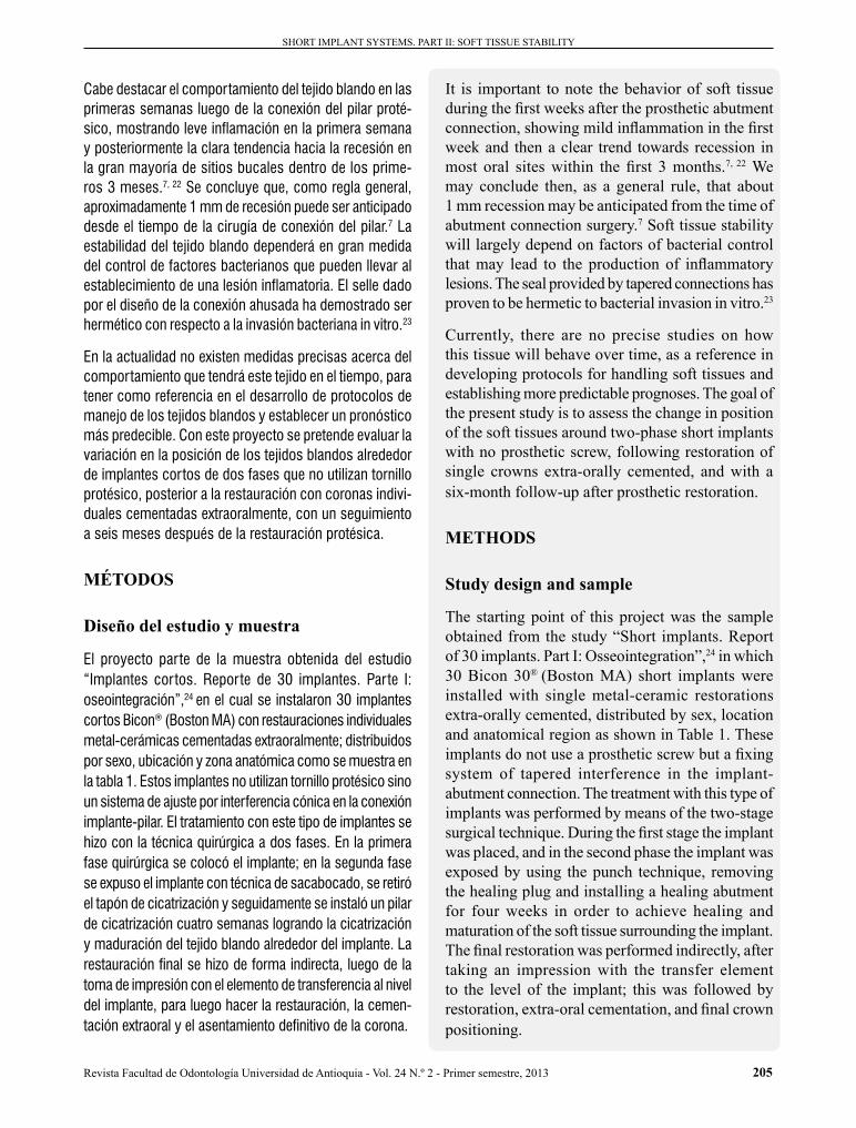
Table 1. Implant distribution by sex, location and mouth region
Variable Implant Implant Total
4.5 * 6.0 mm 5.0 * 6.0 mm
Sex
Male 7 0 7
Female 20 3 23
Location
Maxillary 21 1 22
Mandible 6 2 8
Region
Anterior 20 1 21
Posterior 7 2 9
Tabla 1. Distribución de los implantes por sexo, ubicación y zona
Variable Implante Implante Total
4,5 * 6,0 mm 5,0 * 6,0 mm
Sexo
Masculino 7 0 7
Femenino 20 3 23
Ubicación
Maxilar 21 1 22
Mandíbula 6 2 8
Zona
Anterior 20 1 21
Posterior 7 2 9
SISTEMA DE IMPLANTES CORTOS. PARTE II: ESTABILIDAD DE TEJIDOS BLANDOS
Revista Facultad de Odontología Universidad de Antioquia - Vol. 24 N.º 2 - Primer semestre, 2013 207
Además, se tomó una radiografía periapical a cada uno de los implantes involucrados, inmediatamente después de instalada la restauración definitiva y a los 6 meses, necesaria para evaluar la anatomía y altura de los tejidos óseos adyacentes al implante y establecer los cambios durante el periodo de observación. La radiografía al mo-mento de la instalación de la restauración se consideró como el punto de partida y permitió evaluar los cambios radiográficos posteriores a la carga del implante. La evaluación radiográfica se hizo con la técnica paralela, utilizando un posicionador Rinn® (XCP® Instruments, Elgin, IL) junto con un equipo de rayos X (Trophy CCX Digital®) (f = 3, 70 kV, 8 mA) y se hizo el proceso en un revelador automático (Air Techniques®-Melville, NY). Se utilizó el sofware Axiovision® (Wright, Toronto Canadá) para la evaluación digital de las radiografías donde se estableció la distancia desde la cresta ósea hasta el punto de contacto mesial (CO-PCM) y distal (CO-PCD) (figura 2). Todas las radiografías fueron estandarizadas en cuanto angulación, para lo cual se confeccionó una férula para cada paciente.
Para evitar sesgos en la recolección de la información se utilizaron aparatos previamente calibrados y la lectura de las medidas tomadas fue hecha por un solo examinador. La información recolectada fue ingresada en una base de datos en Excel®, procesada y analizada con el programa SPSS®, versión 17.
In addition, a periapical radiograph of each of the involved implants was taken immediately after insertion of the final restoration and 6 months afterwards, in order to evaluate the anatomy and height of the bone tissues adjacent to the implant and to establish the changes during the observation period. The radiographs at the time of insertion were considered as the starting point and they allowed evaluating the radiographic changes following implant loading. The radiographic evaluation was performed with the parallel technique, by using a Rinn® positioner (XCP® Instruments, Elgin, IL) with an X-ray equipment (Trophy CCX Digital®) (f = 3, 70 kV, 8 mA), and the process was carried out in a developer machine (Air Techniques®, Melville, NY). The Axiovision® software was used (Wright, Toronto Canada) for the digital evaluation of radiographs by establishing the distance from the bone ridge to the mesial contact point (BR-MCP) and to the distalcontact point (BR–DCP) (figure 2). All the radiographswere standardized for angulation, and for that purpose a splint was designed for each patient.
To avoid data collection biases, previously calibrated devices were used and the measurements readings were made by a single examiner. The collected information was entered into an Excel® database, and processed and analyzed by means of SPSS®, version 17.
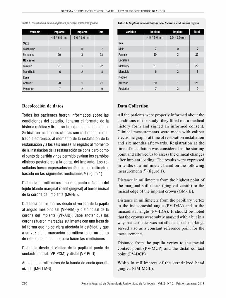
Figura 1. Mediciones clínicas
Figure 1. Clinical measurements
GM-IB: gingival margin-incisal border distance.PV-MCP: papillary vertex-mesial contact point distance.PV-DCP: papillary vertex-distal contact point distance.PV-IMA: papillary vertex-incisomesial angle distance.PV-IDA: papillary vertex-incisodistal angle distance.GM-MGL: gingival margin-mucogingival line distance.
SHORT IMPLANT SYSTEMS. PART II: SOFT TISSUE STABILITY
GM-IB
PV-MCP
PV-DCP
PV-IMA
PV-IDA
GM-MGL
MG-BI: distancia margen gingival-borde incisal.VP-PCM: distancia vértice de papila-punto de contacto mesial.VP-PCD: distancia vértice de papila-punto de contacto distal.VP-AIM: distancia vértice de papila-angulo incisomesial.VP-AID: distancia vértice de papila-angulo incisodistal.MG-LMG: distancia margen gingival-línea mucogingival.
Revista Facultad de Odontología Universidad de Antioquia - Vol. 24 N.º 2 - Primer semestre, 2013208
Análisis de datos
Inicialmente se hizo el análisis exploratorio de las varia-bles cuantitativas que involucraron medidas de tendencia central y de dispersión. De acuerdo con la naturaleza de la variable, se evaluó el cumplimento de los supuestos de normalidad y de homogeneidad de varianzas. Para este propósito se utilizó la prueba de Shapiro-Wilk. Debido a que no se cumplió con el supuesto de normalidad, se utilizaron pruebas no paramétricas, empezando con la prueba de Friedman, con la cual se compararon las medias en los diferentes momentos de medición. Poste-riormente se aplicó la prueba de Wilcoxon, con la cual se comparó la mediana entre dos momentos relacionados con el fin de identificar el momento en el cual aparecen las diferencias estadísticamente significativas. En este estudio se utilizó para todas las pruebas un nivel de significancia de 5%.
RESULTADOS
El grupo poblacional se conformó con 16 mujeres y 6 hombres, con un rango de edad entre 17 y 65 años (promedio: 36 años). Los 30 implantes cortos Bicon® (Boston, MA) colocados en este estudio se encontraron oseointegrados al hacer la cirugía de destape y durante los seis meses de seguimiento después de la restaura-ción protésica, de acuerdo con los parámetros clínicos y radiográficos reportados en la primera parte.
Data Analysis
The exploratory analysis of quantitative variables involving measures of central tendency and disper-sion was performed first. According to the nature of each variable, compliance with the assumptions of normality and homogeneity of variances was assessed. The Shapiro-Wilk test was used for this purpose. Because the assumption of normality was not met, nonparametric tests were used, starting with the Friedman test, which was used to compare the means at different measurement times. Then the Wilcoxon test was applied, which was used to compare the medians between two related moments in order to identify the moment in which statisti-cally significant differences appeared. A 5% level of significance was used for this study.
RESULTS
The study population was made up of 16 women and 6 men, with an age range between 17 and 65 years(mean: 36 years). The 30 short Bicon® implants (Boston, MA) that were inserted during this study were found to be osseointegrated at breakout and during the six-month follow-up period after prosthetic restoration, according to the clinical and radiographic parameters reported in the first part of this article.
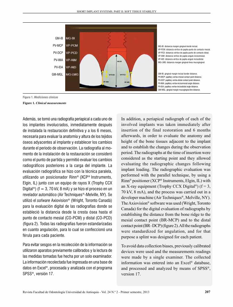
Figura 2. Mediciones radiográficas
Figure 2. Radiographic measurements
BR–MCP: bone ridge-mesial contact point distance.BR–DCP: bone ridge-distal contact point distance.
SISTEMA DE IMPLANTES CORTOS. PARTE II: ESTABILIDAD DE TEJIDOS BLANDOS
BR–MCP
BR–DCP
CO-PCM: distancia cresta ósea-punto de contacto mesial.CO-PCD: distancia cresta ósea-punto de contacto distal.
CO-PCM
CO-PCD
Revista Facultad de Odontología Universidad de Antioquia - Vol. 24 N.º 2 - Primer semestre, 2013 209
SHORT IMPLANT SYSTEMS. PART II: SOFT TISSUE STABILITY
Como evento adverso se registró el desalojo del pilar protésico en tres implantes, y mostró en uno de ellos la presencia de tracto fistuloso vestibular marginal. En estos casos se procedió a la reubicación del aditamento protésico sin complicaciones posteriores y el evento de la fístula se resolvió con irrigación local con agua destilada, sin afectar la integridad del tejido blando, por lo cual, se siguieron teniendo en cuenta como parte de la muestra.
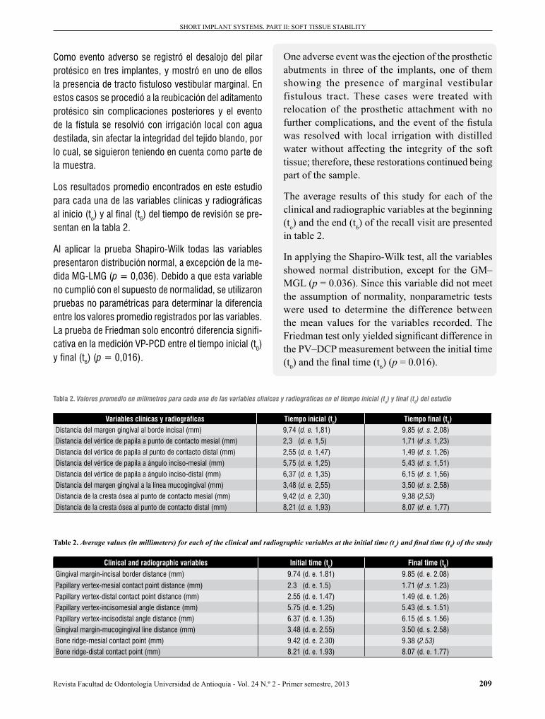
Los resultados promedio encontrados en este estudio para cada una de las variables clínicas y radiográficas al inicio (to) y al final (t6) del tiempo de revisión se pre-sentan en la tabla 2.
Al aplicar la prueba Shapiro-Wilk todas las variables presentaron distribución normal, a excepción de la me-dida MG-LMG (p = 0,036). Debido a que esta variable no cumplió con el supuesto de normalidad, se utilizaron pruebas no paramétricas para determinar la diferencia entre los valores promedio registrados por las variables. La prueba de Friedman solo encontró diferencia signifi-cativa en la medición VP-PCD entre el tiempo inicial (t0) y final (t6) (p = 0,016).
One adverse event was the ejection of the prosthetic abutments in three of the implants, one of them showing the presence of marginal vestibular fistulous tract. These cases were treated with relocation of the prosthetic attachment with no further complications, and the event of the fistula was resolved with local irrigation with distilled water without affecting the integrity of the soft tissue; therefore, these restorations continued being part of the sample.
The average results of this study for each of the clinical and radiographic variables at the beginning (to) and the end (t6) of the recall visit are presented in table 2.
In applying the Shapiro-Wilk test, all the variables showed normal distribution, except for the GM–MGL (p = 0.036). Since this variable did not meet the assumption of normality, nonparametric tests were used to determine the difference between the mean values for the variables recorded. The Friedman test only yielded significant difference in the PV–DCP measurement between the initial time (t0) and the final time (t6) (p = 0.016).
Table 2. Average values (in millimeters) for each of the clinical and radiographic variables at the initial time (to) and final time (t6) of the study
Clinical and radiographic variables Initial time (to) Final time (t6)
Gingival margin-incisal border distance (mm) 9.74 (d. e. 1.81) 9.85 (d. e. 2.08)
Papillary vertex-mesial contact point distance (mm) 2.3 (d. e. 1.5) 1.71 (d .s. 1.23)Papillary vertex-distal contact point distance (mm) 2.55 (d. e. 1.47) 1.49 (d. e. 1.26)Papillary vertex-incisomesial angle distance (mm) 5.75 (d. e. 1.25) 5.43 (d. s. 1.51)Papillary vertex-incisodistal angle distance (mm) 6.37 (d. e. 1.35) 6.15 (d. s. 1.56)Gingival margin-mucogingival line distance (mm) 3.48 (d. e. 2.55) 3.50 (d. s. 2.58)Bone ridge-mesial contact point (mm) 9.42 (d. e. 2.30) 9.38 (2.53)Bone ridge-distal contact point (mm) 8.21 (d. e. 1.93) 8.07 (d. e. 1.77)
Tabla 2. Valores promedio en milímetros para cada una de las variables clínicas y radiográficas en el tiempo inicial (to) y final (t6) del estudio
Variables clínicas y radiográficas Tiempo inicial (to) Tiempo final (t6)Distancia del margen gingival al borde incisal (mm) 9,74 (d. e. 1,81) 9,85 (d. s. 2,08)Distancia del vértice de papila a punto de contacto mesial (mm) 2,3 (d. e. 1,5) 1,71 (d .s. 1,23)Distancia del vértice de papila al punto de contacto distal (mm) 2,55 (d. e. 1,47) 1,49 (d. s. 1,26)Distancia del vértice de papila a ángulo inciso-mesial (mm) 5,75 (d. e. 1,25) 5,43 (d. s. 1,51)Distancia del vértice de papila a ángulo inciso-distal (mm) 6,37 (d. e. 1,35) 6,15 (d. s. 1,56)Distancia del margen gingival a la línea mucogingival (mm) 3,48 (d. e. 2,55) 3,50 (d. s. 2,58)Distancia de la cresta ósea al punto de contacto mesial (mm) 9,42 (d. e. 2,30) 9,38 (2,53)Distancia de la cresta ósea al punto de contacto distal (mm) 8,21 (d. e. 1,93) 8,07 (d. e. 1,77)
Revista Facultad de Odontología Universidad de Antioquia - Vol. 24 N.º 2 - Primer semestre, 2013210
DISCUSIÓN
La oseointegración y su permanencia, posterior a la carga protésica, es el objetivo final cuando se trabaja con implantes en odontología. En este estudio se reporta 100% de oseointegración con 30 implantes Bicon® en un periodo de seguimiento de seis meses posterior a la colocación de la restauración protésica, sin eventos adversos clínicos o radiográficos. Este sistema de im-plantes tiene un protocolo quirúrgico y protésico con variaciones al protocolo Brånemark, evidenciándose su éxito en este estudio y otros en la literatura. Se reporta 100% de éxito y menos de 1 mm de pérdida ósea con 62 implantes Bicon® en seguimiento a cinco años.25 Otro estudio nos reporta 273 implantes con menos de 10 mm de longitud, con seguimiento de 18-60 meses y tasa de supervivencia de 92,82%.26
El sistema Bicon® no utiliza tornillo protésico para la fija-ción de la restauración protésica. El sistema de conexión implante-pilar se basa en la retención por fricción de dos superficies que adaptan perfectamente en un ángulo de 1,5º creando un selle hermético en la interfase de la conexión. El seguimiento a la parte protésica en este estudio reporta el desprendimiento de tres restauraciones protésicas (10%) en un periodo de seis meses. Las com-plicaciones reportadas por la literatura con los sistemas que utilizan tornillo protésico en la conexión del implante con el pilar están en 6-48%.27 El seguimiento a 22 meses reporta la incidencia de aflojamiento del tornillo protésico del 27%28 y en un estudio similar se reportó incidencia de aflojamiento del tornillo del 16%.29 Más recientemente, tras cambios hechos en la conexión pero sin abandonar el uso del tornillo protésico, se reportó 9% de aflojamiento del tornillo después de 3 años.30
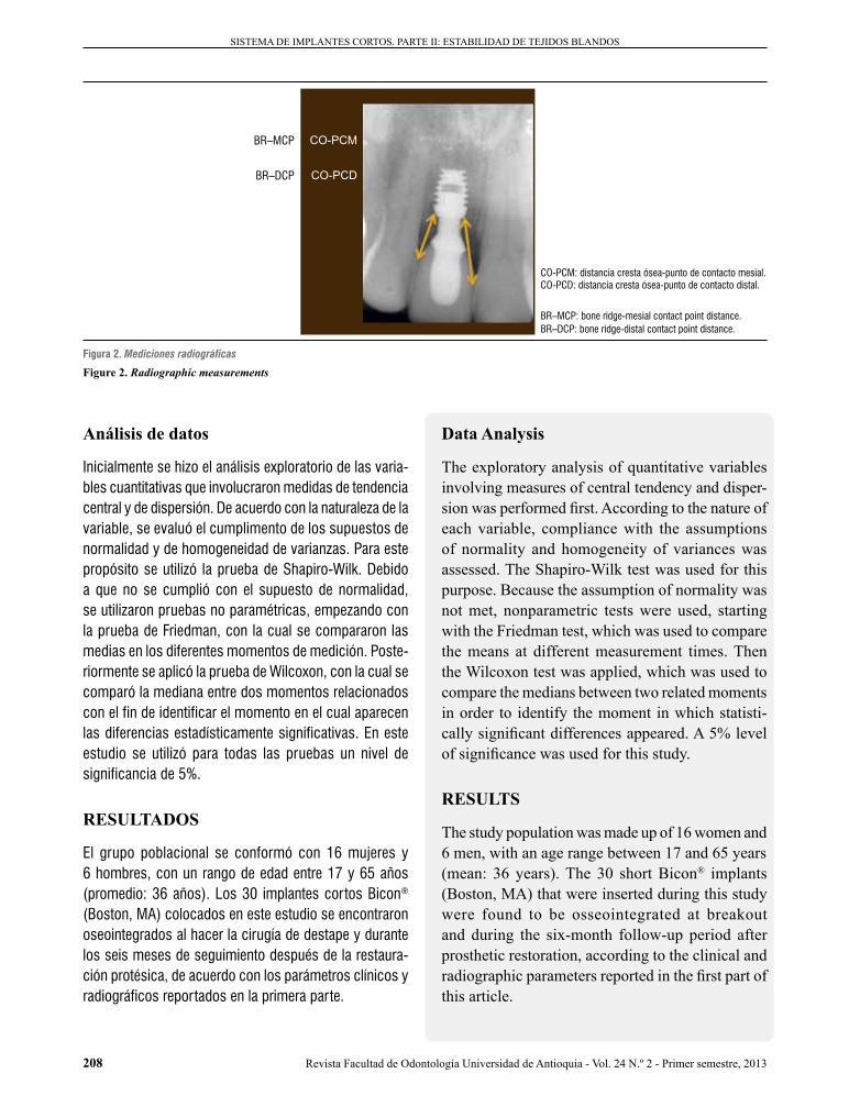
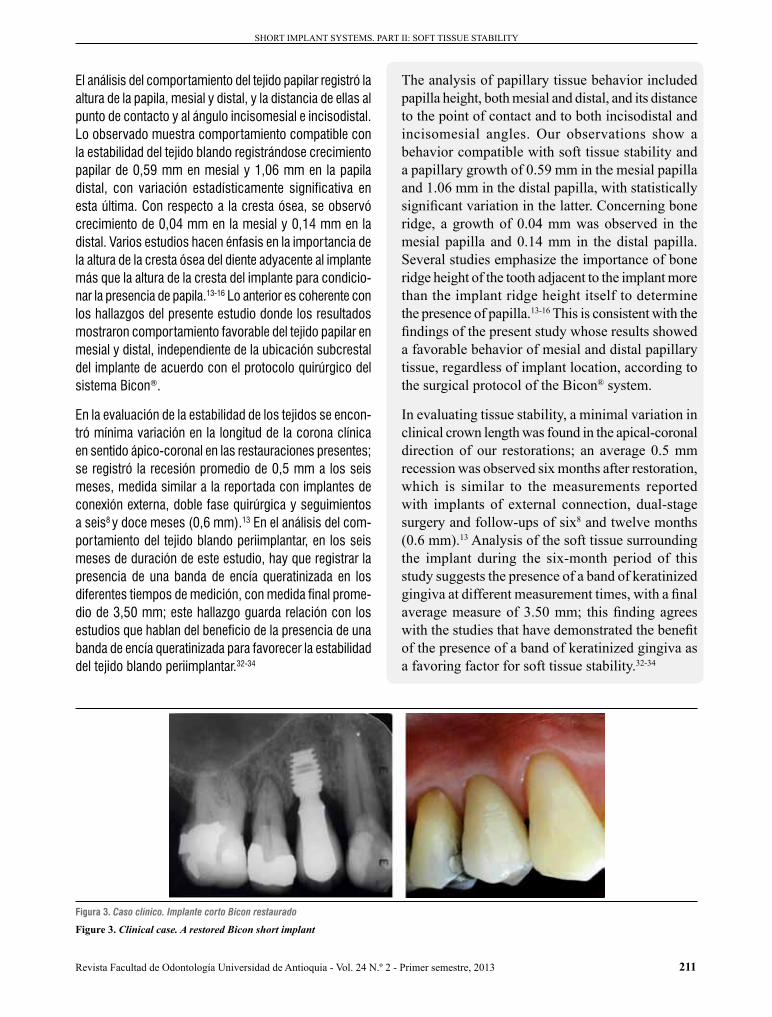
La rehabilitación de la zona estética anterior genera expectativas en el paciente con respecto a un resultado estable en cuanto a la apariencia de la restauración protésica y de los tejidos blandos relacionados con ella. Nuestro estudio utilizó un sistema de rehabilitación que permite la restauración “amigable” con el tejido porque en el protocolo, la cementación de la corona definitiva se hace de forma extraoral, lo que evita llevar cemento hacia los tejidos blandos en su aspecto profundo, disminuyendo al máximo su irritación y posterior inflamación, favore-ciendo la estabilidad de los tejidos blandos (figura 3); un estudio reciente reporta 81% de periimplantitis asociada a excesos de cemento.31
DISCUSSION
Osseointegration and its permanence after prosthetic loading is the ultimate goal when working with dental implants. This study reports 100% osseointegration with 30 Bicon® implants in a six-month follow-up period after placement of the prosthetic restoration, with no clinical or radiographic adverse events. This implant system follows a surgical and prosthetic protocol with variations to the Brånemark protocol, demonstrating its success in this study and in other studies reported in the literature. A 100% success rate and less than 1 mm of bone loss is reported in 62 Bicon® implants in five years.25 Another study reports 273 implants with less than 10 mm in length and a follow-up of 18-60 months with a survival rate of 92.82%.26
The Bicon® system uses no prosthetic screw for fixing the prosthetic restoration. The system of implant-abutment connection is based on the frictional retention of both surfaces to perfectly fit in a 1.5° angle creating a hermetic seal at the connection interface. The monitoring of the prosthetic part in this study reports detachment of three prosthetic restorations (10%) over a six-month period. The complications reported in the literature with screw systems using prosthetic implants in connection with the pillar are between 6 and 48%.27 A 22-month follow-up reports a 27% incidence of prosthetic screw loosening,28 and a related study reported incidence of screw loosening of 16%.29 More recently, after making changes in the connection but still using a prosthetic screw, a screw loosening of 9% was reported after three years.30
Rehabilitation of the anterior esthetic area creates expectations in the patient in relation to a stable result in the appearance of both the prosthetic restoration and the soft tissue connected to it. Our study used a rehabilitation system that allows a restoration which is “friendly” with the tissue as cementation of the final crown is performed extra-orally, avoiding carrying cement into the deep aspect of soft tissues, greatly reducing irritation and inflammation, and favoring soft tissue stability (figure 3); a recent study reported 81% of inflammation associated to an excess in cementation.31
SISTEMA DE IMPLANTES CORTOS. PARTE II: ESTABILIDAD DE TEJIDOS BLANDOS
Revista Facultad de Odontología Universidad de Antioquia - Vol. 24 N.º 2 - Primer semestre, 2013 211
SHORT IMPLANT SYSTEMS. PART II: SOFT TISSUE STABILITY
El análisis del comportamiento del tejido papilar registró la altura de la papila, mesial y distal, y la distancia de ellas al punto de contacto y al ángulo incisomesial e incisodistal. Lo observado muestra comportamiento compatible con la estabilidad del tejido blando registrándose crecimiento papilar de 0,59 mm en mesial y 1,06 mm en la papila distal, con variación estadísticamente significativa en esta última. Con respecto a la cresta ósea, se observó crecimiento de 0,04 mm en la mesial y 0,14 mm en la distal. Varios estudios hacen énfasis en la importancia de la altura de la cresta ósea del diente adyacente al implante más que la altura de la cresta del implante para condicio-nar la presencia de papila.13-16 Lo anterior es coherente con los hallazgos del presente estudio donde los resultados mostraron comportamiento favorable del tejido papilar en mesial y distal, independiente de la ubicación subcrestal del implante de acuerdo con el protocolo quirúrgico del sistema Bicon®.
En la evaluación de la estabilidad de los tejidos se encon-tró mínima variación en la longitud de la corona clínica en sentido ápico-coronal en las restauraciones presentes; se registró la recesión promedio de 0,5 mm a los seis meses, medida similar a la reportada con implantes de conexión externa, doble fase quirúrgica y seguimientos a seis8 y doce meses (0,6 mm).13 En el análisis del com-portamiento del tejido blando periimplantar, en los seis meses de duración de este estudio, hay que registrar la presencia de una banda de encía queratinizada en los diferentes tiempos de medición, con medida final prome-dio de 3,50 mm; este hallazgo guarda relación con los estudios que hablan del beneficio de la presencia de una banda de encía queratinizada para favorecer la estabilidad del tejido blando periimplantar.32-34
The analysis of papillary tissue behavior included papilla height, both mesial and distal, and its distance to the point of contact and to both incisodistal and incisomesial angles. Our observations show a behavior compatible with soft tissue stability and a papillary growth of 0.59 mm in the mesial papilla and 1.06 mm in the distal papilla, with statistically significant variation in the latter. Concerning bone ridge, a growth of 0.04 mm was observed in the mesial papilla and 0.14 mm in the distal papilla. Several studies emphasize the importance of bone ridge height of the tooth adjacent to the implant more than the implant ridge height itself to determine the presence of papilla.13-16 This is consistent with the findings of the present study whose results showed a favorable behavior of mesial and distal papillary tissue, regardless of implant location, according to the surgical protocol of the Bicon® system.
In evaluating tissue stability, a minimal variation in clinical crown length was found in the apical-coronal direction of our restorations; an average 0.5 mm recession was observed six months after restoration, which is similar to the measurements reported with implants of external connection, dual-stage surgery and follow-ups of six8 and twelve months (0.6 mm).13 Analysis of the soft tissue surrounding the implant during the six-month period of this study suggests the presence of a band of keratinized gingiva at different measurement times, with a final average measure of 3.50 mm; this finding agrees with the studies that have demonstrated the benefit of the presence of a band of keratinized gingiva as a favoring factor for soft tissue stability.32-34
Figura 3. Caso clínico. Implante corto Bicon restaurado
Figure 3. Clinical case. A restored Bicon short implant
Revista Facultad de Odontología Universidad de Antioquia - Vol. 24 N.º 2 - Primer semestre, 2013212
Los resultados finales de este estudio a seis meses muestran el comportamiento estable del tejido blando y del soporte óseo alrededor de los implantes, con cambio favorable en la posición de las papilas y la leve modifica-ción hacia apical en la posición del margen gingival y no difieren de lo encontrado en publicaciones preliminares que hacen mención de la estabilidad del tejido blando marginal y papilar alrededor de restauraciones implanto-soportadas.8, 11, 13
CORRESPONDENCIA
Manuel CabralesFacultad de OdontologíaUniversidad de AntioquiaMedellín, ColombiaCorreo electrónico: [email protected]
The final results of this study six months after restoration show a stable behavior of the soft tissue and the supporting bones surrounding the implants with a favorable change in the position of papillae and a slight change in position towards apical to the gingival margin; these results are similar to the ones reported in previous publications, which refer to the stability of marginal soft tissues and the papilla surrounding implant-supported restorations.8, 11, 13
CORRESPONDING AUTHOR
Manuel CabralesFacultad de OdontologíaUniversidad de AntioquiaMedellín, ColombiaEmail address: [email protected]
REFERENCIAS / REFERENCES
1. Adell R, Lekholm U, Rockler B, Brånemark PI. A 15 years study of osseointegrated implants in the treatment of the edentulous jaw. Int J Oral Surgery 1981; 10: 387-416.
2. Schnitman AP, Wohrle SP, Rubenstein EJ, DaSilva DJ, Wang NH. Ten year results for Brånemark implants immediately loaded with fixed prostheses at implant placement. Int J Oral Maxillofac Implants 1997; 12: 495-503.
3. Adell R, Eriksson B, Lekholm, Brånemark P-I and Jemt T. A long-term follow-up study of osseointegrated implants in the treatment of totally edentulous jaws. Int J Oral Maxillofac Implants 1990; 5: 347-359.
4. Wheeler SL, Vogel RE, Casellini R. Tissue preservation and maintenance of optimum esthetics: a clinical report. Int J Oral Maxillofac Implants 2000; 15: 265-271.
5. Holt RL, Rosenberg MM, Zinser PJ, Ganeles J. A concept for a biologically derived, parabolic implant design. Int J Periodontics Restorative Dent 2002; 22: 473-481.
6. Rompen E, Raepsaet N, Domken O, Touati B, Dooren EV. Soft tissue stability at the facial aspect of gingivally converging abutments in the esthetic zone: a pilot clinical study. J Prosthet Dent 2007; 97: S119-S125.
7. Tarnow DP, Cho SC, Wallace SS. The effect of inter-implant distance on the height of inter-implant bone crest. J Periodontol 2000; 71: 546-549.
8. Priest G. Predictability of soft tissue form around single-tooth implant restorations. Int J Periodontics Restorative Dent 2003; 23: 19-27.
9. Meijer HJ, Stellingsma K, Meijndert L, Raghoebar GM. A new index for rating aesthetics of implant-supported single crowns and adjacent soft tissues-the implant crown aesthetic
Index. A pilot study on validation of a new index. Clin Oral Impl Res 2005; 16: 645-649.
10. Jemt T. Regeneration of gingival papillae after single-implant treatment. Int J Periodontics Restorative Dent 1997; 17: 327-333.
11. Belser UC, Schmid B, Higginbottom F, Buser D. Outcome analysis of implant restorations located in the anterior maxilla: a review of the recent literature. Int J Oral Maxillofac Implants 2004; 19 sup l: 30-42.
12. Covani U, Barone A, Cornelini R, Crespi R. Soft tissue healing around implants placed immediately after tooth extraction without incision: a clinical report. Int J Oral Maxillofac Implants 2004; 19: 549-553.
13. Grunder U. Stability of the mucosal topography around single-tooth implants and adjacent teeth: 1-year results. Int J Periodontics Restorative Dent 2000; 20: 11-17.
14. Zetu L, Wang HL. Management of inter-dental/inter-implant papilla. J Clin Periodontol 2005; 32: 831-839.
15. Kinsel RP, Lamb RE. Development of gingival esthetics in the edentulous patient with immediately loaded, single-stage, implant-supported fixed prostheses: a clinical report. Int J Oral Maxillofac Implants 2000; 15: 711-721.
16. Jemt T. Restoring the gingival contour by means of provisional resin crowns after single implant treatment. Int J Periodontics Restorative Dent 1999; 19: 21-29.
17. Kan J, Rungcharassaeng K, Umezu K, Kois JC. Dimensions of peri-implant mucosa: an evaluation of maxillary anterior single implants in humans. J Periodontol 2003; 74: 557-562.
SISTEMA DE IMPLANTES CORTOS. PARTE II: ESTABILIDAD DE TEJIDOS BLANDOS
Revista Facultad de Odontología Universidad de Antioquia - Vol. 24 N.º 2 - Primer semestre, 2013 213
SHORT IMPLANT SYSTEMS. PART II: SOFT TISSUE STABILITY
18. Lee D, Park K, Moon I. Dimension of keratinized mucosa and the interproximal papilla between adjacent implants. J Periodontol 2005; 76: 1856-1860.
19. Choquet. V, Hermans M, Adriaenssens P, Daelemans P, Tarnow DP, Malevez C. Clinical and radiographic evaluation of the papilla level adjacent to single-tooth dental implants. A retrospective study in the maxillary anterior region. J Periodontol 2001; 72: 1364-1371.
20. Ryser MR, Block MS, Mercante DE. Correlation of papilla to crestal bone levels around single tooth implants in immediate or delayed crown protocols. J Oral Maxillofac Surg 2005; 63: 1184-1195.
21. Novaes AB, Papalexiou V, Muglia V, Taba M. Influence of interimplant distance on gingival papilla formation and bone resorption: clinical-radiographic study in dogs. Int J Oral Maxillofac Implants 2006; 21: 45-51.
22. Small PN, Tarnow DP. Gingival recession around implants: a 1 year longitudinal prospective study. Int J Oral Maxillofac Implants 2000; 15: 527-532.
23. Dibart S, Warbington M, Su FM, Skobe Z. In vitro evaluation of the implant-abutment bacterial seal: the locking taper system. Int J Oral Maxillofac Implant 2005; 20: 732-737.
24. Arismendi JA, Cabrales M, Duque JA, Castañeda WR. Implantes cortos. Reporte de 30 implantes. Parte 1: oseointegración. Rev Fac Odontol Univ Antioq 2011; 22 (2): 198-204.
25. Venuleo C, Chuang SK, Weed M, Dibart S. Long term bone level stability on short implants: a radiographic follow-up study. J Maxillofac Oral Surg 2008; 7: 340-345.
26. Sánchez MA, Costa X, Gay C. Short Implants. A descriptive study of 273 implants. Clin Implant Dent Relat Res 2012; 14(4): 508-516.
27. Binon PP. Implants and components: entering the new millennium. Int J Oral and Maxillofac Implants 2000; 15: 76-93.
28. Walton JN, McEntee MI. A prospective study on the maintenance of implant prostheses in private practice. Int J Prosthodont 1997; 10: 453-458
29. Mangano C, Bartolucci EG. Single tooth replacement by morse taper connection implants: A retrospective study of 80 implants. Int J Oral Maxillofac Implants 2001; 16: 675-680.
30. Boggan RS, Strong JR, Misch CE, Bidez MW. Influence of hex geometry and prosthetic table width on static and fatigue strength of dental implants. J Prosthet Dent 1999; 82: 436-440.
31. Wilson T. The positive relationship between excess cement and peri-implant disease: a prospective clinical endoscopic study. J Periodontol 2009; 80: 1388- 1392.
32. Lavelle, CL. Mucosal seal around endosseous dental implants. J Oral Implant 1981; 9: 357-371.
33. Berglundh T, Lindhe J. Dimension of the peri-implant mucosa. Biological width revisited. J Clin Periodontol 1996; 23: 971-973.
34. Rapley JW, Mills MP, Wylam J. Soft tissue management during implant maintenance. Int J Periodontics Restorative Dent 1992; 12: 373-381.