Neoplasia lecture 5 - JU Medicine€¦ · Neoplasia 2018 lecture 4 Dr Heyam Awad MD, FRCPath. ILOS...
60
Neoplasia 2018 lecture 4 Dr Heyam Awad MD, FRCPath
Transcript of Neoplasia lecture 5 - JU Medicine€¦ · Neoplasia 2018 lecture 4 Dr Heyam Awad MD, FRCPath. ILOS...

Neoplasia 2018 lecture 4
Dr Heyam Awad
MD, FRCPath

ILOS
• To understand the concept of the hallmarks of cancer and that they are phenotypic changes needed in all cancer cells.
• To list the tumor enablers
• To understand the role of host cells in carcinogenesis
• In depth understanding of the first hallmark: self sufficiency in growth signals.

Intro
• Cancer occurs due to successful breach of anticancer mechanisms, i:e of regulatory mechanisms in our cells and tissues that prevent uncontrolled proliferation.
• Each cancer cell needs to acquire certain phenotypic characteristics to become malignant.
• Of course these phenotypic characteristics reflect genetic mutations ( or alterations)
• The phenotypic characteristics needed for cancer cells to survive are called : the hallmarks of cancer.

Hallmarks of Cancer
• Hallmarks of cancer: changes in cell physiology resulting from genetic changes that result in cancer.
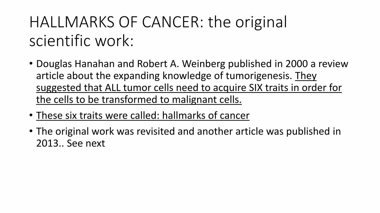
HALLMARKS OF CANCER: the original scientific work:• Douglas Hanahan and Robert A. Weinberg published in 2000 a review
article about the expanding knowledge of tumorigenesis. They suggested that ALL tumor cells need to acquire SIX traits in order for the cells to be transformed to malignant cells.
• These six traits were called: hallmarks of cancer
• The original work was revisited and another article was published in 2013.. See next
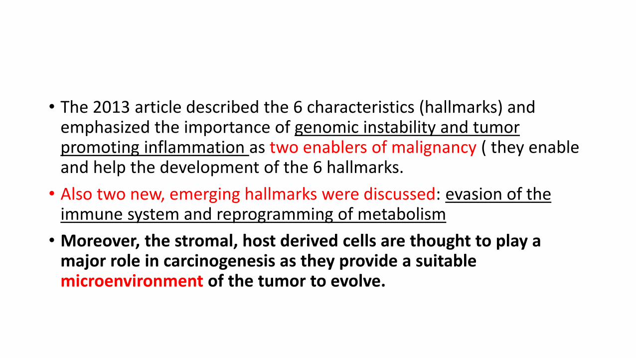
• The 2013 article described the 6 characteristics (hallmarks) and emphasized the importance of genomic instability and tumor promoting inflammation as two enablers of malignancy ( they enable and help the development of the 6 hallmarks.
• Also two new, emerging hallmarks were discussed: evasion of the immune system and reprogramming of metabolism
• Moreover, the stromal, host derived cells are thought to play a major role in carcinogenesis as they provide a suitable microenvironment of the tumor to evolve.
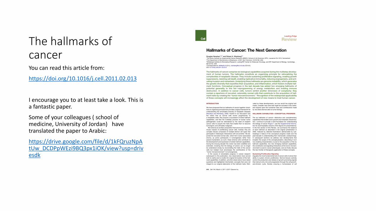
The hallmarks of cancerYou can read this article from:
https://doi.org/10.1016/j.cell.2011.02.013
I encourage you to at least take a look. This is a fantastic paper.
Some of your colleagues ( school of medicine, University of Jordan) have translated the paper to Arabic:
https://drive.google.com/file/d/1kFQruzNpAtUw_DCDPpWEzi9BQ3px1iOK/view?usp=drivesdk
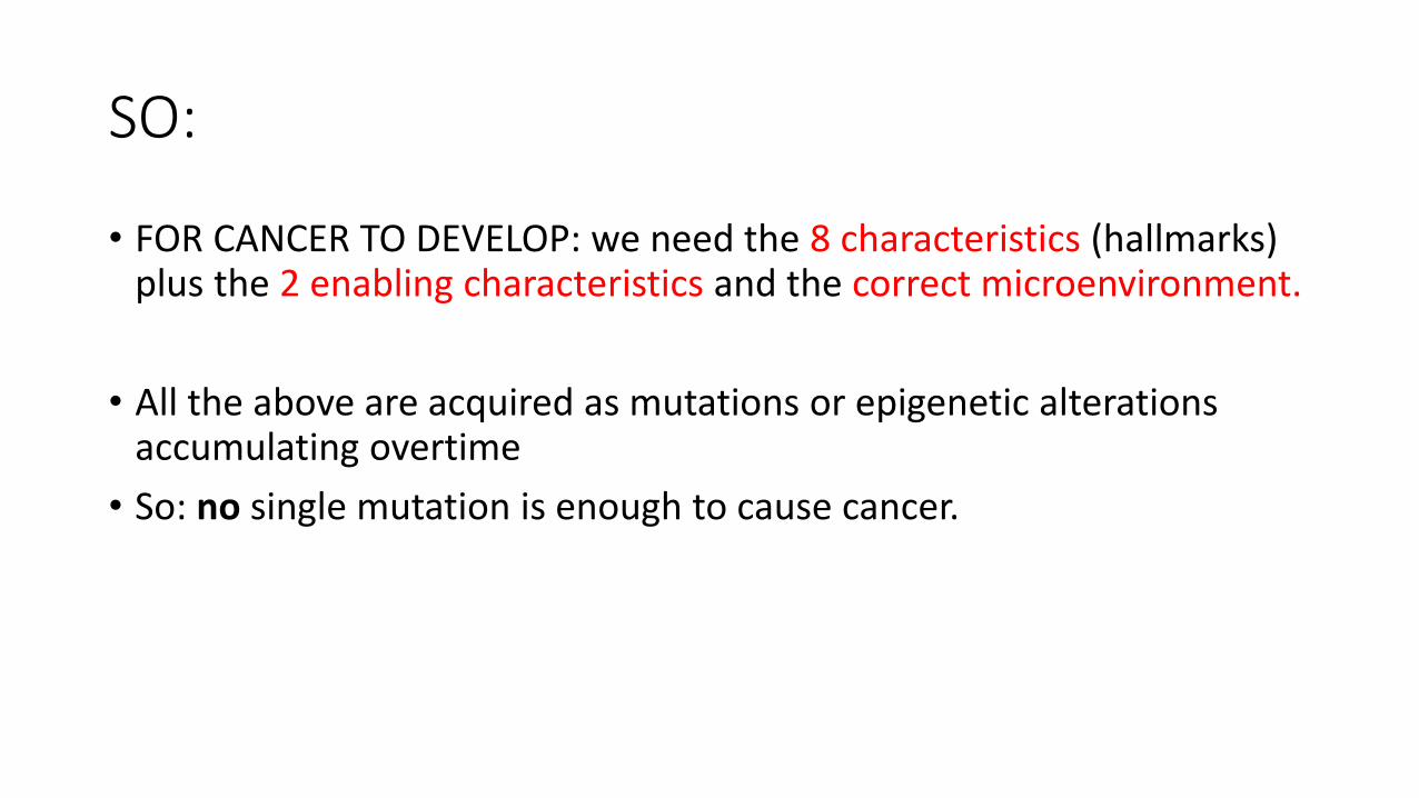
SO:
• FOR CANCER TO DEVELOP: we need the 8 characteristics (hallmarks) plus the 2 enabling characteristics and the correct microenvironment.
• All the above are acquired as mutations or epigenetic alterations accumulating overtime
• So: no single mutation is enough to cause cancer.
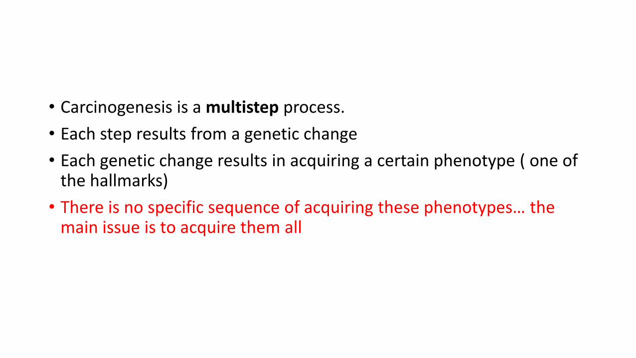
• Carcinogenesis is a multistep process.
• Each step results from a genetic change
• Each genetic change results in acquiring a certain phenotype ( one of the hallmarks)
• There is no specific sequence of acquiring these phenotypes… the main issue is to acquire them all
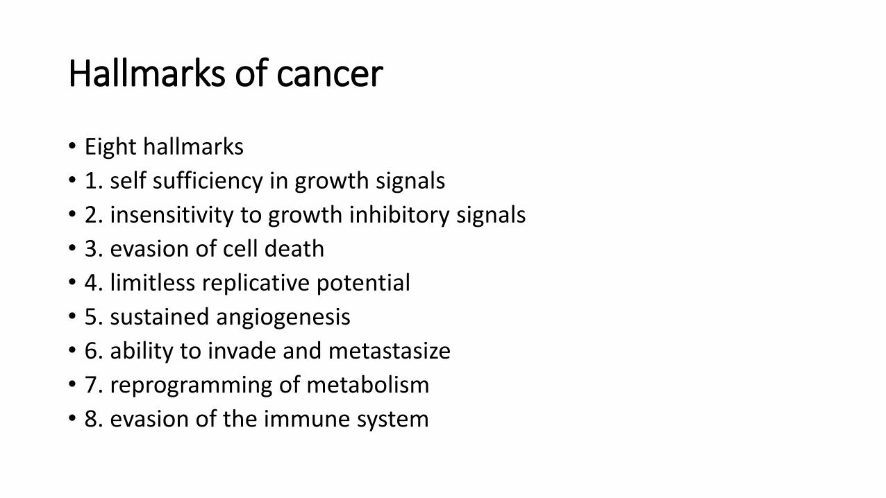
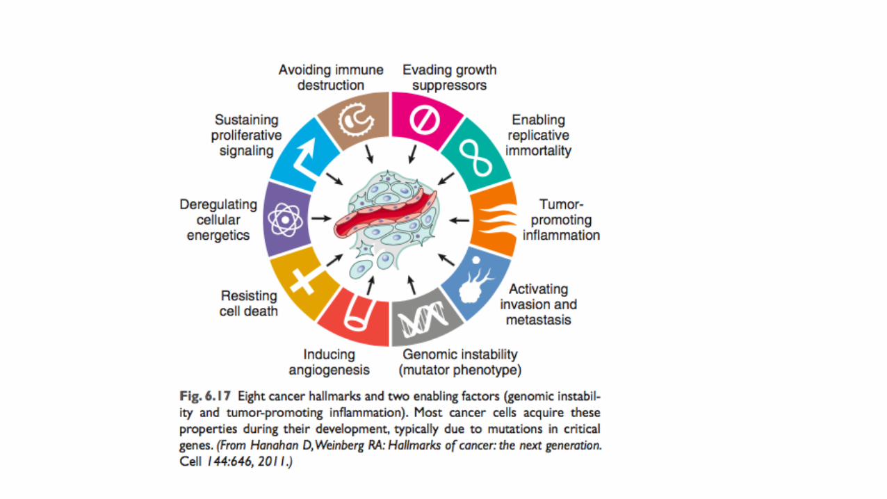
Hallmarks of cancer
• Eight hallmarks
• 1. self sufficiency in growth signals
• 2. insensitivity to growth inhibitory signals
• 3. evasion of cell death
• 4. limitless replicative potential
• 5. sustained angiogenesis
• 6. ability to invade and metastasize
• 7. reprogramming of metabolism
• 8. evasion of the immune system

Two enablers
• Enablers ( help in the tumorigenesis process and set the right conditions for genetic changes to develop)
• 1. genomic instability
• 2. inflammation

• Tumor microenvironment
• Tumors need a suitable environment to flourish.
• This is obtained from the host cells: Stromal cells, mainly fibroblasts and pericytes
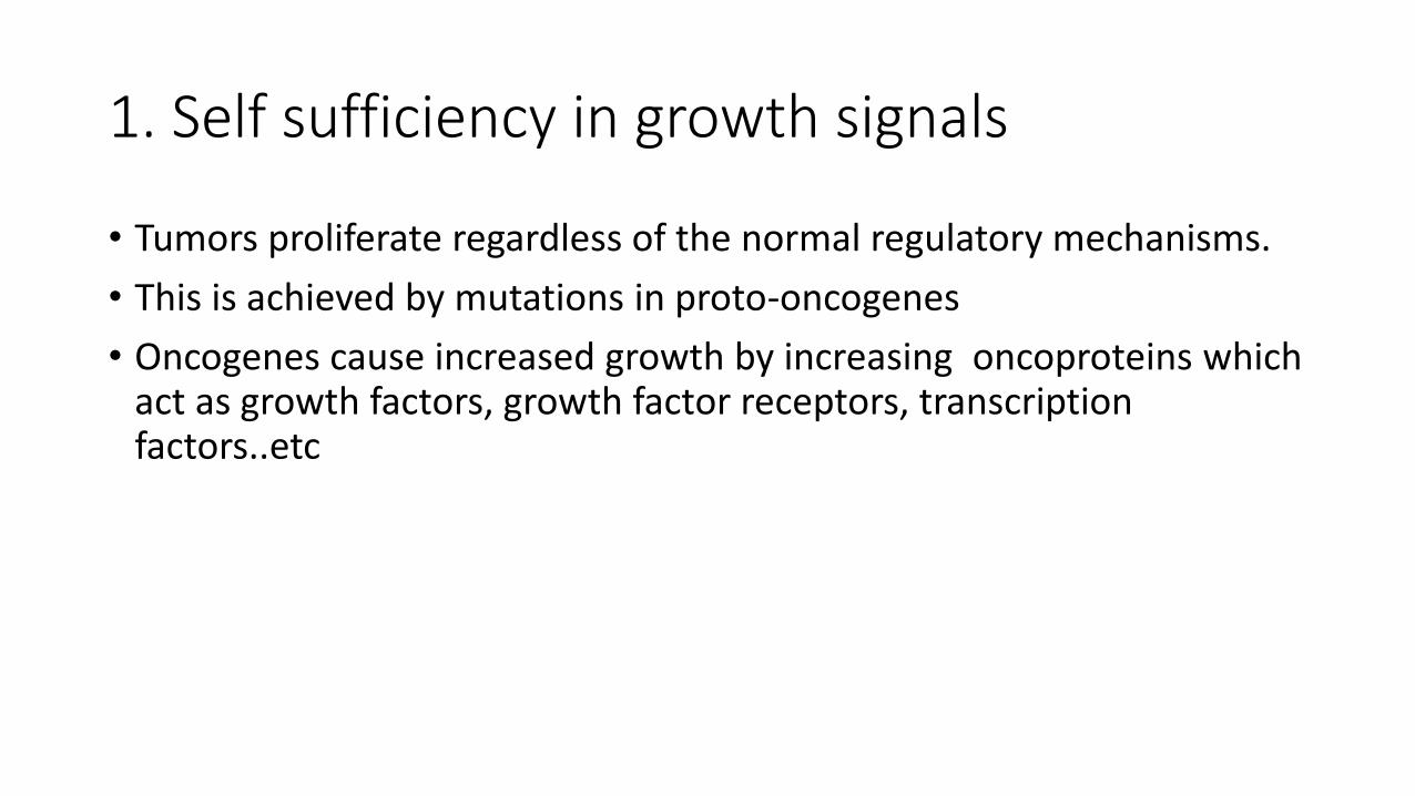
1. Self sufficiency in growth signals
• Tumors proliferate regardless of the normal regulatory mechanisms.
• This is achieved by mutations in proto-oncogenes
• Oncogenes cause increased growth by increasing oncoproteins which act as growth factors, growth factor receptors, transcription factors..etc
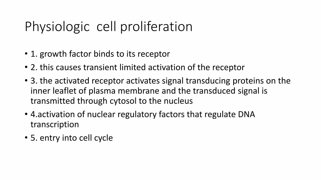
Physiologic cell proliferation
• 1. growth factor binds to its receptor
• 2. this causes transient limited activation of the receptor
• 3. the activated receptor activates signal transducing proteins on the inner leaflet of plasma membrane and the transduced signal is transmitted through cytosol to the nucleus
• 4.activation of nuclear regulatory factors that regulate DNA transcription
• 5. entry into cell cycle
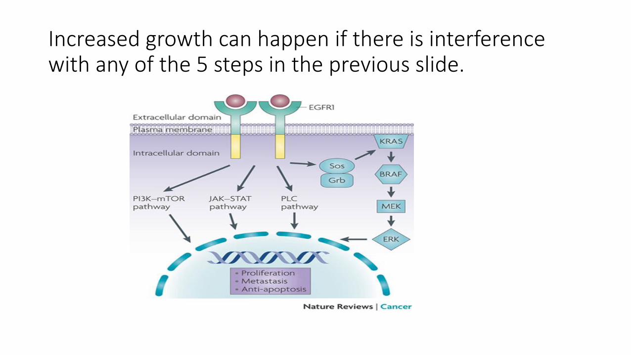
Increased growth can happen if there is interference with any of the 5 steps in the previous slide.

1. Increased growth facors
2. Increased receptors or increase in their function
3. Increased signal transduction
4. Increased transcription factors5. Cell cycle entry

Growth factors
• Some tumors produce their own growth factors, ie factors they respond to.
• This creates an autocrine loop
• Eg glioblastomas produce PDGF and express its receptor
Growth factors
• Tumor cells can interact with their stroma to produce growth factors.
• This is an example of how microenvironment ( interaction with host cells including stromal cells) can help tumors to grow.

GF receptors
• GF receptors can increase cell proliferation in two situations:• 1. Overexpression of the receptor .. So, Receptor is hyper responsive to GF
even in levels that don’t normally trigger proliferation.• 2. Mutant receptor proteins. So the receptor itself is mutated and acquires
a configuration that is always stimulated. This causes continuous mitogenic signal even in the absence of GF.
Remember that when a GF ( or indeed any ligand) binds to its receptor, this causes conformational changes in the receptor proteins, so its shape changes and this change is what activates the receptor.If there is a mutation that causes conformational change resulting in the receptor’s shape being in the active state, then we don’t need any stimulator for the receptor to function! It’s in the alert, functioning state continuously.
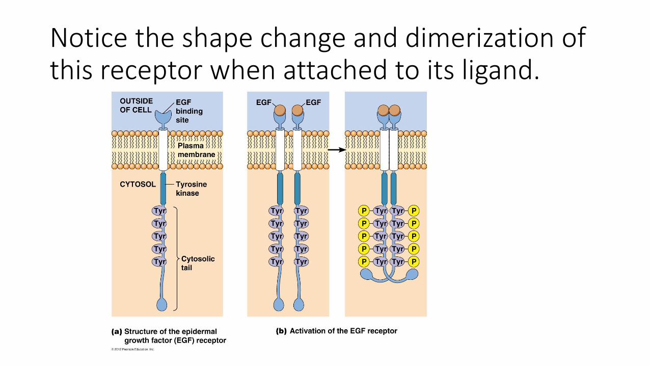
Notice the shape change and dimerization of this receptor when attached to its ligand.
Amplification of GF receptor/ example
• Gene encoding her2/neu is amplified in 25-30% of breast cancers and adenocarcinoma of lung, ovary and salivary gland
• High level of HER2/Neu protein in breast cancer worsens prognosis ( results in more proliferation)
• If her 2 receptor blocked.. By anti her2 antibodies.. Treatment used ( refer to details in previous lecture: the case study)

Signal transducing proteins
• Main signal transducing proteins involved: RAS and ABL
RAS
• RAS is the most commonly mutated oncogene in humans
• 30% of human cancers contain RAS mutation
• Even higher incidence in colon and pancreatic carcinomas

How does RAS function normally?
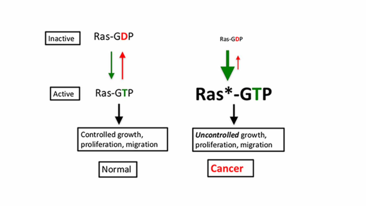
• RAS is a small protein that binds GDP and GTP ( it’s a G protein)
• It is inactive with GDP binding.
• If a growth factor binds to a receptor.. GDP is phosphorylated to GTP..
• This Actives RAS
• Activation is short lived as GTPase hydrolyses GTP to GDP
• GTPase activating proteins (GAPs ) prevent over activation of RAS, so they act as brakes of RAS activation.
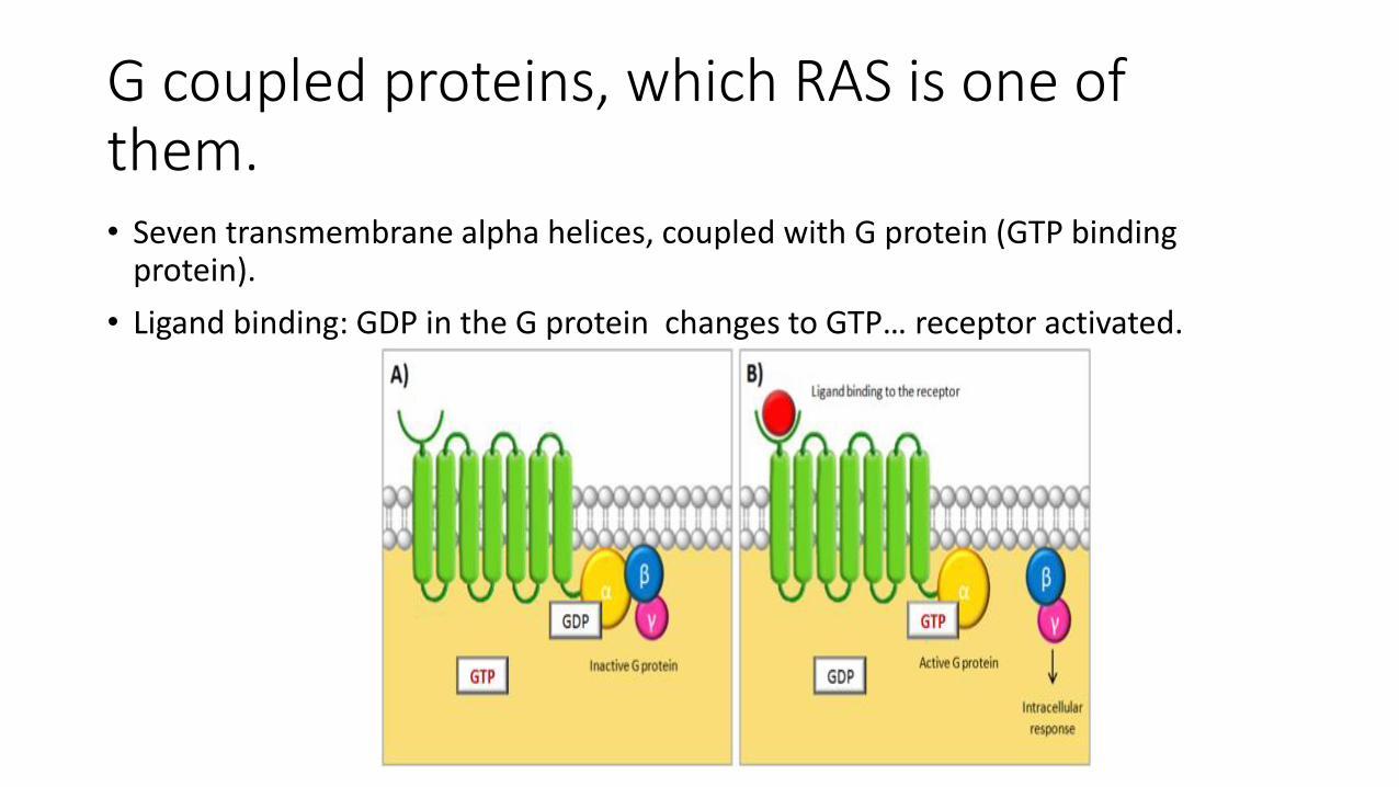
G coupled proteins, which RAS is one of them.• Seven transmembrane alpha helices, coupled with G protein (GTP binding
protein).
• Ligand binding: GDP in the G protein changes to GTP… receptor activated.
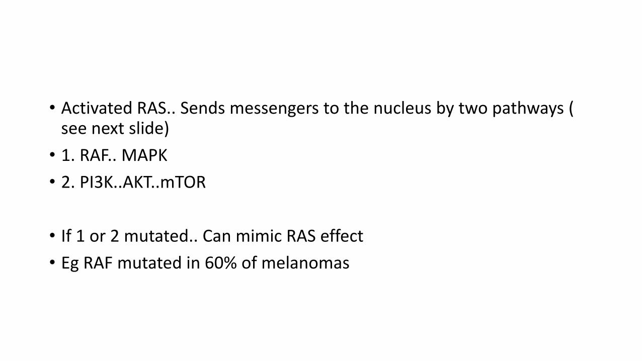
• Activated RAS.. Sends messengers to the nucleus by two pathways ( see next slide)
• 1. RAF.. MAPK
• 2. PI3K..AKT..mTOR
• If 1 or 2 mutated.. Can mimic RAS effect
• Eg RAF mutated in 60% of melanomas
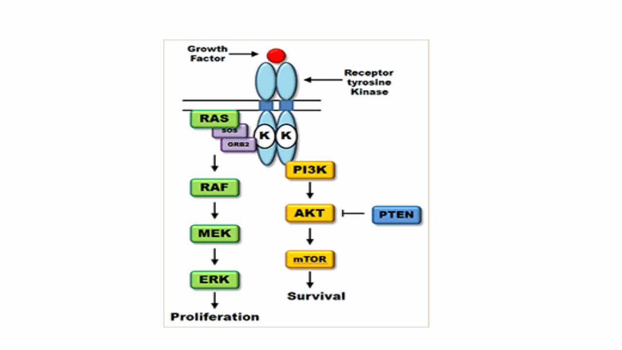
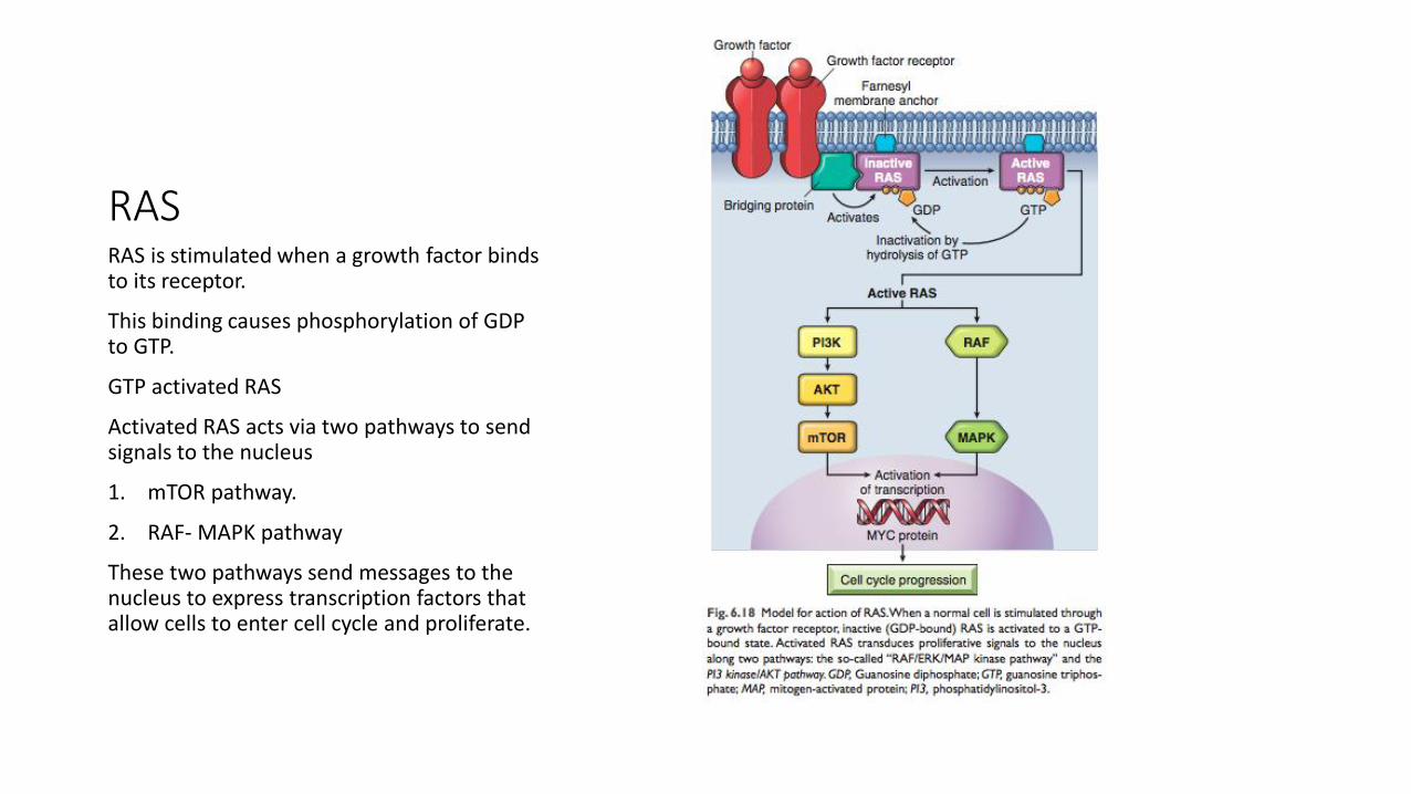
RASRAS is stimulated when a growth factor binds to its receptor.
This binding causes phosphorylation of GDP to GTP.
GTP activated RAS
Activated RAS acts via two pathways to send signals to the nucleus
1. mTOR pathway.
2. RAF- MAPK pathway
These two pathways send messages to the nucleus to express transcription factors that allow cells to enter cell cycle and proliferate.
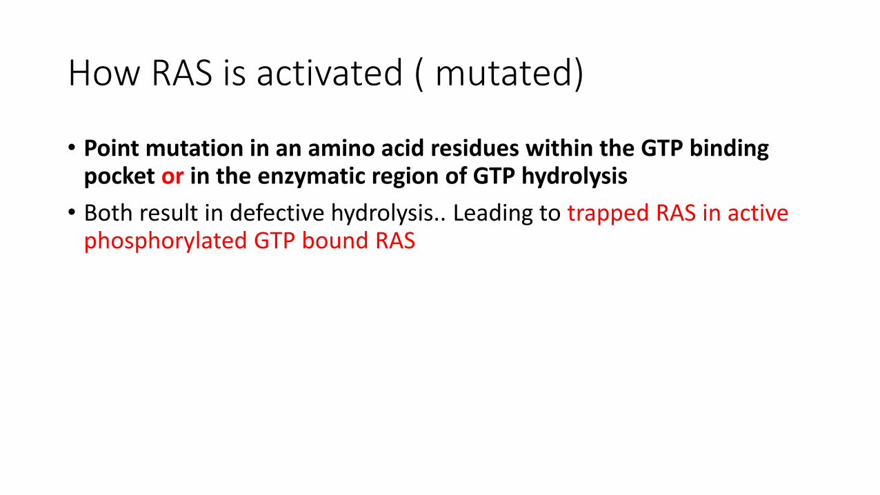
How RAS is activated ( mutated)
• Point mutation in an amino acid residues within the GTP binding pocket or in the enzymatic region of GTP hydrolysis
• Both result in defective hydrolysis.. Leading to trapped RAS in active phosphorylated GTP bound RAS
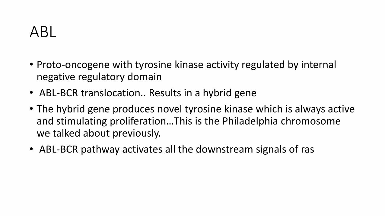
ABL
• Proto-oncogene with tyrosine kinase activity regulated by internal negative regulatory domain
• ABL-BCR translocation.. Results in a hybrid gene
• The hybrid gene produces novel tyrosine kinase which is always active and stimulating proliferation…This is the Philadelphia chromosome we talked about previously.
• ABL-BCR pathway activates all the downstream signals of ras
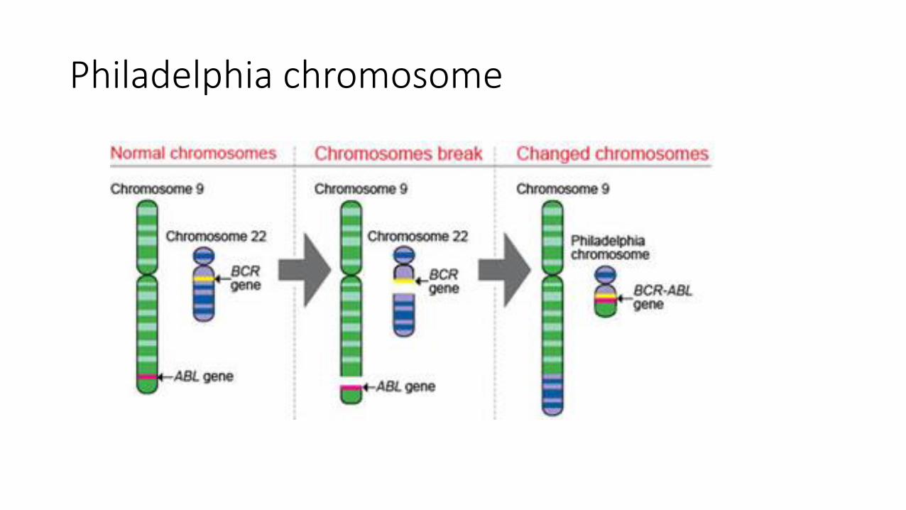
Philadelphia chromosome

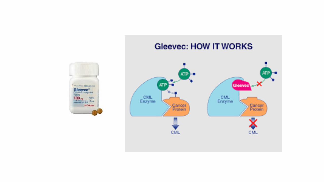
• CML =chronic myelogenous leukemia : t(9;22)= Philadelphia chromosome. ABL-BCR fusion gene….tyrosine kinase which is always turned on.
• Kinase inhibitors can be used to treat CML
• Imatinab/gleevec .. Inhibits the kinase and treats the leukemia
• = targeted therapy
Nuclear transcription factors
• Most commonly involved in human cancers: MYC
myc
• Can activate or repress expression of other genes
• Activates cyclins and cyclin dependent kinases CDK ( so cells enter cell cycle and divide)
• Myc also inhibits cyclin dependent kinase inhibitors (CDKI)
• These cyclins, CDK, CDKI play a role in entry to cell cycle, which we will discuss in detail in this lecture.
• MYC amplified in breast, colon, lung and other carcinomas
• T(8; 14) in Burkitt dysregulates myc activate
Entry into cell cycle
• All the stimuli mentioned till now aim for quiescent cells to enter the cell cycle .
• Each phase in the cell cycle depends on successful completion of the previous one
• Cycle stops when essential gene function is lost

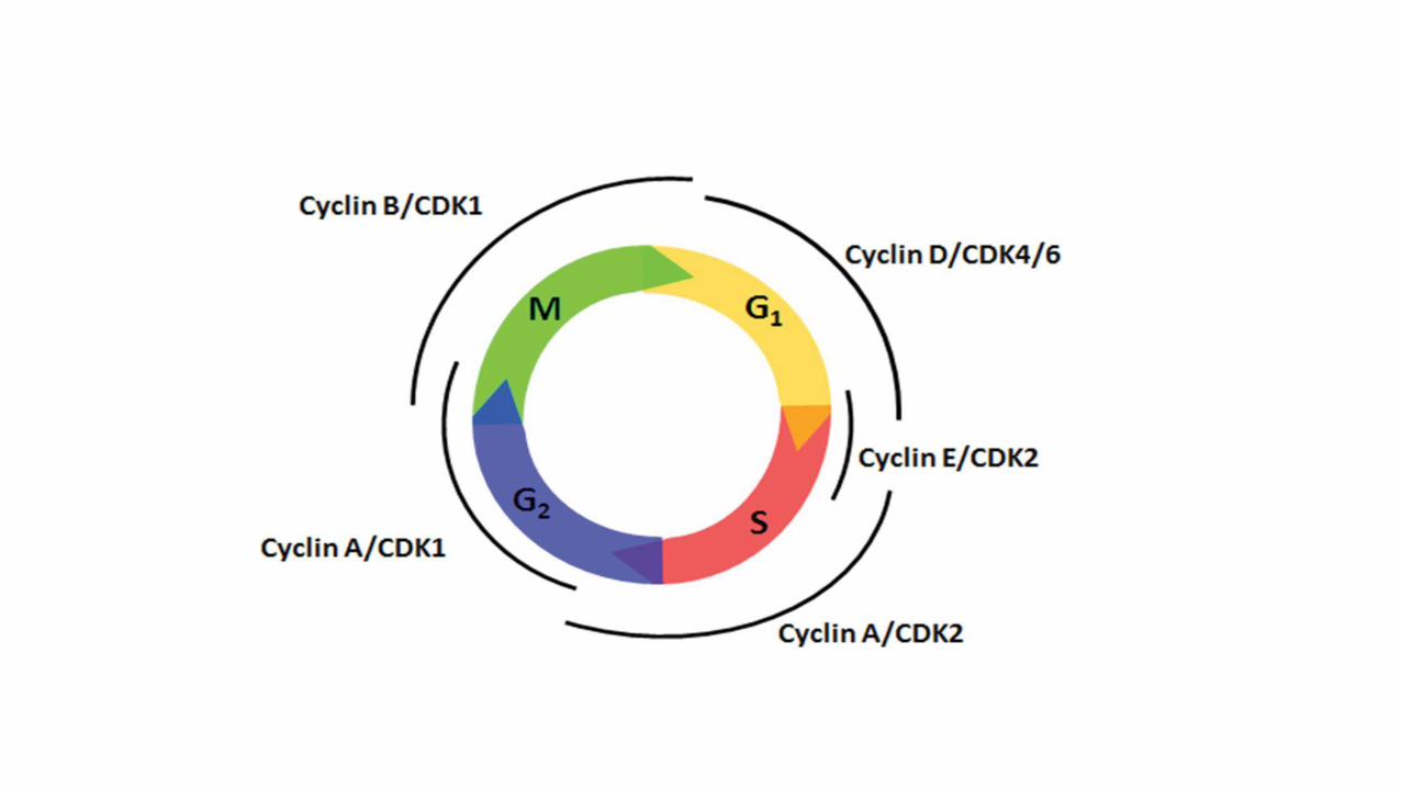
cyclins
• Progression through cell cycle, especially G1-S is regulated by proteins called cyclins
• Cyclins activate kinases CDK (cyclin dependent kinases)
• Cyclin and CDK form complexes that phosphorylate target proteins that drive the cell through the cell cycle.
• Cyclins : D, E, A , B (they appear in this sequence)

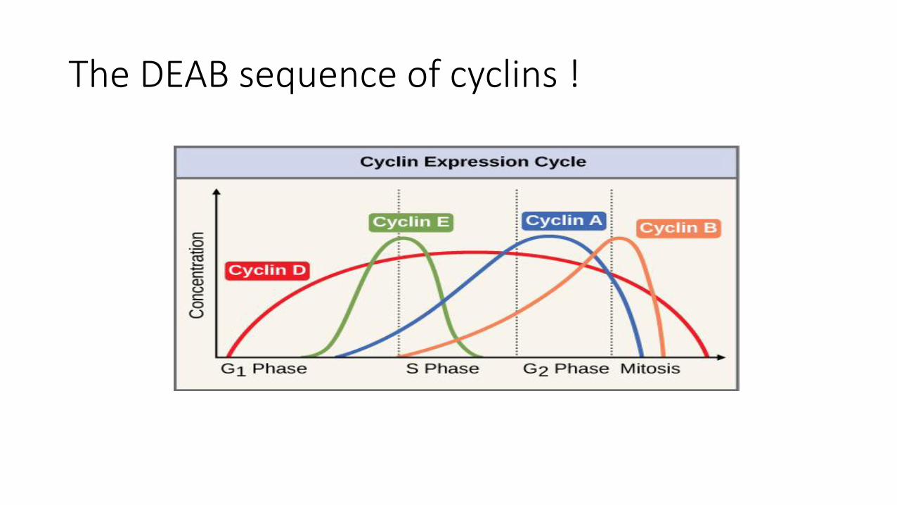
The DEAB sequence of cyclins !
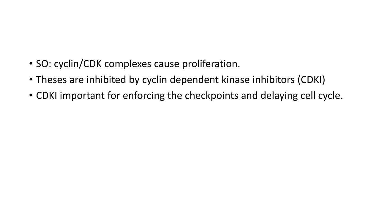
• SO: cyclin/CDK complexes cause proliferation.
• Theses are inhibited by cyclin dependent kinase inhibitors (CDKI)
• CDKI important for enforcing the checkpoints and delaying cell cycle.

• Mutations causing increased cyclins or CDK cause self sufficiency in growth signals.
• Mutations inhibiting CDKI will cause increased growth.
• Examples: cyclin D is activated in several tumors mainly lymphomas.
Summary 1/3
• Transformed cells acquire several mutations resulting in several phenotypes.
• There are 8 phenotypes needed in each transformed cell and these are called the hall marks of cancer.
• All these hallmarks must be gained through mutations, but there is no specific sequence for acquiring them.
• We don’t need 8 mutations for the 8 phenotypes as one mutation might cause more than phenotype.
• these 8 are: self sufficiency of growth factors, insensitivity to growth inhibitory signals, evasion of cell death and the immune system, changes in metabolism, immortality , ustained angiogenesis and ability to invade and metastasize.
• Genomic instability and inflammation act as enablers of malignancy; they set the proper environment for mutations to occur.
• Tumors also need a proper microenvironment provided by host stromal cells.
Summary 2/3
• Self sufficiency in growth signals can occur through increased growth factors, growth factor receptors, signal transduction proteins, transcription factors or cell cycle stimulators.
• GF can be synthesized by tumor cells or host stromal cells.
• GF receptors can be activated via overexpression ( HER 2) or changes in their architecture that makes them active even without binding to GF.
• Signal transduction can increase via increase in any protein involved in second messengers that convey growth signals to the nucleus. These include RAS and ABL plus their downstream protein pathways ( BRAF- MAP kinase and MTOR pathways)
• RAS is the most commonly mutated oncogene in humans.
• RAS is a G protein that is stimulated upon phosphorylation of GDP to GTP.
• Point mutations that cause entrapment of RAS in the activated state cause cell transformation.
Summary 3/3
• ABL is a protooncogene that can be stimulated via translocation ( 9;22) with formation of ABL-BCR fusion gene that encodes a kinase which is active and causes cell proliferation. This causes a leukemia that be treated by blocking the kinase.
• Cell cycle is stimulated by cyclins/ Cyclin dependent kinase(CDK) complexes. Increase in Cyclins or CDK can transform cells.
• Cyclin/CDK complexes are normally regulated by CDK inhibitors.. A decrease in the inhibitors can also transform cells.

Test yourself
• The questions to follow are from webpath website, it’s a very good source of pathology MCQ questions:
https://library.med.utah.edu/WebPath/EXAM/EXAMIDX.html
- My aim here is to show you how long, seemingly difficult questions are actually very easy. Just know your concepts and practice how to deal with these questions: let’s try
• A 73-year-old man has an episode of hematemesis. Upper GI endoscopy reveals an irregular 4 cm gastric antral ulceration. Biopsies are performed and microscopically reveal adenocarcinoma. Molecular analysis shows DNA hypermethylation of the CDKN2 complex. Through which of the following mechanisms has this abnormal gene expression most likely occurred?
• A Amplification
• B Epigenetic alteration
• C Growth factor binding
• D Point mutation
• E Reduced miRNA expression

answer• A 73-year-old man has an episode of hematemesis. Upper GI endoscopy
reveals an irregular 4 cm gastric antral ulceration. Biopsies are performed and microscopically reveal adenocarcinoma. Molecular analysis shows DNA hypermethylation of the CDKN2 complex ( this is the only relevant piece of information. If you know that hypermwthylation is an epigenetic change then you solved the whole issue.. Through which of the following mechanisms has this abnormal gene expression most likely occurred?
• A Amplification • B Epigenetic alteration • C Growth factor binding • D Point mutation • E Reduced miRNA expression
• Columnar epithelial cells from the colonic mucosa are studied to identify abnormalities in cell signaling pathways. Abnormal epithelial cells from colonic adenocarcinoma are shown to have a mutation that blocks hydrolysis of GTP-bound active RAS. Normal columnar cells have active RAS protein that undergoes hydrolysis to the inactive GDP-bound form. Which of the following signaling pathways is most likely abnormally stimulated in the carcinoma cells?
• A ADP
• B BCR-ABL
• C Cyclic AMP
• D Cyclin D1
• E MAP kinase
• F p53
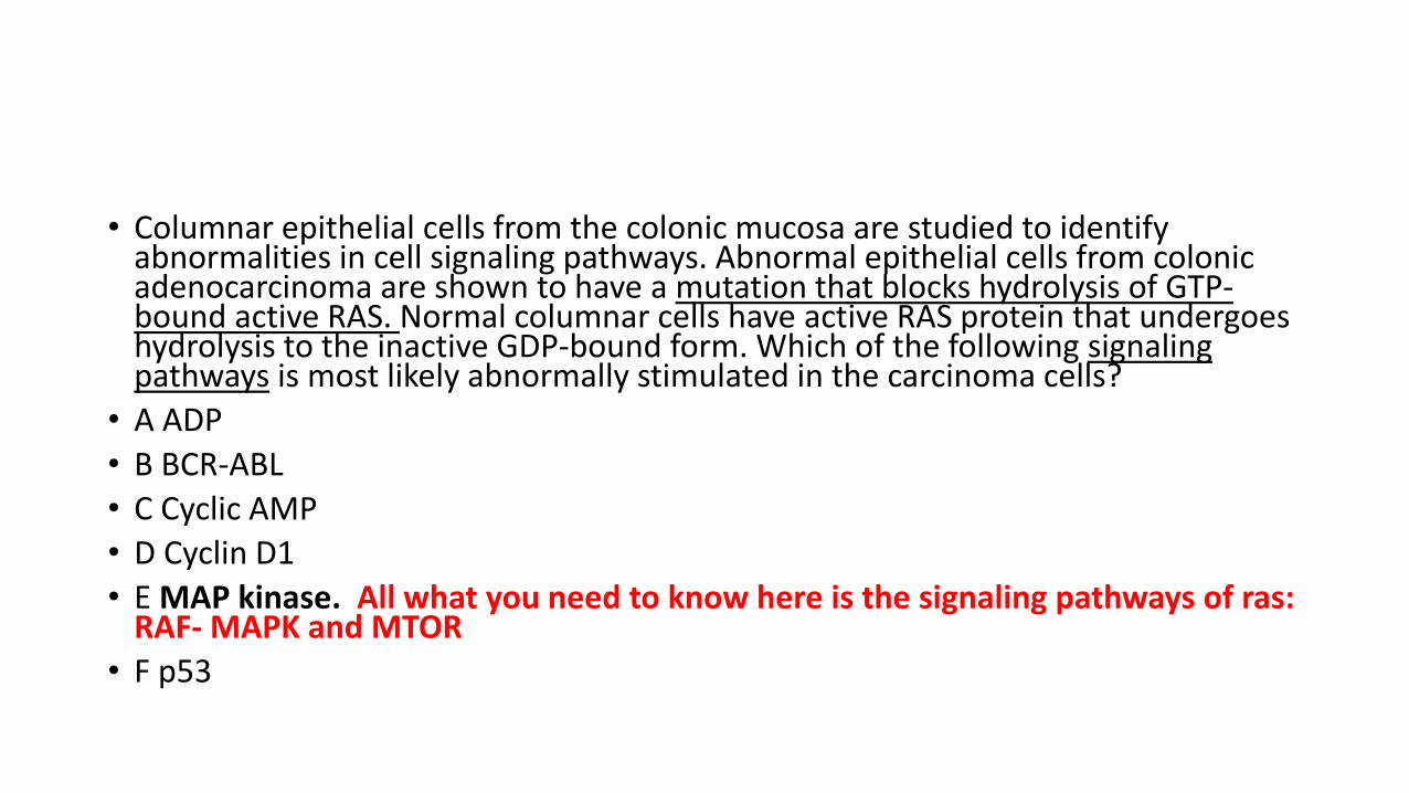
• Columnar epithelial cells from the colonic mucosa are studied to identify abnormalities in cell signaling pathways. Abnormal epithelial cells from colonic adenocarcinoma are shown to have a mutation that blocks hydrolysis of GTP-bound active RAS. Normal columnar cells have active RAS protein that undergoes hydrolysis to the inactive GDP-bound form. Which of the following signaling pathways is most likely abnormally stimulated in the carcinoma cells?
• A ADP
• B BCR-ABL
• C Cyclic AMP
• D Cyclin D1
• E MAP kinase. All what you need to know here is the signaling pathways of ras: RAF- MAPK and MTOR
• F p53
• A 50-year-old woman notes a lump in her breast. Her physician's assistant palpates a 2 cm firm mass in her left breast. A fine needle aspiration biopsy is performed, and on microscopic examination a carcinoma is present. Molecular analysis shows HER2 positivity but estrogen receptor negativity in these malignant cells. Through which of the following mechanisms has this abnormal expression most likely occurred?
• A Amplification
• B Epigenetic alteration
• C Growth factor binding
• D Point mutation
• E Reduced miRNA expression
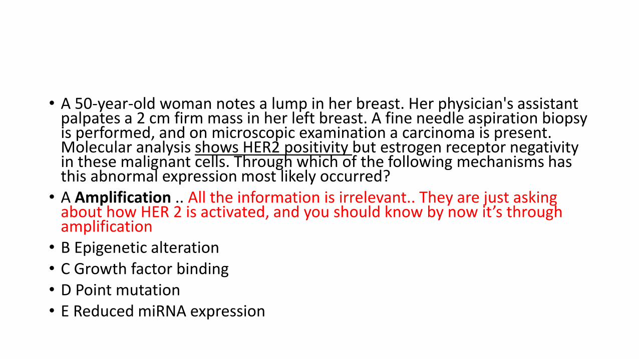
• A 50-year-old woman notes a lump in her breast. Her physician's assistant palpates a 2 cm firm mass in her left breast. A fine needle aspiration biopsy is performed, and on microscopic examination a carcinoma is present. Molecular analysis shows HER2 positivity but estrogen receptor negativity in these malignant cells. Through which of the following mechanisms has this abnormal expression most likely occurred?
• A Amplification .. All the information is irrelevant.. They are just asking about how HER 2 is activated, and you should know by now it’s through amplification
• B Epigenetic alteration • C Growth factor binding • D Point mutation • E Reduced miRNA expression

• A 66-year-old man has noted darker urine for the past 2 weeks. A urine alysis shows hematuria. Cystoscopy is performed and there is a 3 cm mass in the dome of the bladder. Biopsies of the mass are taken and on microscopic examination show a urothelial carcinoma. Cells of this neoplasm demonstrate a single mutation causing cellular inability to hydrolyze GTP, thus resulting in cellular transformation. Which of the following oncogenes is most likely implicated in this case?
• A ABL • B ERBB2 • C SIS • D RAS • E N-MYC
• A 66-year-old man has noted darker urine for the past 2 weeks. A urine alysisshows hematuria. Cystoscopy is performed and there is a 3 cm mass in the dome of the bladder. Biopsies of the mass are taken and on microscopic examination show a urothelial carcinoma. Cells of this neoplasm demonstrate a single mutation causing cellular inability to hydrolyze GTP, thus resulting in cellular transformation. Which of the following oncogenes is most likely implicated in this case?
• A ABL
• B ERBB2
• C SIS
• D RAS …. You only need to know that RAS is a G protein and its function is related to GDP/GTP. You should be able to answer the question even if you have no idea what the rest of the choices are.
• E N-MYC
• A 52-year-old man has had increasing fatigue for the past 6 months. On physical examination he has a palpable spleen tip. Laboratory studies show a WBC count of 189,000/microliter. The peripheral blood smear shows many mature and immature myeloid cells present. Cytogenetic analysis of cells obtained via bone marrow aspiration reveals a t(9:22) translocation. This translocation leads to formation of a hybrid gene that greatly increases tyrosine kinase activity. Which of the following genes is most likely translocated to cause these findings?
• A p53 • B RB • C ABL • D NF-1 • E RAS

• A 52-year-old man has had increasing fatigue for the past 6 months. On physical examination he has a palpable spleen tip. Laboratory studies show a WBC count of 189,000/microliter. The peripheral blood smear shows many mature and immature myeloid cells present. Cytogenetic analysis of cells obtained via bone marrow aspiration reveals a t(9:22) translocation. This translocation leads to formation of a hybrid gene that greatly increases tyrosine kinase activity. Which of the following genes is most likely translocated to cause these findings?
• A p53
• B RB
• C ABL .. This is very easy, you only need to practice to pick the relevant information in these questions
• D NF-1
• E RAS

• THANK YOU
• AND PLEASEDO NOT HESITATE TO ASK IF YOU NEED HELP WITH THE QUESTIONS.