Neck muscle mass index as a predictor of post laryngectomy …orca.cf.ac.uk › 114451 › 1 ›...
20
This is an Open Access document downloaded from ORCA, Cardiff University's institutional repository: http://orca.cf.ac.uk/114451/ This is the author’s version of a work that was submitted to / accepted for publication. Citation for final published version: Bozkurt, Gulpembe, Elhassan, Hassan, Soydan, Abdullah, Celebi, Irfan, Mcleod, Robert, Soytas, Pinar, Nur Erol, Zeynep and Sozen, Esra 2018. Neck muscle mass index as a predictor of post laryngectomy wound complications. Annals of Otology, Rhinology and Laryngology 127 (11) , pp. 841-847. 10.1177/0003489418798660 file Publishers page: https://doi.org/10.1177/0003489418798660 <https://doi.org/10.1177/0003489418798660> Please note: Changes made as a result of publishing processes such as copy-editing, formatting and page numbers may not be reflected in this version. For the definitive version of this publication, please refer to the published source. You are advised to consult the publisher’s version if you wish to cite this paper. This version is being made available in accordance with publisher policies. See http://orca.cf.ac.uk/policies.html for usage policies. Copyright and moral rights for publications made available in ORCA are retained by the copyright holders.
Transcript of Neck muscle mass index as a predictor of post laryngectomy …orca.cf.ac.uk › 114451 › 1 ›...

This is an Open Access document downloaded from ORCA, Cardiff University's institutional
repository: http://orca.cf.ac.uk/114451/
This is the author’s version of a work that was submitted to / accepted for publication.
Citation for final published version:
Bozkurt, Gulpembe, Elhassan, Hassan, Soydan, Abdullah, Celebi, Irfan, Mcleod, Robert, Soytas,
Pinar, Nur Erol, Zeynep and Sozen, Esra 2018. Neck muscle mass index as a predictor of post
laryngectomy wound complications. Annals of Otology, Rhinology and Laryngology 127 (11) , pp.
841-847. 10.1177/0003489418798660 file
Publishers page: https://doi.org/10.1177/0003489418798660
<https://doi.org/10.1177/0003489418798660>
Please note:
Changes made as a result of publishing processes such as copy-editing, formatting and page
numbers may not be reflected in this version. For the definitive version of this publication, please
refer to the published source. You are advised to consult the publisher’s version if you wish to cite
this paper.
This version is being made available in accordance with publisher policies. See
http://orca.cf.ac.uk/policies.html for usage policies. Copyright and moral rights for publications
made available in ORCA are retained by the copyright holders.

1
Neck muscle mass index as a predictor of post laryngectomy wound complications
Gülpembe Bozkurt1, Hassan Ahmed Elhassan2, A dullah “oyda Mah utoğlu3, İ fa Çele i4,
Robert WJ Mcleod5, Pı a “oytaş6, Zeynep Nur Erol7, Esra Sözen8
1MD, Department of Otorhinolaryngology, Acıbadem University Hospital, Istanbul, Turkey,
ORCID ID: 0000-0003-2100-023X
2FRCS DOHNS, Lewisham University Hospital, London, ORCID ID: 0000-0001-5378-8709,
3MD, Department of Radiology, Istanbul Education and Research Hospital, Istanbul, Turkey,
ORCID ID: 0000-0001-7987-0006, [email protected]
4MD, Department of Radiology, Sisli Hamidiye Etfal Education and Research Hospital,
Istanbul, Turkey, ORCID ID: 0000-0003-2562-2482, [email protected]
5MRCS PhD, Department of Otolaryngology, University Hospital of Wales, Cardiff, UK,
6MD, Department of Otorhinolaryngology, Sisli Hamidiye Etfal Education and Research
Hospital, Istanbul, Turkey, [email protected]
7MD, Department of Otolaryngology, Hopa State Hospital, Artvin, Turkey, ORCID ID: 0000-
0002-3680-4911, [email protected]
8MD, Department of Otorhinolaryngology, Sisli Hamidiye Etfal Education and Research
Hospital, Istanbul, Turkey, [email protected]
Corresponding author for proofs and reprints: Gulpembe Bozkurt MD, Department of
Otolaryngology-Head and Neck Surgery, School of Medicine, Acibadem University, Özel
A ı ade Atake t Hasta esi, Ista ul, Turkey
Tel: +90 5325586990
Fax: +90 2124044445
manuscript word count: 2437

2
Objective
We investigated the relationship between paravertebral muscle cross-sectional area (PVM
CSA) at the third vertebra (C3) level using CT neck images, and its relationship with
complications after total laryngectomy.
Design
Retrospective analysis of 60 advanced laryngeal cancer patients who underwent total
laryngectomy was performed. The cross-sectinal areas of paravertebral neck muscles using
neck computerized tomography (CT) at C3 level images obtained preoperatively were
analysed.
Results
A significant difference in PVM CSA between complication and no complication groups
[F(1,53=4.319, p=0.043] was identified by ANCOVA. There were no significant differences in
between-subject effects: T-stage [F=1.652, p=0.204], BMI [F=0.889, p=0.35], Albumin
[F0.359, p=0.552], Age [F=1.623 p=0.208], Smoking [F 4.319, p=0.41].
Conclusion
PVM CSA measured at C3 level on pretreatment CT may help identify patients at higher risk
of postoperative wound complications after total laryngectomy and who may particularly
benefit from pre-operative optimization of nutritional status.
Keywords: Total laryngectomy, lean muscle mass, sarcopenia, pharyngocutaneous fistula,
cross-sectional area

3
Neck muscle mass index as a predictor of post laryngectomy wound complications
Introduction
Patients with laryngeal cancer experience loss of major weight and skeletal muscle mass (SMM),
prior to and during therapy, which is associated with poor functional and survival outcomes.1 30–60% of head and neck cancer patients have malnutrition, related to the tumour and treatment.2
These patients have a high incidence of complications after major head and neck surgery that can
delay the start of adjuvant therapy and add to the cost of health care.3 Pharyngocuteous fistule (PCF)
and hematomas are the most common complications following total laryngectomy (TL).4,5 The PCF
incidence varies from 2.6% to 65.5%.6 Factors associated with PCF formation include low body mass
index (BMI), poor nutritional status as well as pre-operative radiotherapy and chemotherapy, male
gender, increased age, tobacco and alcohol history, tumour site, size and stage, intra-operative flap
reconstructions, tracheoesophageal puncture and pre-operative tracheostomy.7-10
In head and neck cancer, indicators of nutritional status including serum protein concentrations and
serum albumin, when low, are associated adverse outcomes.1,11 Due to their variability these
fa to s i di idually a e u a le to dete t a patie t s ut itio al status. As su h, the e is a o e away from these biomarkers and a focus towards patient energy intake, anthropometric
measurements (BMI, body composition, weight) and functional measures.13,14
The loss of skeletal muscle mass (SMM) leads to objective weakness, is associated with functional
impairment and disability15, a longer hospital stay16, higher economic burden, higher risk of
nosocomial infections17, and decreased survival in both nonmalignant15,18 and malignant
conditions19-21 and predicts chemotherapy toxicity22 and may be valuable in assessing patients
prior to surgery. Although patients who lost more than 10% of their ideal body weight are at
increased risk of postoperative complications23, weight loss alone poorly predicts outcome in head
and neck cancer (HNC) patients when compared with depleted SMM.24 Low SMM is an independent
predictive factor for chemotherapy toxicity in patients with advanced HNC treated with primary
chemoradiotherapy. 25 Grossberg et al24 found that BMI and SMM depletion were powerful
prognostic indicators of mortality. Nevertheless, BMI alone is not a reliable indicator of SMM.26
Sarcopenia was initially defined as muscle mass two standard deviations below that of a healthy
adults27, there is now no clear definition of sarcopenia or one cut-off value for SMM that is valid for
all oncological patients.28
The use of functional imaging techniques such as dual-energy X-ray absorption (DEXA) and magnetic
resonance imaging (MRI), has improved the measurement of SMM. Studies using CT/MR imaging of
the psoas muscle crosssectional area (CSA) at the level of the third lumbar (L3) vertebra28,29 have
been used to estimate overall SMM. Assessing paravertebral muscle cross-sectional area (PVM CSA)
on CT-scan at the third cervical (C3) vertebra appears to be a good alternative to abdominal CT-scans,
based on the strong correlation between skeletal muscle cross sectional area at C3 and L3
vertebrae.30 The C3 level is preferred as it captures only the neck muscles. Higher cervical images
include the tongue muscles while lower levels capture the trapezius muscle. The C3 level includes the
following muscles, longus capitis, longus colli muscles located anterior to vertebrae and spinalis
cervicis, semispinalis cervicis, semispinalis capitis, splenius capitis, and levator scapulae muscles.
4
There is currently little literature on the effect of sarcopenia on laryngeal cancer outcomes and no
practical diagnostic tools for detecting sarcopenia. As laryngeal cancer staging includes cross
sectional imaging of the neck as routine and a skeletal muscle mass estimate using C3 level CT-scans
is practical and can be used to reliably to estimate SMM, this may be a cost effective and widely
available tool to preoperatively assess patients prone to postoperative complications among HNC
patients. Abdominal CT scans that include the third lumber vertebra (L3) and DEXA-scans are not
routinely performed in HNC patients.
As sarcopenia and low SMM are associated with increased postoperative wound complications in
patients with HNC31, we investigated the relationship between PVM CSA at C3 using CT neck images,
and complications in advanced laryngeal cancer patients.
Methods
A single-center-retrospective analysis of 77 laryngeal cancer patients who underwent total
laryngectomy between January 1, 2010, to January 31, 2017 was performed. Data was collected from
January 1, 2010, to March 31, 2017. Patients with wide tumor resections including skin, extended
pharyngeal resection requiring flap reconstruction (n=3) and patients with positive surgical margins
(n = 7) were excluded as these are risk factors for PCF32, 33, patients with missing pre-operative CT
images (n=7) were excluded. Patients who had head and neck CT scans with a maximum interval of
two months (n=2) prior to total laryngectomy surgery were included (n = 60). All patients were
discussed at a multidisciplinary tumor board. Total laryngectomy was preferred in cases where lung
capacity was insufficient for partial surgery (FEV1 < 60%), > 1 cm subglottic tumor extension,
interaritenoid and postcricoid involvement, extralaryngeal extension, and vocal cord fixation due to
cricoarytenoid joint invasion. The laryngectomies were performed with an open technique and
simultaneous selective neck dissection (levels II-IV +/- IV); prophylactic (n=21) or therapeutic (n=39).
Informed consent was obtained from 52 patients, 5 patients died and no contact information was
available for 3. The institutional ethics committee approved the study.
Patient demographics
Data including age, hospital stay time, BMI, smoking status, preoperative albumin levels, and co-
existing morbidity (Charlson Comorbidity Index (CCI) scores34 were collected, as well as stage and
prior treatment history including chemotherapy and radiation. The need of a pedicled or free flap for
reconstruction was also included. Disease was staged per the American Joint Committee on Cancer
using the TNM system.35 Advanced-stage laryngeal squamous cell carcinoma (LSCC) was defined as
T3–T4 and any TN+, M+. Surgery-related wound complications (including infection,
pharyngocutaneous fistula, dehiscence, hematoma, and seroma) defined as occurring within 30
postoperative days were recorded.
Cross-sectional measurement of skeletal muscle index at C3
By calculating the cross-sectional area (CSA) of neck muscles using staging CT images obtained
preoperatively and using the method devised by Swartz et al30 we estimated SMM. Swartz et al.
recently presented a method to estimate skeletal muscle mass in routinely performed head and neck
CT-scans; CSA at C3 strongly correlated with L3 muscle CSA. The middle of the C3 vertebrae was

5
chosen as the reference point on sagittal reformatted image and CSA measurement undertaken on
the axial image at this level.
“keletal us le as ua tified y the use of sta da d Hou sfield u it HU th esholds − to Separately, the cross-sectional area of pixels that had a radiodensity between _29 and +150 HU was
ret ie ed fu the efe ed to as HU us le a ea . Lo gus apitis, lo gus olli us les, spi alis cervicis, semispinalis cervicis, semispinalis capitis, splenius capitis, and levator scapulae muscles were
evaluated. The muscles were outlined using an image-processing system (PACS, Infinit Healthcare,
South Korea, Guro-gu) by a single head and neck radiologist. The cross-sectional areas within the
limits of the drawn boundaries was calculated using Xelis 3D software (V1.0.6.1, Infinit Healthcare,
South Korea, Guro-gu ) were measured (Figure 2). The sum of the cross-sectional areas (mm2) of all
muscles was calculated for each patient; the total area was then normalised for patient height (Cross
Sectional Area (CSA) mm2/m2) to give a C3 skeletal muscle index.37
Exclusion of sternocleidomastoid muscles
Swartz et al. presented two methods to estimate skeletal muscle mass in routinely performed head
and neck CT-scans. They assessed the correlation between CSA at L3 and PVM only and between L3
and the PVM and only a single SCM muscle. Swartz et al found significant correlation between the
sum of the PVM and SCM muscles and L3 (r = 0.785, p < 0.001) and the PVM muscles only and L3 (r =
. , p < . . Ho e e as the fo e as the st o ge o elatio they o ly performed further
analysis on the sum of the PVM and SCM muscles.
This approach had several limitations that they alluded to in their section sub-titled Cli i ally issi g data . I thei study C“A assess e t of o e o oth “CM us les as i pai ed y lymph node
metastases in 8 SCMs in 6 patients (7.6% of all HNC patient SCMs, 11.5% of all HNC patients, 17.6% of
patients with neck metastases).
As such we agreed with Scwartz et al that CSA L3 correlated with CSA PVM alone, but disagreed with
them that the PVM and SCM method of measuring neck CSA was best for further analysis. Stronger
correlation is not reason enough to ignore the fact that in a significant proportion of patients data is
not available, as the SCM muscles is invaded by lymph node metastases (Figure 1).
To minimise measurement inconsistency for our study we elected to use Schwartz et als first method
that excluded the SCM muscles from the CSA calculations. Our study included 12 patients with N2c
(bilateral) nodal involvement.
6
Statistical analysis
A one-way analysis of covariance (ANCOVA) was performed using SPSS statistical package for Mac
version 23 (SPSS Inc., Chicago, Illinois) to investigate if there was a difference in LMM in complication
and no complication groups. Statistical significance was defined as p < 0.05. Covariates included were
BMI, albumin, age, T-stage (categorized as 2, 3 or 4) and smoking status. Complication or no
complication groups were defined as the independent variable. This allowed the creation of 95%
confidence intervals (CI) for the LMM dependent variable.
Results
Patient demographics
Table 1 summarizes the clinical characteristics of 60 patients; all were men with a mean (SD) age of
59.4 (8.4) years. Hypoalbuminemia was identified preoperatively in 25 (41.6 %) patients. Among
these study patients, 46 (76.6%) had locally advanced disease (T3–4), 14 (23.4 %) had T2 disease.
Twenty one (35%) patients had N0 neck; 17 (28%), 5 (8%), 3 (13%), 12 (20%) and 2 (3%) patients had
N1, N2a, N2b, N2c and N3 nodal involvement, respectively. None of the patients had detected
distant metastasis. Nine patients (15%) had a history of prior chemo/RT.
Wound Complications
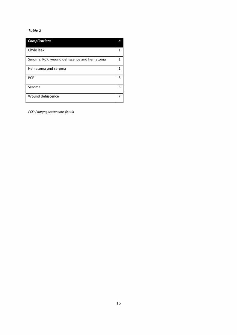
Wound complications occurred in 22 patients out of 60 (37%). One patient developed chyle leak, 1
had seroma, PCF, wound dehiscence and hematoma together, 1 had hematoma and seroma
together, 8 had PCF, 3 had seroma, and 7 experienced dehiscence of their neck incision in the
absence of a recognized salivary fistula (Table 2). PCFs occurred in 10 patients (16%); six (60%) were
managed conservatively and four required surgical closure with pectoralis major flap. Regarding
wound dehiscence (n=8), spontaneous closure with local wound care was noted in 6 (75%) patients
whereas a surgical closure was necessary in two.
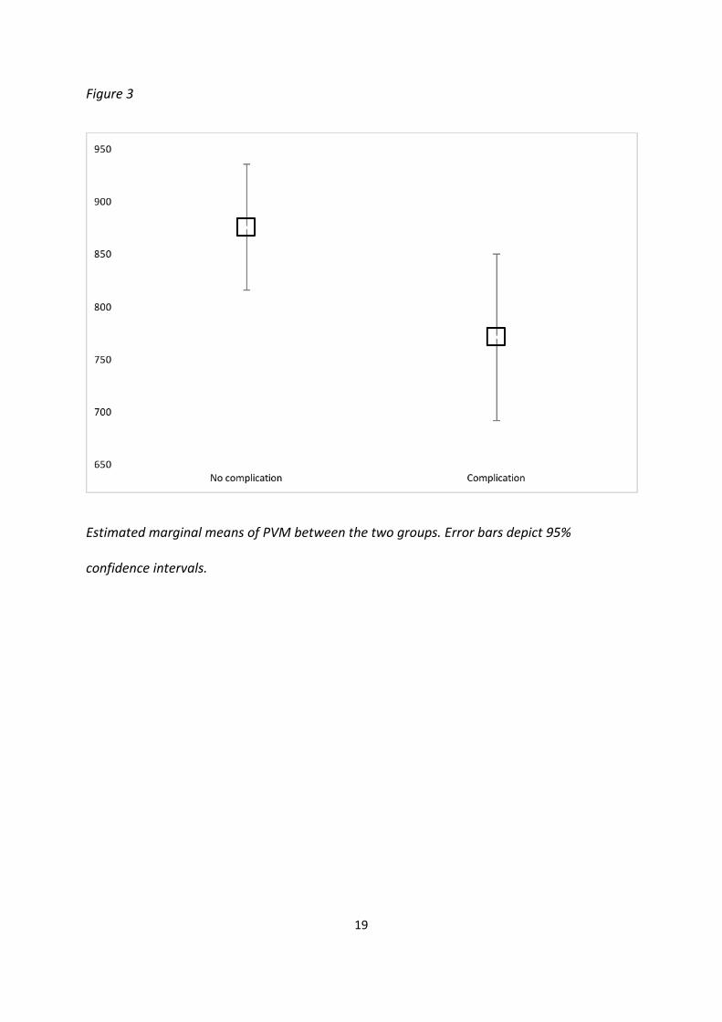
Cross-sectional skeletal muscle index and postoperative complications
Of the 60 patients 38 22 were categorized in the complications group and 22 38 in the no
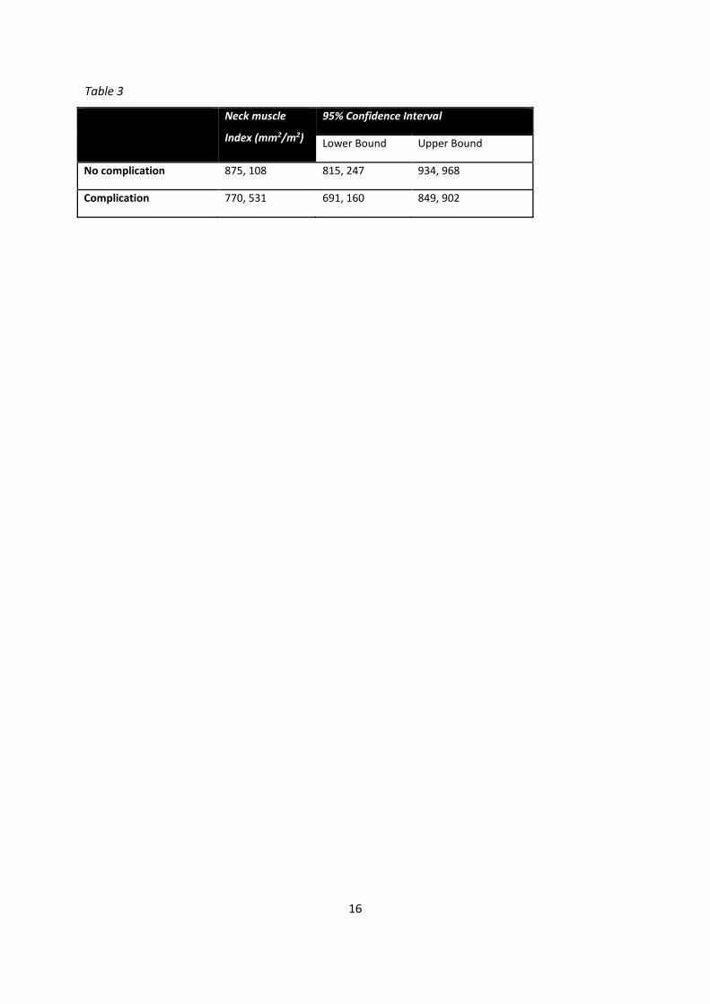
complications group. ANCOVA identified a significant difference in PVM CSA between complication
and no complication groups [F (1,53=4.319, p=0.043] (Table 3). There were no significant differences
in between-subject effects: T-stage [F=1.652, p=0.204], BMI [F=0.889, p=0.35], Albumin [F0.359,
p=0.552], Age [F=1.623 p=0.208], Smoking [F 4.319, p=0.41]. Estimated marginal means accounting
for covariates are shown in figure 3. Covariates in the model are evaluated at the following values: T
stage = 3.0167, BMI = 23.5787, Albumin = 3.6492, Age = 59.3667, smoking = .50
Postoperative complications related to other variables
The focus of this study was the relationship between SMM, estimated by PVM CSA, and post
operative complications. The relationship between postoperative complications and variables BMI
and T-stage are widely known and we did not concentrate on within this study. However we analysed
the data using t-tests looking for differences between the complication and non-complication within
this study. Only BMI showed a significant difference between groups as an independent variable. This
difference between groups is corrected for in the ANCOVA and not explored further.

7
Discussion
Postoperative wound complications account for most complications after major head and neck
surgery.38 Adequate nutritional status is necessary to support wound healing and surgical
recovery.39
Our study shows that lean muscle mass estimated by PVM CSA at C3 may be a potentially useful
predictor of post-operative wound complications in in laryngeal cancer patients. We found that PVM
CSA level below 815 mm2/m2 is a potential prognostic indicator of postoperative wound
complication independent of other factors, such as BMI, hypoalbunemia, smoking history, and
previous chemo/radiotherapy.
Grossberg24 et al reported that patients with HNSCC and low SMM at presentation or after
treatment exhibit decreased overall and cancer-specific survival. Weight loss itself poorly predicts
outcome in HNC patients when compared with depleted SMM. Therefore, weight loss alone cannot
be used reliably to stratify by risk patients with larynx cancer undergoing laryngectomy. In our study,
we also did not find a statistically significant difference between the complication and no
complication groups using PVM CSA with regards to T-or N-stages, or low preoperative albumin.
Radiologically assessed muscle mass has been suggested as a surrogate marker of functional
status p e ıously. , Pu lished studies use ite ia that a e study spe ifi , ith so e studies usi g imaging modalities such as CT or DEXA scans and others basing their criteria on muscle strength due
to the cost effectiveness of the measurements.26-29,37 In the present study, Image analysis was
performed as described in a previous report.16 For the purpose of comparing to literature values,
measurements for skeletal muscle measured by CT were o alized to ea h patie t s height.
Limitations of our study include, retrospective data collection, lack of female patients and sample
size. Another limitation in our study in a few (n=2) patients, the interval between CT and
laryngectomy was up to two months, and arguably there may have been significant weight loss in
that time. The number of independent variables included in the ANCOVA was limited by the sample
size, and the number of wound complications and PCF. This may explain why we did not have a
significant difference between complications and disease stage. A larger and more homogeneous
patient cohort is warranted.
This study, to our knowledge, is the first to investigate the role of PVM CSA measured at the C3
vertebrate level and postoperative wound complication rate among larynx cancer patients. The lower
boundary of the 95% CI no complication rate is 815 mm2/m2 and the upper boundary of the 95% for
the complication group is 850 mm2/m2. We propose that a neck muscle index below 815 mm2/m2 is
considered as a risk factor for postoperative wound complications in head and neck patients that can
be used as additional factor for stratifying low muscle mass and subsequent postoperative
complication risk. We suggest this figure to research colleagues in head and neck surgery for future
validation studies.

8
Conclusion
Paravertebral muscle cross-sectional area (PVM CSA) at the third vertebra (C3) level may
help predict patients at high risk of postoperative wound complications before total
laryngectomy and who would particularly benefit from pre-operative optimization of
nutritional status. The incorporation of CT-assessed muscle mass into routine pre-surgical
assessment may aid nutritional status assesment.
Abbreviations: LMM: Lean muscle mass, SMM: Skeletal muscle mass, PCF:
Pharyngocutaneous fistule, TL: Total laryngectomy, SCM: Sternocleidomastoid muscle, BMI:
body mass index, DEXA: Dual-energy X-ray absorption, MRI: Magnetic resonance imaging,
CT: Computed tomography, HNC: Head and neck cancer, PVM CSA: Paravertebral muscle
cross sectional area, SM: Skeletal muscle, LSCC: Laryngeal squamous cell carcinoma
Compliance with Ethical Standards
CONFLICTS OF INTEREST: None.
ACKNOWLEDGEMENTS: None
ETHICAL APPROVAL: All procedures performed in studies involving human participants were
in accordance with the ethical standards of the institutional and/or national research
committee and with the 1964 Helsinki declaration and its later amendments or comparable
ethical standards.

9
References
1. Alshadwi A, Nadershah M, Carlson ER, Young LS, Burke PA, Daley BJ. Nutritional
considerations for head and neck cancer patients: a review of the literature. J Oral
Maxillofac Surg. 2013;71:1853-60.
2. Ravasco P, Monteiro-Grillo I, Vidal PM, Camillo ME. Nutritional deterioration in
cancer: the role of disease and diet. Clin Oncol. 2003;15:443–50.
3. Cavalot AL, Gervasio CF, Nazionale G, Albera R, Bussi M, Staffieri A, et al.
Pharyngocutaneous fistula as a complication of total laryngectomy: review of the
literature and analysis of case records. Otolaryngol Head Neck Surg. 2000;123:587-
92.
4. Herranz J, Sarandeses A, Fernandez MF, Barro CV, Vidal JM, Gavilan J. Complications
after total laryngectomy in nonradiated laryngeal and hypopharyngeal carcinomas.
Otolaryngol Head Neck Surg. 2000;122:892-98.
5. Ganly I, Patel S, Matsuo J, Singh B, Kraus D, Boyle J, et al. Postoperative
complications of salvage total laryngectomy. Cancer. 2005;103:2073-81.
6. Paydarfar JA, Birkmeyer NJ. Complications in head and neck surgery: a meta-analysis
of postlaryngectomy pharyngocutaneous fistula. Arch Otolaryngol Head Neck Surg.
2006;132:67-72
7. Erdag MA, Arslanoglu S, Onal K, Songu M, Tuylu AO. Pharyngocutaneous fistula
following total laryngectomy: multivariate analysis of risk factors. Eur Arch
Otorhinolaryngol. 2013;270:173-79.
8. Qureshi SS, Chaturvedi P, Pai PS, Chaukar DA, Deshpande MS, Pathak KA, et al. A
prospective study of pharyngocutaneous fistulas following total laryngectomy. J
Cancer Res Ther. 2005;1:51-6.
9. Patel UA, Moore BA, Wax M, Rosenthal E, Sweeny L, Militsakh ON, et al. Impact of
pharyngeal closure technique on fistula after salvage laryngectomy. JAMA
Otolaryngol Head Neck Surg. 2013;139:1156-62.
10. Timmermans AJ, Lansaat L, Theunissen EA, Hamming-Vrieze O, Hilgers FJ, van den
Brekel MW. Predictive factors for pharyngocutaneous fistulization after total
laryngectomy. Ann Otol Rhinol Laryngol. 2014;123:153-61.
10
11. Danan D, Shonka DC Jr, Selman Y, Chow Z, Smolkin ME, Jameson MJ. Prognostic value
of albumin in patients with head and neck cancer. Laryngoscope. 2016;126:1567-71.
12. Skipper A, Ferguson M, Thompson K, Castellanos V, Porcari J. Nutrition screening
tools: an analysis of the evidence. JPEN J Parenter Enteral Nutr. 2012;36:292-98.
13. Rosenbaum K, Wang J, Pierson RN Jr, Kotler DP. Time-dependent variation in weight
and body composition in healthy adults. JPEN J Parenter Enteral Nutr. 2000;24:52-5.
14. White JV, Guenter P, Jensen G, Malone A, Schofield M. Academy of Nutrition and
Dietetics Malnutrition Work Group; ASPEN Malnutrition Task Force; ASPEN Board of
Directors. Consensus statement of the Academy of Nutrition and Dietetics/American
Society for Parenteral and Enteral Nutrition: characteristics recommended for the
identification and documentation of adult malnutrition (undernutrition). J Acad Nutr
Diet. 2012;112:730-8.
15. Morley JE, Baumgartner RN, Roubenoff R, Mayer J, Nair KS. Sarcopenia. J Lab Clin
Med. 2001;137:231–43.
16. Pichard C, Kyle UG, Morabia A, Perrier A, Vermeulen B, Unger P. Nutritional
assessment: lean body mass depletion at hospital admission is associated with an
increased length of stay. Am J Clin Nutr. 2004;79:613–8.
17. J R Lieffers, O F Bathe, K Fassbender, M Winget, V E Baracos. Sarcopenia is associated
with postoperative infection and delayed recovery from colorectal cancer resection
surgery. Br J Cancer. 2012;107:931–36.
18. Montano-Loza AJ, Meza-Junco J, Prado CM, Lieffers JR, Baracos VE, Bain VG, et al.
Muscle wasting is associated with mortality in patients with cirrhosis. Clin
Gastroenterol Hepatol. 2012;10:166–73.
19. Garth AK, Newsome CM, Simmance N, Crowe TC. Nutritional status, nutrition
practices, and postoperative complications in patients with gastrointestinal cancer. J
Hum Nutr Diet. 2010;23:393–401.
20. Tan BH, Birdsell LA, Martin L, Baracos VE, Fearon KC. Sarcopenia in an overweight or
obese patient is an adverse prognostic factor in pancreatic cancer. Clin Cancer Res.
2009;15:6973–9.

11
21. Pacelli F, Bossola M, Rosa F, Tortorelli AP, Papa V, Doglietto GB. Is malnutrition still a
risk factor of postoperative complications in gastric cancer surgery? Clin Nutr.
2008;27:398–407.
22. Prado CM, Baracos VE, McCargar LJ, Reiman T, Mourtzakis M, Tonkin K, Mackey
JR, Koski S, Pituskin E, Sawyer MB. Sarcopenia as a determinant of chemotherapy
toxicity and time to tumor progression in metastatic breast cancer patients receiving
capecitabine treatment. Clin Cancer Res. 2009;15:2920-6.
23. Van Bokhorst-de van der Schueren MA, van Leeuwen PA, Sauerwein HP, Kuik
DJ, Snow GB, Quak JJ. Assessment of malnutrition parameters in head and neck
cancer and their relation to postoperative complications. Head Neck. 1997;19:419-
25.
24. Grossberg AJ, Chamchod S, Fuller CD, Mohamed AS, Heukelom J, Eichelberger H, et
al. Association of body composition with survival and locoregional control of
radiotherapy-treated head and neck squamous cell carcinoma. JAMA Oncol.
2016;2:782-9.
25. Wendrich AW, Swartz JE, Bril SI, Wegner I, Graeff A, Smid EJ, et al. Low skeletal
muscle mass is a predictive factor for chemotherapy dose-limiting toxicity in patients
with locally advanced head and neck cancer Oral oncology. 2017;71:26-33
26. Martin L, Birdsell L, Macdonald N, Reiman T, Clandinin MT, McCargar LJ et al. Cancer
cachexia in the age of obesity: skeletal muscle depletion is a powerful prognostic
factor, independent of body mass index. J Clin Oncol. 2013;31:1539-47.
27. Rizzoli R, JY Reginster, JF Arnal, I Bautmans, C Beuadart, H Bischoff-Ferrari, et al.
Quality of Life in Sarcopenia and Frailty. Calcified Tissue International. 2013;93: 101–
20.
28. Shen W, Punyanitya M, Wang Z, Gallagher D, St-Onge M-P, Albu J, et al. Total body
skeletal muscle and adipose tissue volumes: estimation from a single abdominal
cross-sectional image. J Appl Physiol. 2004;97:2333–8.
29. Jones KI, Doleman B, Scott S, Lund JN, Williams JP. Simple psoas cross sectional area
measurement is a quick and easy method to assess sarcopenia and predicts major
surgical complications. Colorectal Dis. 2015;17:O20-6.
https://www.ncbi.nlm.nih.gov/pubmed/?term=Baracos%20VE%5BAuthor%5D&cauthor=true&cauthor_uid=19351764

12
30. Swartz JE, Pothen AJ, Wegner I, Smid EJ, Swart KM, de Bree R, Leenen LP, Grolman W.
Oral Oncol. Feasibility of using head and neck CT imaging to assess skeletal muscle
mass in head and neck cancer patients. 2016;62:28-33.
31. Achim V, Bash J, Mowery A, Guimaraes AR, Li R, Schindler J, Wax M, Andersen
P, Clayburgh D. Prognostic Indication of Sarcopenia for
Wound Complication After Total Laryngectomy. JAMA Otolaryngol Head Neck Surg.
2017;doi: 10.1001/jamaoto.2017.0547. [Epub ahead of print]
32. Dedivitis RA, Aires FT, Cernea CR, Brandão LG. Pharyngocutaneous fistula after total
laryngectomy: A systematic review of risk factors. Head Neck. 2015;37:1691–7.
33. Timmermans AJ, Lansaat L, Theunissen EA, Hamming-Vrieze O, Hilgers FJ, van den
Brekel MW. Predictive factors for pharyngocutaneous fistulization after total
laryngectomy. Ann Otol Rhinol Laryngol. 2014;123:153–61.
34. Charlson ME, Pompei P, Ales KL, MacKenzie CR. A new method of classifying
prognostic comorbidity in longitudinal studies: development and validation. J Chronic
Dis. 1987;40:373–83.
35. Edge S, Byrd DR, Compton CC, Fritz AG, Greene FL, Trotti A. American joint committee
on cancer—AJCC cancer staging manual. 7th ed. New York (2010): Springer.
36. Mitsiopoulos N, Baumgartner RN, Heymsfield, SB, Lyons W, Gallagher D, Ross R.
Cadaver validation of skeletal muscle measurement by magnetic resonance imaging
and computerized tomography. J Appl Physiol (1985). 1998; 85:115-22.
37. Mourtzakis M, Prado CMM, Lieffers JR, Reiman T, McCargar LJ, Baracos VE. A
practical and precise approach to quantification of body composition in cancer
patients using computed tomography images acquired during routine care. Appl
Physiol Nutr Metab. 2008;33:997-1006.
38. McMahon JD, MacIver C, Smith M, Stathopoulos P, Wales C, McNulty R, et al.
Postoperative complications after major head and neck surgery with free flap
repair—prevalence, patterns, and determinants: a prospective cohort study. Br J Oral
Maxillofac Surg. 2013;51:689-95.
https://www.ncbi.nlm.nih.gov/pubmed/?term=Andersen%20P%5BAuthor%5D&cauthor=true&cauthor_uid=28448668

13
39. Huckleberry Y. Nutritional support and the surgical patient. Am J Health Syst Pharm.
2004;61:671–82.
40. Prado CM, Wells JC, Smith SR, Stephan BC, Siervo M. Sarcopenic obesity: a critical
appraisal of the current evidence. Clin Nutr. 2012;31:583–601.
41. Baumgartner, RN. Body composition in healthy aging. Ann. N.Y. Acad.Sci.
2000;904:437-48.
Summary
In larynx cancer patients, skeletal muscle mass can be estimated using neck muscle index
from the PVM CSA at the C3 level and this tool may help predict patients at high risk of
postoperative wound complications before total laryngectomy.
Table Legends
Table 1: Patient characteristics (n = 60).
Table 2: Complications for surgical site (n = 22).
Table 3: Neck muscle index means for no complication and complications groups.
Figure Legends
Figure 1: Asymmetric enlargement of sternocleidomastoid muscle. Right sternocleidomastoid
muscle shows asymmetric enlargement (white arrow) due to invasion by metastatic lymph
node.
Figure 2: Paravertebral Muscles at C3 Depicted in green. Cross-sectional areas of the muscles
outlined in green can be seen on axial CT image at C3 vertebrae level.
Figure 3: Box plot of PVM means between the two groups. Error bars depict 95% confidence
intervals. Estimated marginal means of PVM between the two groups. Error bars depict 95%
confidence intervals.

14
Table 1
Abbreviations: BMI, body mass index (calculated as weight in kilograms divided by height in meters squared); XRT, prior
radiation history; CCI, Charlson comorbidity index; PVM CSA, Paravertebral muscle cross-sectional area
a Staging based on AJCC, American Joint Committee on Cancer (7th edition).
b Based on WHO BMI classification.
Characteristics Datum (n, %)
Total no. of patients 60
Age (year) (mean) 59.37 ± 8.40 (range 40–77)
Flap 6 (10 %)
Complication 22 (36,6 %)
Prior Chemo/XRT 9 (15 %)
T-staginga
T2 14 (23.4 %)
T3 30 (50 %)
T4 16 (26.6 %)
Preoperative albumin 3,64±0.59
<3,5 g/dL 25 (41.6 %)
≥3,5 g/dL 35 (58.33 %)
CCI
<5/ ≥5 37/23 (61.6 % / 38.4 %)
BMIb, (kg/m2) 23.57±5.13
Underweight (<18.5) 5 (8.4 %)
Normal weight (18.5–24.9) 41 (68.3 %)
Overweight (25.0–29.9) 8 (13.3 %)
Obese (>30.0) 6 (10 %)
PVM CSA (mm2/m2) (mean) 836.76±187.79
Smoking status
Current/former/never 28/30/2( 46,6% / 50% / 3,4%)
Hospital stay time (day) (mean) 19.01±10.22
15
Table 2
Complications n
Chyle leak 1
Seroma, PCF, wound dehiscence and hematoma 1
Hematoma and seroma 1
PCF 8
Seroma 3
Wound dehiscence 7
PCF: Pharyngocutaneous fistula
16
Table 3
Neck muscle
Index (mm2/m2)
95% Confidence Interval
Lower Bound Upper Bound
No complication 875, 108 815, 247 934, 968
Complication 770, 531 691, 160 849, 902

17
Figure 1
Right sternocleidomastoid muscle shows asymmetric enlargement (white arrow) due to
invasion by metastatic lymph node.

18
Figure 2
Cross-sectional areas of the muscles outlined in green can be seen on axial CT image at C3
vertebrae level.
19
Figure 3
Estimated marginal means of PVM between the two groups. Error bars depict 95%
confidence intervals.