SARAWAK. 2014.08.07. Una Nova Aportació Documental a La Història de La Cova de Las Graderas

RESEARCH Open Access
Leptospirosis infections among hospitalpatients, Sarawak, MalaysiaKing-Ching Hii1†, Emily R. Robie2,3†, Izreena Saihidi1, Antoinette Berita1, Natalie A. Alarja2,3, Leshan Xiu2,3,4,James A. Merchant5, Raquel A. Binder2,3, Johnny Keh-Tun Goh1, Vanina Guernier-Cambert6, Diego Galán3,Michael J. Gregory7 and Gregory C. Gray2,3,8,9*
Abstract
Background: Leptospirosis diagnoses have increased in Sarawak, Malaysia in recent years.
Methods: To better understand the burden of disease and associated risk factors, we evaluated 147 patientspresenting with clinical leptospirosis to local hospitals in Sarawak, Malaysia for the presence of Leptospira andassociated antibodies. Sera and urine specimens collected during the acute illness phase were assessed via acommercially available rapid diagnostic test (Leptorapide, Linnodee Ltd., Antrim, Northern Ireland), an ELISA IgMassay (Leptospira IgM ELISA, PanBio, Queensland, Australia) and a pan-Leptospira real-time PCR (qPCR) assay toestimate disease prevalence and diagnostic accuracy of each method. Microagglutination testing was performed ona subset of samples.
Results: Overall, 45 out of 147 patients (30.6%) showed evidence of leptospires through qPCR in either one or bothsera (20 patients) or urine (33 patients), and an additional ten (6.8%) were considered positive through serologicaltesting, for an overall prevalence of 37.4% within the study population. However, each diagnostic methodindividually yielded disparate prevalence estimates: rapid test 42.2% for sera and 30.5% for urine, ELISA 15.0% forsera, qPCR 13.8% for sera and 23.4% for urine. Molecular characterization of a subset of positive samples byconventional PCR identified the bacterial species as Leptospira interrogans in 4 specimens. A multivariate risk factoranalysis for the outcome of leptospirosis identified having completed primary school (OR = 2.5; 95 CI% 1.0–6.4) andweekly clothes-washing in local rivers (OR = 10.6; 95 CI% 1.4–214.8) with increased likelihood of leptospirosis whencompared with those who had not.
Conclusion: Overall, the data suggest a relatively high prevalence of leptospirosis in the study population. The lowsensitivities of the rapid diagnostic test and ELISA assay against qPCR highlight a need for better screening tools.
Keywords: Leptospirosis, Sarawak, Malaysia, Diagnostics
© The Author(s). 2021 Open Access This article is licensed under a Creative Commons Attribution 4.0 International License,which permits use, sharing, adaptation, distribution and reproduction in any medium or format, as long as you giveappropriate credit to the original author(s) and the source, provide a link to the Creative Commons licence, and indicate ifchanges were made. The images or other third party material in this article are included in the article's Creative Commonslicence, unless indicated otherwise in a credit line to the material. If material is not included in the article's Creative Commonslicence and your intended use is not permitted by statutory regulation or exceeds the permitted use, you will need to obtainpermission directly from the copyright holder. To view a copy of this licence, visit http://creativecommons.org/licenses/by/4.0/.The Creative Commons Public Domain Dedication waiver (http://creativecommons.org/publicdomain/zero/1.0/) applies to thedata made available in this article, unless otherwise stated in a credit line to the data.
* Correspondence: [email protected]†King-Ching Hii and Emily R. Robie contributed equally to this work.2Duke Global Health Institute, Duke University, Durham, NC, USA3Division of Infectious Diseases, Duke University School of Medicine, DUMCBox 102359, Durham, NC 27710, USAFull list of author information is available at the end of the article
Hii et al. Tropical Diseases, Travel Medicine and Vaccines (2021) 7:32 https://doi.org/10.1186/s40794-021-00154-2
BackgroundLeptospirosis has been on the rise in Malaysia in recentyears [1, 2], where it has been designated a notifiable dis-ease since 2010. The disease is biphasic in nature, withacute flu-like symptoms at the onset lasting for about aweek, followed by antibody production and excretion ofleptospires in the urine in the second week of illness.Mild cases may present with little to no symptoms, butsevere infections can damage vital organs, trigger cyto-kine storms, and lead to death [3, 4]. In Malaysia, a peakof 7806 confirmed cases and 92 deaths was reported in2014 [2], which suggests that leptospirosis is a significantpublic health threat locally [1, 2, 5], particularly consid-ering that many cases may be under-reported or mis-diagnosed [5, 6].Leptospirosis is a zoonotic bacterial infection that
causes widespread morbidity and disproportionately im-pacts people in regions with warm, humid climates inwhich the bacteria can easily proliferate [3, 6]. It iscaused by a gram-negative spirochete of the genus Lep-tospira, which has 64 known genomic species and 300identified serovars [7, 8]. Early identification and treat-ment of leptospirosis is challenging as symptoms of in-fection are non-specific and easily confused with othermore common causes of acute febrile illness.Given limited capacity for culture and molecular test-
ing in many clinical settings, leptospirosis is commonlydiagnosed by serology. Microscopic agglutination test-ing (MAT) is considered the reference test for lepto-spirosis serological diagnosis, but it has low sensitivityin the early stage of infection until measurable levels ofantibodies develop in the blood [9]. Such delays in diag-nosis may lead to missing the critical window for ad-ministering antibiotics. In addition, MAT requires apanel of live leptospires as well as highly-trainedpersonnel, and only a few laboratories in Malaysia areable to offer this technique [5]. Alternatively, commer-cially available enzyme linked immunosorbent assays(ELISAs) can be used to detect antibodies in sera, withthe added advantage that they can be standardized [10].Studies have found some ELISA assays to be more sen-sitive than MAT at identifying IgM presence duringearly stages of leptospirosis, particularly when pairedsera are not available [3, 10].When molecular testing is available, evidence of lepto-
spires can be seen in sera samples before antibody levelsbecome noticeably elevated [3, 10]. The bacteria can alsobe detected in cerebrospinal fluid and urine specimensas the infection progresses [3, 4, 11]; some studies haveidentified Leptospira excreted in urine samples within aslittle as seven days of symptom onset [11, 12]. Polymer-ase chain reaction (PCR) assays have been successful indetecting leptospires in both sera and urine, however,the specialized equipment and training required for this
method make it impractical in lower-resourced labora-tory settings where such assets are often not available.Serological rapid diagnostic tests (RDTs) have been
developed as user-friendly alternatives to these morerigorous laboratory diagnostics and are now commonlyused to screen for suspected leptospirosis [10]. This isstandard practice in Malaysia, where RDT-positive sam-ples are further sent to a central laboratory for confirma-tory MAT [2, 5, 7, 13, 14].In this study, we sought to assess the diagnostic
accuracy of commercially available RDT and ELISAassays against a newly developed pan-Leptospira real-time PCR molecular reference test [15] while estimatingthe prevalence of leptospirosis in Sarawak, Malaysia.We additionally tested the feasibility of using urinesamples to diagnose suspected leptospirosis patients,and characterized disease presentation among the studypopulation.
MethodsStudy subject enrollmentWritten consent was obtained from a parent and/or legalguardian of all participants under the age of 18. Childrenaged 7–18 were additionally asked to assent to studyparticipation. Between February and September 2019, li-censed medical officers at three primary hospitals inSarawak, Malaysia identified patients that matched theU.S. Center for Disease Control and Prevention (CDC)case definition for clinical leptospirosis infection. Pa-tients were invited to enroll in this cross-sectional studyif they presented with either fever (axillary temperaturemeasurement of more than 37.5°C by digital thermom-eter) or known exposure to Leptospira contaminatedwater, accompanied by at least two commonly associatedsymptoms of leptospirosis: myalgia, headache, jaundice,conjunctivitis, or rash [16]. Hospitals were located inboth urban (Sibu Hospital) and rural settings (KapitHospital and Sarikei Hospital). Kapit, Sibu and Sarikeiare located in a triangular point with intercity distancesof 66 km to 222 km. Altogether, these three divisionscover 51,544.7 km2 and account for 42.4% of the landarea of Sarawak.Once informed consent was obtained from the patient
and/or their legal guardian, trained study staff collecteda blood sample, and instructed the participant in mid-stream urine collection procedures. Participants wereadditionally asked to provide basic demographic, symp-tomatology, and exposure risk information through thecompletion of a brief questionnaire.
Sample processingIn the clinical laboratory, blood samples were left to co-agulate for up to 60 min at room temperature beforecentrifugation at 2000×g for 10 min. Serum was
Hii et al. Tropical Diseases, Travel Medicine and Vaccines (2021) 7:32 Page 2 of 13

aliquoted into three 2.0 ml cryovial tubes, with one im-mediately used for RDT screening, as described below.This aliquot was later sent for confirmatory testing if theRDT was found to be positive. Of the remaining serum,one aliquot was used to run an ELISA, and the other fornucleic acid extraction and real-time PCR (qPCR)analysis.Urine samples were transferred to new sterile tubes
and centrifuged at 800×g for 10 min to form a visiblepellet before remaining supernatant was removed; if avisible pellet did not form, an estimated 100 μL of thesample was retained in the base of the tube. The urinesample was resuspended to a final volume of 300 μL inphosphate-buffered saline solution (PBS). A portion ofthe resuspended urine sample was used for the RDTassay and the remaining volume was aliquoted into 2.0ml cryovial tubes for nucleic acid extraction and qPCRanalysis.All specimens were stored at − 80 °C until remaining
tests were performed.
Rapid diagnostic testInitial RDT screening was performed by a laboratorytechnician at the enrolling hospital using a commerciallatex agglutination test currently used for leptospirosisscreening in Sarawak, Malaysia (Leptorapide, LinnodeeLtd., Antrim, Northern Ireland). This test is designed todetect IgM antibodies for the rapid serological diagnosisof human leptospirosis [17]. This screening was doneseparately using sera and urine specimens. Even thoughthis RDT was designed for use with serum, both serumand urine samples were tested using the same procedureas per manufacturer instructions. Briefly, 5 μl each of theLeptorapide reagent and specimen were applied to theprovided agglutination card and then mixed. The agglu-tination card was rotated gently for 2–3 min and thepositive/negative result was interpreted by visual com-parison with the included score card. Specimens withambiguous results were run a second time, and consid-ered inconclusive if still unclear.
Microscopic agglutination testIn accordance with the Malaysian Ministry of Healthprotocol, acute sera samples with a positive RDT resultwere shared with the Institute for Medical Research inKuala Lumpur, Malaysia for confirmatory testing. Apositive MAT was defined as a single antibody titer≥400, an equivocal MAT was defined as a single anti-body titer between 1:50 and 1:200, and a negative MATwas defined as a single antibody titer < 1:50. In ourstudy, convalescent sera samples were not available forMAT analysis.
ELISA assayOnce acute sera samples were received, technicians per-formed a Leptospira IgM ELISA assay (PanBio, Queens-land, Australia) per the manufacturer’s instructions.Briefly, patient sera and controls were diluted 1:100 indiluent reagent then added to Leptospira antigen coatedmicrowells (serovar: Patoc). The plate was incubated for30 min at 37 °C. After washing the plate with PBS, 100 μlof horseradish peroxidase-conjugated anti-human IgMwas added and incubated further for 30 min at 37 °C.After a second PBS wash, 100 μl of the tetramethylbenzi-dine substrate was added and the plate was incubatedfor 10 min at room temperature. The reaction wasstopped with 100 μl of 1M phosphoric acid. The absorb-ance value of each well was read at 450 nm wave lengthand interpreted in terms of Pan-Bio units, calculatedusing the absorbance of positive control serum, negativecontrol serum, and cut-off of calibrators provided by themanufacturer. A Pan-Bio unit ≥11 was considered posi-tive for the presence of Leptospira IgM antibodies.
qPCRTotal DNA was extracted from 140 μl each of the ali-quoted sera and urine specimen according to the manu-facturer’s instructions, using the QIAamp RNA Mini Kit(QIAGEN, Hilden, Germany) as recommended by ElBali, et al. [18]. The extracted DNA was then added to amaster mix, containing 10 ul Sso Advanced UniversalProbes Supremix (Bio-Rad, Hercules, California, USA),4.4 µl ultra-pure water, and 0.2 µm each of 20 µMprimers and probes targeting the 16S Leptospira gene, asdescribed by Mohd Ali, et al. [15] in their pan-Leptos-pira qPCR assay, specifically designed for the Malaysiancontext. The amplification protocol consisted of 95 °Cfor 3 min followed by 50 cycles of 95 °C for 30 s and61.3 °C for 30 s. As the qPCR results were not used forclinical diagnosis, a conservative approach was used toconsider specimens with threshold levels (Cq) below40.0 positive for infection. Positive and negative controlswere included in each run.
Molecular characterizationExtracted nucleic acid from specimen were shipped ondry ice to Duke University in North Carolina, USA forfurther characterization. At Duke, samples werescreened by conventional PCR and, when positive (bandat ~ 550 bp), Sanger sequencing (Eton Bioscience, Inc.,Research Triangle, NC, USA) was done to determine theLeptospira genetic diversity at the specific level targetingthe secY gene. Primers secY-F (5′-ATGCCGATCATTTTTGCTTC-3′) and secY-R (5′-CCGTCCCTTAATTTTAGACTTCTTC-3′) [19] were used to amplify 549-bpfragments. The PCR conditions consisted of a transcrip-tion step at 50 °C for 30 min and an initial denaturation
Hii et al. Tropical Diseases, Travel Medicine and Vaccines (2021) 7:32 Page 3 of 13

step at 95 °C for 15 min, followed by 45 cycles of de-naturation at 94 °C for 30 s, annealing between 50 and58 °C for 30 s, and extension at 72 °C for 1 min, with afinal extension at 72 °C for 10 mins, see standard operat-ing procedure in supplemental information.
Statistical analysesQuestionnaire data and laboratory results were enteredinto Microsoft Excel and then analyzed using SPSS Sta-tistics version 21 (IBM, 2012) and R 3.5.1 (R Core Team,2018), with epitools (Aragon, 2020), epiR (Stevenson,2020), ggplot2 (Wickham, 2016) and caret (Kuhn, 2020)packages.Participants were considered positive for leptospirosis
if (i) either sera or urine showed evidence of Leptospiraby qPCR, or (ii) at least two out of three serological tests(RDT, MAT, or ELISA) were positive. Sensitivity, specifi-city, and positive and negative predictive values (PPV,NPV) of the Leptorapide RDT and ELISA were calcu-lated against both the combined and specimen-specificqPCR results. Correlation between tests was assessedusing the phi coefficient.Bivariate analysis using a student t-test and Fisher’s
exact test were used to compare participant characteris-tics and their laboratory assays against the qPCR out-comes. Characteristics determined to be predictive ofleptospirosis infection (p-value < 0.10) were incorporatedinto a generalized linear model, then further refinedthrough stepwise, backward-elimination to identify sta-tistically significant (p-value < 0.10) adjusted odds ratios.
ResultsParticipant characteristicsA total of 147 patients (88 male, 59 female) were en-rolled across all three hospital sites (Table 1). Participantages ranged from 2 to 78 years, with 56 participants(38.1%) under 12 years of age, 13 (8.8%) between 12 and18 years, 71 (48.3%) between 18 and 65 years, and 7(4.8%) above 65 years of age. Five different ethnicitieswere represented in the study, with the majority of par-ticipants at each location and in the overall study (122,83.0%) identifying as Iban.Many participants lived in longhouses (78, 53.1%),
with 49 (33.3%) reporting residency in houses, and 20(13.6%) in wooden huts. Households frequently reportedobtaining drinking water from multiple sources; 65(44.2%) collected water from the river, 72 (49.0%) hadwater piped into their homes, 17 (11.6%) used rainwater, and 10 (6.8%) had gravity feed water systems.River water was the most common drinking watersource for participants enrolled at Kapit Hospital (55,53.9%), while participants from Sarikei and Sibu Hospitalmore commonly reported using piped water (14 per site,58.3 and 66.7%, respectively).
Among the study population, 34 (23.1%) had not com-pleted any level of formal schooling, while 57 (38.8%)had completed primary and 49 (33.3%) completed sec-ondary schooling. Only 7 (4.7%) participants had goneon to complete college or further professional studies.Among the 78 participants greater than 18 years of age,46 (59.0%) were involved in occupations associated withhigh risk of leptospirosis infections, including agriculture(n = 29), fishing (n = 17), hunting (n = 11), and logging(n = 9). Many participants participated in multiple suchactivities. Within the study population, 52.6% of individ-uals greater than 18 years of age reported wearingclosed-toe shoes for work (n = 41), while 30.8% woreopen-toed shoes or sandals. Other reported personalprotective equipment (PPE) usage included gloves(14.1%), eye protection (5.1%), and face masks (9.0%).
Leptospirosis prevalenceSerological RDT and ELISA results were available for all147 patients, with calculated prevalences of 42.2% and15.0% respectively. Out of 62 positive RDT sera speci-mens, 53 were sent for confirmatory MAT results, alongwith 5 inconclusive RDT samples and 1 negative. Re-gardless of RDT outcome, 15/59 (25.4%) sera samplesreturned positive MAT results. Per our positivity criteria(at least two out of three serological tests positive), atotal of 23 participants (15.6%) were consideredserology-positive for leptospirosis, of which thirteen par-ticipants also presented molecular evidence of lepto-spires in either sera or urine (Fig. 1).Two sera samples did not have sufficient volume to
complete qPCR testing. Among 145 remaining sera and141 matched urine specimens, a total of 45 leptospirosisinfections were identified through qPCR (positive for ei-ther sera and/or urine). Evidence of Leptospira wasfound in 20 (13.8%) sera and 33 (23.4%) urinespecimens.Taken together, serological and molecular testing
identified 55 patients with leptospirosis, resulting in anestimated prevalence of 37.4% (55 out of 147) within ourstudy population (Table 2). At Kapit Hospital, 34 of 102patients (33.3%) were considered positive for leptospir-osis, as compared to 9 of 24 (37.5%) at Sarikei Hospital,and 12 of 21 (57.1%) at Sibu Hospital.
Diagnostic accuracyLeptorapide RDT analysis on patient sera samples iden-tified 62 (42.2%) as positive, 67 (45.6%) as negative, and18 (12.2%) inconclusive. RDT analysis on urine identified43 specimens (30.5%) as positive and 98 (69.5%) as nega-tive. Sensitivity, specificity, PPV, and NPV were assessedfor each set of RDT analyses, using combined qPCR re-sults for sera and urine as the gold standard (Table 3).MAT results were primarily available for RDT positive
Hii et al. Tropical Diseases, Travel Medicine and Vaccines (2021) 7:32 Page 4 of 13

samples, and thus excluded from the diagnostic accuracycalculations to avoid bias; of note, 45.8% of MAT resultswere inconclusive, as compared to 25.4% positive and28.8% negative findings (Supplemental Fig. 1). Results ofthe Leptorapide RDT were further compared tospecimen-specific qPCR results.The IgM ELISA assay identified 22 positive (15.0%),
113 negative (76.9%), and 12 equivocal specimens (8.2%).Sensitivity, specificity, PPV, and NPV of the IgM ELISAassay were calculated in comparison to combined qPCRresults for sera and urine, as well as for sera-specific
qPCR results (Table 4). As before, MAT results were ex-cluded from this analysis.
Molecular characterizationSecY sequencing was performed on samples showingPCR amplification and a clean band of the expected size(549 bp) on gel electrophoresis. Samples successfully se-quenced (n = 4) originated from urine specimen col-lected within five days of fever onset. After cleaning andtrimming, we compared the 410-bp fragments to secYsequences available from the MLST database (https://
Table 1 Demographic and household characteristics of patients with febrile illness enrolled across three hospitals in Sarawak,Malaysia, between February and October 2019 (n = 147)
Characteristics Kapit Hospitaln (%)
Sarikei Hospitaln (%)
Sibu Hospitaln (%)
Totaln (%)
Total 102 (69.4) 24 (16.3) 21 (14.3) 147 (100)
Gender
Female 37 (36.3) 7 (29.2) 15 (71.4) 59 (40.1)
Male 65 (63.7) 17 (70.8) 6 (28.6) 88 (59.9)
Age
Under 5 6 (5.9) 1 (4.2) 1 (4.8) 8 (5.4)
5–11 35 (34.3) 11 (45.8) 2 (9.5) 48 (32.7)
12–17 11 (10.8) 0 (0.0) 2 (9.5) 13 (8.8)
18–64 45 (44.1) 11 (45.8) 15 (71.4) 71 (48.3)
65 + 5 (4.9) 1 (4.2) 1 (4.8) 7 (4.8)
Education completed
None 23 (21.6) 6 (25.0) 5 (23.8) 34 (23.1)
Primary 39 (38.2) 12 (50.0) 6 (28.6) 57 (38.8)
Secondary 36 (35.3) 5 (20.8) 8 (38.1) 49 (33.3)
College 2 (2.0) 0 (0.0) 2 (9.5) 4 (2.7)
Post-college 2 (2.0) 1 (4.2) 0 (0.0) 3 (2.0)
Ethnicity
Iban 86 (84.3) 22 (91.7) 14 (66.7) 122 (83.0)
Chinese 2 (2.0) 1 (4.2) 2 (9.5) 5 (3.4)
Malay 4 (3.9) 1 (4.2) 1 (4.8) 6 (4.1)
Other 10 (9.8) 0 (0.0) 4 (19.0) 14 (9.5)
Type of housing
House 37 (36.3) 5 (20.8) 7 (33.3) 49 (33.3)
Hut / Wood house 11 (10.8) 4 (16.7) 5 (23.8) 20 (13.6)
Longhouse 54 (52.9) 15 (62.5) 9 (42.9) 78 (53.1)
Primary drinking water source*
River 55 (53.9) 8 (33.3) 2 (9.5) 65 (44.2)
Piped 44 (43.1) 14 (58.3) 14 (66.7) 72 (49.0)
Rain 10 (9.8) 4 (16.7) 3 (14.3) 17 (11.6)
Gravity feed 8 (7.8) 0 (0.0) 2 (9.5) 10 (6.8)
Other 1 (1.0) 0 (0.0) 1 (4.8) 2 (1.4)
* Some respondents gave multiple answers
Hii et al. Tropical Diseases, Travel Medicine and Vaccines (2021) 7:32 Page 5 of 13
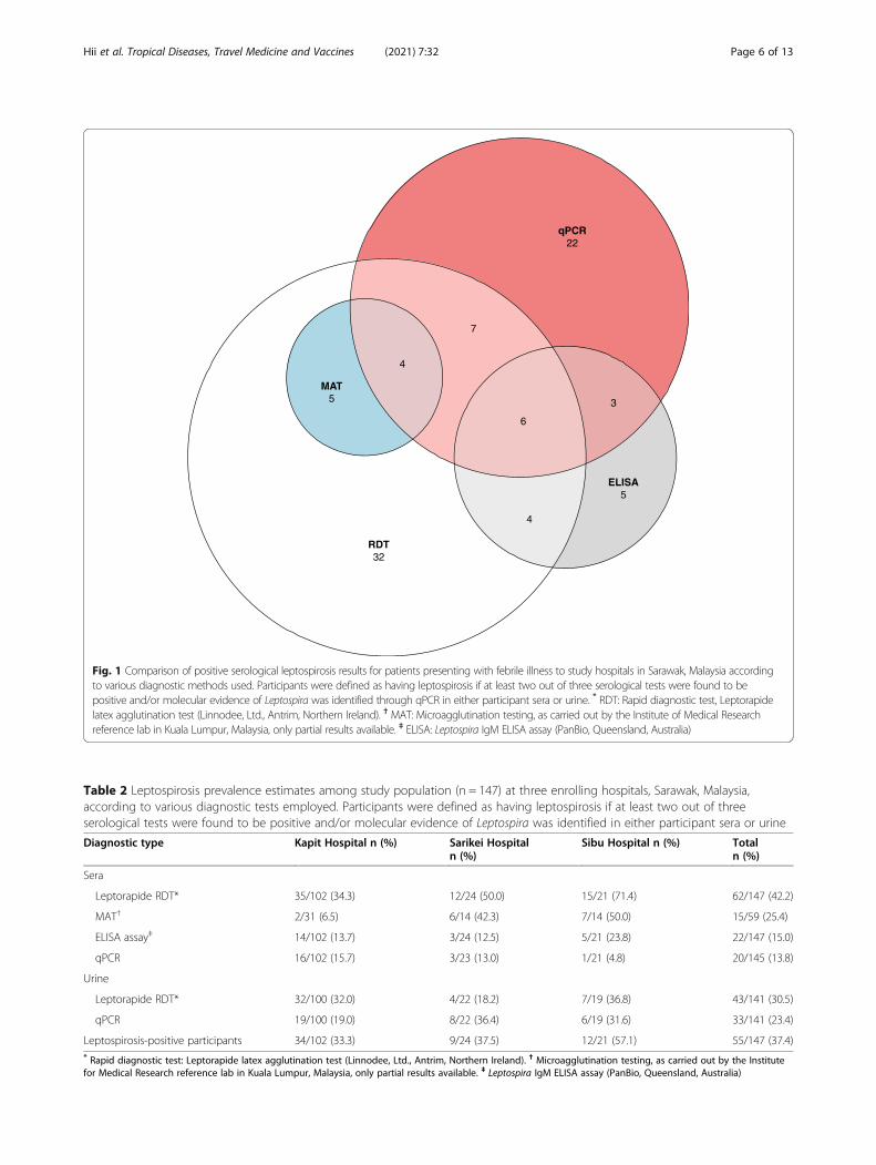
Fig. 1 Comparison of positive serological leptospirosis results for patients presenting with febrile illness to study hospitals in Sarawak, Malaysia accordingto various diagnostic methods used. Participants were defined as having leptospirosis if at least two out of three serological tests were found to bepositive and/or molecular evidence of Leptospira was identified through qPCR in either participant sera or urine. * RDT: Rapid diagnostic test, Leptorapidelatex agglutination test (Linnodee, Ltd., Antrim, Northern Ireland). † MAT: Microagglutination testing, as carried out by the Institute of Medical Researchreference lab in Kuala Lumpur, Malaysia, only partial results available. ‡ ELISA: Leptospira IgM ELISA assay (PanBio, Queensland, Australia)
Table 2 Leptospirosis prevalence estimates among study population (n = 147) at three enrolling hospitals, Sarawak, Malaysia,according to various diagnostic tests employed. Participants were defined as having leptospirosis if at least two out of threeserological tests were found to be positive and/or molecular evidence of Leptospira was identified in either participant sera or urine
Diagnostic type Kapit Hospital n (%) Sarikei Hospitaln (%)
Sibu Hospital n (%) Totaln (%)
Sera
Leptorapide RDT* 35/102 (34.3) 12/24 (50.0) 15/21 (71.4) 62/147 (42.2)
MAT† 2/31 (6.5) 6/14 (42.3) 7/14 (50.0) 15/59 (25.4)
ELISA assay‡ 14/102 (13.7) 3/24 (12.5) 5/21 (23.8) 22/147 (15.0)
qPCR 16/102 (15.7) 3/23 (13.0) 1/21 (4.8) 20/145 (13.8)
Urine
Leptorapide RDT* 32/100 (32.0) 4/22 (18.2) 7/19 (36.8) 43/141 (30.5)
qPCR 19/100 (19.0) 8/22 (36.4) 6/19 (31.6) 33/141 (23.4)
Leptospirosis-positive participants 34/102 (33.3) 9/24 (37.5) 12/21 (57.1) 55/147 (37.4)* Rapid diagnostic test: Leptorapide latex agglutination test (Linnodee, Ltd., Antrim, Northern Ireland). † Microagglutination testing, as carried out by the Institutefor Medical Research reference lab in Kuala Lumpur, Malaysia, only partial results available. ‡ Leptospira IgM ELISA assay (PanBio, Queensland, Australia)
Hii et al. Tropical Diseases, Travel Medicine and Vaccines (2021) 7:32 Page 6 of 13

pubmlst.org/organisms/leptospira-spp) and identifiedLeptospira interrogans as the infecting species (GenBankaccession numbers are provided in SupplementalTable 1). When compared to a worldwide database in-cluding 134 secY sequences of Leptospira interrogans[20], our four sequences were identical or closely relatedto leptospires identified in Malaysia, Indonesia, India,the Philippines and China.
Disease presentationAmong the 55 defined leptospirosis patients, 9.1% (n =5) presented with severe disease, characterized by death(n = 1), septic shock (n = 4) and respiratory distress re-quiring invasive ventilation support (n = 3). Anotherseven (12.7%) presented with moderate disease requiringnon-invasive ventilation support (n = 4), upper gastro-intestinal bleeding (n = 1), atrial fibrillation (n = 1) andthrombocytopenia (n = 2). The remaining 43 patients(78.2%) presented with mild disease. Acute kidney injuryand transaminitis were observed at all disease severitylevels (34.5% and 32.7% respectively).The most commonly self-reported symptoms among
the leptospirosis-positive patients were fever (52, 94.5%),nausea or vomiting (39, 70.9%), headache (37, 67.3%),and chills (36, 65.5%) (Supplemental Table 2). Severalpatients reported experiencing myalgia (29, 52.7%), jointpain (26, 47.3%), or conjunctivitis (17, 30.9%), while leth-argy and diarrhea were uncommon (3, 5.5% and 9,
16.4%, respectively). Notably, jaundice was not observedin any of the leptospirosis positive patients.Of note, two additional study participants died during
the course of their hospitalization, but we found no de-tectable evidence of Leptospira infection in either oftheir samples.
Symptom onsetLeptospirosis-positive patients generally presented in theearly phase of the disease, indicating that they werelikely in the acute stage of infection at the time of sam-ple collection (Fig. 2). Overall, 47 of 55 defined positivepatients (85.4%) were enrolled within 6 days post symp-tom onset (DPSO; self-reported), with five (9.1%) pre-senting on day 7 and one (1.8%) on day 8. Two patientsdid report having experienced joint pain and myalgia,among other symptoms, for 14 days.Comparatively, patients considered negative for lepto-
spirosis reported having experienced non-specific febrilesymptoms from one to 28 days prior to samplecollection.Correlation calculations identified sera and urine spe-
cimen qPCR analysis as complementary (SupplementalTable 3). Of the 45 participants with molecular evidencefor leptospirosis, 12 (26.7%) were qPCR positive onlythrough sera analysis, 25 (55.6%) only through urine,and 8 (17.8%) had Leptospira positivity in both sampletypes (Table 5). Sera-based qPCR results primarilypicked up evidence of Leptospira presence between 2
Table 3 Diagnostic accuracy of Leptorapide latex agglutination test (Linnodee, Ltd., Antrim, Northern Ireland) using acute sera andurine specimen, in comparison to real-time PCR (qPCR)
Diagnostic Type Serum-based RDT* Urine-based RDT*
RDT vs qPCRon sera or urine (95% CI†)
RDT vs qPCRon sera(95% CI†)
RDT vs qPCRon sera or urine (95% CI†)
RDT vs qPCRon urine(95% CI†)
Apparent prevalence 48.1 (39.2–57.0) 48.0 (39.1–57.1) 30.5 (23.0–38.8) 30.5 (23.0–38.8)
Sensitivity 47.5 (31.5–63.9) 36.8 (16.3–61.6) 22.2 (11.2–37.1) 18.2 (7.0–35.5)
Specificity 51.7 (40.8–62.4) 50.0 (40.2–59.8) 65.6 (55.2–75.0) 65.7 (56.0–74.6)
Positive predictive value 30.6 (19.6–43.7) 11.5 (4.7–22.2) 23.3 (11.8–38.6) 14.0 (5.3–27.9)
Negative predictive value 68.7 (56.2–79.4) 81.8 (70.4–90.2) 64.3 (54.0–73.7) 72.4 (62.5–81.0)
* Rapid diagnostic test. † Calculated confidence interval
Table 4 Diagnostic accuracy of Leptospira IgM ELISA assay (PanBio, Queensland, Australia) using acute sera specimen, in comparisonto real-time PCR (qPCR)
Diagnostic Type ELISA assay vs qPCR on sera or urine(95% CI)
ELISA assay vs qPCR on sera only(95% CI)
Apparent prevalence 16.3 (10.5–23.6) 16.4 (10.6–23.8)
Sensitivity 24.5 (13.3–38.9) 21.1 (6.1–45.6)
Specificity 88.4 (79.7–94.3) 84.3 (76.4–90.5)
Positive predictive value 54.5 (32.2–75.6) 18.2 (5.2–40.3)
Negative predictive value 67.3 (57.8–75.8) 86.6 (78.9–92.3)
Hii et al. Tropical Diseases, Travel Medicine and Vaccines (2021) 7:32 Page 7 of 13

and 6 DPSO, with one patient testing positive with nodocumented fever (Fig. 3). Urine-based qPCR resultswere positive between 1 and 8 DPSO, with three patientstesting positive with no documented fever (Fig. 4).Participants with serological evidence for leptospirosis
(n = 23) were identified from 1 to 8 DPSO, with oneidentified at 14 DPSO and one reporting no fever.Among these, the ten patients with no molecular evi-dence of Leptospira were primarily identified between 2and 7 DPSO, with one patient presenting at 14 DPSO.Independently of other serological assessments, the
Leptorapide RDT provided positive results for patients 1to 20 DPSO using sera samples and 1 to 14 DPSO usingurine samples. The majority of the urine-based RDTpositive results were within 1 to 7 DPSO, with 1 speci-men positive at 10 DPSO and another at 14.
The IgM ELISA assay detected antibody evidence inpatients 2 to 8 DPSO, and in one participant presentingwithout fever. MAT positives were identified primarilyin participants 2 to 7 DPSO, as well as in one participantat day 10, one at day 14, and one presenting withoutfever.
Risk factor analysisBivariate analyses demonstrated the participant’s com-pleted level of education, type of housing, and frequencyof washing clothes in the local river as being associatedwith leptospirosis diagnosis. Participant ethnicity, occu-pation, use of PPE, primary water source, and exposureto cats, dogs, pigs, poultry, or rodents were not found tobe significantly associated with leptospirosis diagnosis,nor was the enrollment site, recent flooding, engaging in
Fig. 2 Days post symptom onset (DPSO) at the time of enrollment, as self-reported by leptospirosis patients (diagnosis determined by molecularand/or serological evidence). The acute phase typically takes place in the first week of illness, followed by antibody development 5–7 days postsymptom onset
Table 5 Likely phase of disease progression for leptospirosis qPCR-positive patients as defined by sample positivity in sera and/orurine specimens. Participants with Leptospira evidence only in sera were classified to be in the onset phase, those with evidence inboth sera and urine were classified in the dissemination phase, and those with evidence only in urine were classified in theexcretion phase
Phase No. of Patients DPSO* Range Sera Mean Cq† (Range) Urine Mean Cq† (Range)
Onset 12 2–6 37.16 (35.36–39.45) –
Dissemination 8 0–5 36.57 (34.80–39.60) 35.77 (27.17–39.85)
Excretion 25 0–8 – 37.38 (29.23–39.96)
*Days post-symptom onset (DPSO): fever duration at time of enrolment and sampling as self-reported by the patient. †Real-time PCR-determined cyclethreshold (Cq)
Hii et al. Tropical Diseases, Travel Medicine and Vaccines (2021) 7:32 Page 8 of 13

other river-based activities, household income, or pri-mary mode of transportation (Supplemental Table 4).Multivariate analysis identified completing primary
school and weekly washing of clothes in the local riveras most predictive of a positive leptospirosis result insera and/or urine. In this joint model, participants whohad completed primary school were more than twice aslikely to test positive for leptospirosis infection whencompared to those with no education (OR 2.5, 95% CI1.0–6.4). Those who completed secondary school werenominally less likely to show evidence of infection thanthose with no education (OR 0.9, 95% CI 0.4–2.5)(Table 6).Washing clothes in the local rivers on a daily basis re-
sulted in similar odds of having a leptospirosis diagnosiswhen compared to those who reported never washingclothes in the river (OR 0.9, 95% CI 0.3–2.3). In contrast,individuals who washed clothes weekly in the rivers hada tenfold increase in the likelihood of leptospirosis
diagnosis, albeit with very low specificity (OR 10.6, 95%CI 1.4–214.8).Multicollinearity across the model was not identified
as a concern, with a mean variance inflation factor of1.06 for all included variables.
DiscussionOur study data document the poor performance of theLeptorapide RDT assay compared to qPCR diagnosis.The screening tool displayed low sensitivity and specifi-city for both sera and urine specimen; over half the truecases would have gone unidentified through using onlythe RDT screening followed by MAT confirmation, ascommonly prescribed in Malaysia. Insufficient data wasavailable to provide effective comparison between theRDT and validated MAT results; the study of pairedacute and convalescent sera samples would providemore conclusive MAT results and allow for better as-sessment of the antibody detection accuracy of the
Fig. 3 Evidence of leptospirosis in sera specimens from different diagnosis methods and according to patient-reported time of fever duration attime of sampling. Diagnosis determined by real-time PCR analysis, Leptorapide latex agglutination test (Linnodee, Ltd., Antrim, Northern Ireland),or Leptospira IgM ELISA assay (PanBio, Queensland, Australia)
Fig. 4 Evidence of leptospirosis in urine specimens from different diagnosis methods and according to patient-reported time of fever duration attime of sampling. Diagnosis determined by real-time PCR analysis or Leptorapide latex agglutination test (Linnodee, Ltd., Antrim,Northern Ireland)
Hii et al. Tropical Diseases, Travel Medicine and Vaccines (2021) 7:32 Page 9 of 13

Leptorapide RDT. Nevertheless, the present results areconsistent with other studies carried out in SoutheastAsia [21, 22] which have likewise shown that sensitivityand specificity of RDTs for Leptospira infections areoften low when compared to PCR.When using qPCR as the gold standard test, the
serum-based ELISA assay produced higher specificitythan the RDT, but much lower sensitivity. Results forthe ELISA test may be improved by testing paired serasamples rather than only acute stage samples [23], how-ever this is more labor intensive and delays diagnosis.There is a great need for more accurate, yet simple to
perform diagnostic tests for leptospirosis in SoutheastAsia (and other resource-restricted settings). PCR canconfirm diagnosis in the early phase of the disease beforeantibody titers are at detectable levels, but appropriatelaboratory support is lacking in many areas [24]. MATtesting is similarly resource intensive and restricted toreference laboratories, and misses a critical window asantibodies are lacking during the acute phase of the dis-ease [5].Our estimated prevalence data suggest that 37.4% (55
out of 147) of the patients who present with clinicalsigns and symptoms consistent with leptospirosis receivea positive laboratory confirmation. We recognize thatthis estimate has a number of limitations and may ormay not represent true prevalence. However, our resultssupport our position that leptospirosis is a frequentcause of illness in Sarawak, as reported in previous stud-ies conducted in the region. From 2011 to 2013, LelaSuut et al. likewise found a population-based leptospir-osis seroprevalence of 37.4% among 508 subjects amongrural communities in the Rejang Basin of Sarawak, usingELISA and MAT [25]. They found evidence of three
main serogroups detected in all three divisions: 22.1%Djasiman, 13.2% Shermani, and 7.9% Pomona [25]. Astudy conducted in urban areas of Sarawak describingthe diversity of Leptospira spp. in rats and the environ-ment identified four pathogenic species: L. interrogans,L. weilii, L. borgpetersenii and L. noguchii [26], while an-other study focusing on various rodent species in urban,suburban, and rural Sarawak identified L. interrogansand L. borgpetersenii [27]. In the present study, we didnot perform serological typing but were able tocharacterize a subset of positive samples as infected byL. interrogans, a pathogenic species previously identifiedin human leptospirosis in the region [1, 28] and com-monly reported worldwide.To our knowledge, this is the first clinical study on hu-
man leptospirosis in Sarawak, East Malaysia. Most of theleptospirosis patients were enrolled during the first sixdays of fever onset, suggesting they were in the acutestage of the disease. Study data were remarkable in thatno patients with laboratory evidence of leptospirosis in-fection exhibited jaundice, a finding that is inconsistentwith previous reports from West Malaysia that reportedhigher prevalences (22.5% [29] and 4.9% [30]) of jaun-dice among their patients. As icteric leptospirosis is as-sociated with higher mortality rates [3, 4], it isunsurprising that low patient mortality was attributed toacute infections in our patients, despite having been re-ported in other case series [29, 30].Our data (Supplemental Fig. 2) support other studies
that have also been able to detect Leptospira in urineduring the acute phase (first week) of infection [11, 12].Our study identified more urine samples as qPCR posi-tive for leptospirosis than sera, which was unexpected asour samples were typically collected within a week post
Table 6 Predictive factors for positive leptospirosis diagnosis (having either molecular or serological evidence of leptospirosis) in thestudy population (n = 147), as calculated through bivariate and multivariate analyses
Risk Factor Positive cases (%) Negative cases (%) Unadjusted OR*(95% CI†)
Adjusted OR*(95% CI†)
Education completed
None (n = 34) 10 (29.4) 24 (70.6) Ref‡ Ref‡
Primary (n = 57) 28 (49.1) 29 (50.9) 2.28 (0.95–5.86) 2.47 (1.00–6.41)
Secondary / College (n = 56) 17 (30.4) 39 (69.6) 1.04 (0.41–2.74) 0.95 (0.36–2.53)
Type of Housing
House (n = 49) 23 (46.9) 26 (53.1) Ref‡ –
Hut / Wood house (n = 20) 9 (45.0) 11 (55.0) 0.93 (0.32–2.67) –
Longhouse (n = 78) 23 (29.5) 55 (70.5) 0.48 (0.22–1.00) –
Washing clothes in river
Daily (n = 24) 7 (29.2) 17 (70.8) 0.70 (0.25–1.78) 0.86 (0.30–2.27)
Weekly (n = 5) 4 (80.0) 1 (20.0) 6.03 (0.80–168.44) 10.56 (1.44–214.82)
None (n = 118) 44 (37.3) 74 (62.7) Ref‡ Ref‡
* Odds ratio. † Calculated confidence interval. ‡ Reference category
Hii et al. Tropical Diseases, Travel Medicine and Vaccines (2021) 7:32 Page 10 of 13

symptom onset. During the early stage of infection, Lep-tospira are most commonly investigated and identifiedwithin the blood stream [3, 7]. Bacterial presence canlater be seen in both sera and urine samples as the infec-tion disseminates and Leptospira colonize the kidneys.Finally, leptospires are cleared from the bloodstreamduring the convalescent phase, while still present inurine. As described by Esteves, et al., different sampletypes may indeed prove more effective for identifyingLeptospira infection at varying stages of disease [31],with the inclusion of urine permitting a wider window ofdetection [3]. By testing paired sera and urine specimens,we were able to identify 25 additional leptospirosis cases,which would have been missed had only sera beenanalyzed.Risk factor data were interesting in that, while weekly
washing of clothes in the river greatly increased diseaserisk when compared to those not using the river for suchactivities, doing so daily had minimal effect on this samerisk. This finding was fairly imprecise, given the lownumber of observances of weekly clothes washing, how-ever it may reflect longer exposure time in Leptospira-contaminated river water, if one assumes that once-weekly washing correlates with a larger amount of
clothes to process. Despite taking place in the same bodyof water, other river-based activities such as swimming,bathing, fishing, or commuting were not significantly as-sociated with disease risk within the study population,regardless of frequency.Completing primary school appeared to greatly in-
crease the risk of leptospirosis in comparison to thosewith either no formal education or with more advancedtraining. Although household income and known occu-pational risk factors for leptospirosis were not found tosignificantly impact leptospirosis outcomes in our studypopulation, the increased risk associated with havingcompleted primary school may serve as a proxy for othersocioeconomic exposures not studied here. Though theMalaysian Ministry of Health has reported greater ratesof leptospirosis within 25–60 year old individuals [2], agewas surprisingly not found to increase disease risk in ourbivariate analysis and thus not incorporated into ourmultivariate modeling.The study had a number of limitations. First it was a
short-term study with limited geographical spread; wesampled for only a short time and only at three hospitalsamong the 22 hospitals in Sarawak so our findings maynot be representative of the entire state of Sarawak.
Fig. 5 There are multiple potential sources of Leptospira infections in the region. Much of the area around Kapit Hospital has dense tropical forestand riverine systems rich in wildlife. This wildlife may introduce Leptospira into water systems. Most people live in longhouses or farm huts on theoutskirts of the town of Kapit. Transportation is chiefly via boats on rivers. While Kapit is supplied with treated water, the source of the water inrural areas is either from river water or gravity fed rain-water-systems. Images here illustrate the living and transportation environments in whichthis study took place (photos provided by Dr. King-Ching Hii)
Hii et al. Tropical Diseases, Travel Medicine and Vaccines (2021) 7:32 Page 11 of 13

Further, in accordance with local clinical diagnostic pro-cedures, MAT assays were only performed on a subsetof the acute sera specimens that had positive RDT find-ings. Hence, our prevalence estimates may have underes-timated true prevalence. Additionally, occupationalexposure was difficult to assess, as only half the partici-pants were of working age and few self-reported en-gaging with previously recognized high-risk occupations(e.g. fishing and agriculture). As people from rural Sara-wak have many environmental water exposures whichcould serve as the source of Leptospira infection (Fig. 5),it is challenging to implicate specific risks and plans tostudy particular interventions. Finally, risk factor andsymptom data were self-reported and could not be veri-fied, offering the possibility of bias.
ConclusionThis study confirms leptospirosis endemicity among thestudy population in Sarawak, Malaysia and identified lowdiagnostic performance of the commercial rapid diag-nostic test and IgM ELISA assay studied when used dur-ing the acute phase of the disease. We conclude thatthere is a critical need for more sensitive, inexpensive,and easy to use rapid diagnostics for leptospirosis in thisregion. Until such diagnostics are available, clinicians inareas where molecular testing is unavailable will be wiseto continue to treat empirically if the clinical suspicionof leptospirosis is high and RDTs are negative. Finally,our study found that in areas where PCR-based diagnos-tics are possible, testing both sera and urine can increasethe probability of detecting leptospirosis during theacute phase.
AbbreviationsCDC: U.S. Center for Disease Control and Prevention; IgG: Immunoglobulin G;IgM: Immunoglobulin M; ELISA: Enzyme-linked immunosorbent assay;MAT: Microscopic agglutination testing; NPV: Negative predictive value;PBS: Phosphate-buffered saline solution; PCR: polymerase chain reaction;PPV: positive predictive value; RDTs: Serological rapid diagnostic tests;rPCR: real-time polymerase chain reaction
Supplementary InformationThe online version contains supplementary material available at https://doi.org/10.1186/s40794-021-00154-2.
Additional file 1.
AcknowledgementsWe thank the Director General of Health Malaysia for his permission topublish this article. This work was conducted in partnership with DukeUniversity, the Duke Global Health Institute, Sibu Hospital Clinical ResearchCenter, SEGi University Sibu Clinical Campus, and the Ministry of HealthMalaysia. This study was supported by Vysnova and Naval Medical ResearchUnit 2. Special thanks to Lee Nung Kion from Faculty of Cognitive Sciencesand Human Development, University Malaysia Sarawak for contributingstatistical analyses. Special thanks, also, to Jakie Ting and Tiing-Tiing Chua forcollaboration in handling specimens between Sibu, Sarikei and Kapit Hos-pital. We appreciate Ivan Jan-Shui Vun, Huong-Nai Law, Khiu Fu Lung, Pra-vind Narayanan, Ing-Tien Wong, Toh-Mee Wong for supervising at the
respective study sites. We thank the medical officers Jia-Yun Chuang, Wei-Yieng Tang, Fong-Kean Lim, Shen-Ly Lai, Syn-Hwan Wong, Awang EmirNaim, Eng-Seng Tiew, Chiong-Kim Law, Jeffrey Soon-Yit, Lee, Jun-Wei Liew,Rashmika Nambiar, Edmund Kwong-Yuen Wong, Bing-En Chia for recruitingstudy participants at Kapit, Sarikei, and Sibu Hospitals. We thank laboratorytechnicians Hieng-Hua Goh of Kapit Hospital and the laboratory techniciansfrom Sibu and Sarikei for preparing clinical specimens for study. We also ap-preciate the drivers and those who helped with transporting specimens.
Authors’ contributionsK.H. and E.R. wrote the manuscript with support from V.G., M.G., R.B., and G.G.All authors contributed to editing and review of the manuscript. I.S., A.B., andJ.G. carried out primary laboratory analyses. N. A and L.X. validated laboratoryfindings and facilitated sequencing. E.R. performed principle statisticalanalysis in association with V.G. and J.M. K.H., D.G., and G.G. conceptualizedand designed the study. K.H. and G.G. supervised the project.
FundingThis study was funded by the United States of America Naval MedicalResearch Unit No. TWO (NAMRU-2) via task order Vysnova/Duke SC-N6264518D5058-DUKE-007 FSS Malaysia, awarded to GCG. MJG is a militaryservice member. This work was prepared as part of their official duties. Title17, U.S.C., §105 provides that copyright protection under this title is not avail-able for any work of the U.S. Government. Title 17, U.S.C., §101 defines a U.S.Government work as a work prepared by a military service member or em-ployee of the U.S. Government as part of that person’s official duties. Theviews expressed in this article are those of the authors and do not necessar-ily reflect the official policy or position of the U.S. Department of the Navy,the U.S. Department of Defense or the US government. The funders had norole in study design, data collection and analysis, decision to publish, orpreparation of the manuscript.
Availability of data and materialsThe data collected during the current study are available from thecorresponding author on reasonable request.
Declarations
Ethics approval and consent to participateThe study was conducted in accordance with the ethical standards of theHelsinki Declaration and received a scientific review by and ethical approvalfrom the Medical Research and Ethics Committee of the Ministry of Health,Malaysia (NMRR-18-1022-40334) and Duke University Institutional ReviewBoard (Pro00100246), with extramural research review in accordance withthe U.S. Navy Human Research Protection Program(HPRO.NAMRU2.2019.0003), in compliance with all applicable federalregulations governing the protection of human subjects.
Consent for publicationNot applicable.
Competing interestsThe authors have no financial interests to disclose. Dr. Gray serves oneditorial board for Tropical Diseases, Travel Medicine and Vaccines.
Author details1Department of Pediatrics, Kapit Hospital, Ministry of Health Malaysia, Kapit,Sarawak, Malaysia. 2Duke Global Health Institute, Duke University, Durham,NC, USA. 3Division of Infectious Diseases, Duke University School of Medicine,DUMC Box 102359, Durham, NC 27710, USA. 4National Health CommissionKey Laboratory of Systems Biology of Pathogens, Institute of PathogenBiology, Chinese Academy of Medical Sciences and Peking Union MedicalCollege, Beijing, China. 5Department of Biostatistics and Bioinformatics, DukeUniversity, Durham, NC, USA. 6Present address: Agricultural Research Service,National Animal Disease Center, United States Department of Agriculture,Ames, IA, USA. 7United States Naval Medical Research Center- Asia,Singapore, Singapore. 8Emerging Infectious Disease Program, Duke-NUSMedical School, Singapore, Singapore. 9Global Health Center, Duke KunshanUniversity, Kunshan, China.
Hii et al. Tropical Diseases, Travel Medicine and Vaccines (2021) 7:32 Page 12 of 13
Received: 30 June 2021 Accepted: 7 September 2021
References1. Garba B, Bahaman AR, Khairani-Bejo S, Zakaria Z, Mutalib AR. Retrospective
study of leptospirosis in Malaysia. Ecohealth. 2017;14(2):389–98. https://doi.org/10.1007/s10393-017-1234-0.
2. Zainudin A, editor Epidemiology and Current Situation of Leptospirosis inMalaysia. Persidangan Kesihatan Persekitaran Pihak Berkuasa Tempatan; 2015September 8–9, 2015; Labuan: Ministry of Health Malaysia.
3. Levett PN. Leptospirosis. Clin Microbiol Rev. 2001;14(2):296–326. https://doi.org/10.1128/CMR.14.2.296-326.2001.
4. Haake DA, Levett PN. Leptospirosis in humans. Curr Top Microbiol Immunol.2015;387:65–97. https://doi.org/10.1007/978-3-662-45059-8_5.
5. Garba B, Bahaman AR, Bejo SK, Zakaria Z, Mutalib AR, Bande F. Majorepidemiological factors associated with leptospirosis in Malaysia. Acta Trop.2018;178:242–7. https://doi.org/10.1016/j.actatropica.2017.12.010.
6. Costa F, Hagan JE, Calcagno J, Kane M, Torgerson P, Martinez-Silveira MS,et al. Global morbidity and mortality of leptospirosis: a systematic review.PLoS Negl Trop Dis. 2015;9(9):e0003898. https://doi.org/10.1371/journal.pntd.0003898.
7. Picardeau M. Diagnosis and epidemiology of leptospirosis. Med Mal Infect.2013;43(1):1–9. https://doi.org/10.1016/j.medmal.2012.11.005.
8. Vincent AT, Schiettekatte O, Goarant C, Neela VK, Bernet E, Thibeaux R, et al.Revisiting the taxonomy and evolution of pathogenicity of the genusLeptospira through the prism of genomics. PLoS Negl Trop Dis. 2019;13(5):e0007270. https://doi.org/10.1371/journal.pntd.0007270.
9. Courdurie C, Le Govic Y, Bourhy P, Alexer D, Pailla K, Theodose R, et al.Evaluation of different serological assays for early diagnosis of leptospirosisin Martinique (French West Indies). PLoS Negl Trop Dis. 2017;11(6):e0005678.https://doi.org/10.1371/journal.pntd.0005678.
10. WHO. Human leptospirosis : guidance for diagnosis, surveillance andcontrol. World Health Organization. 2003.
11. Bal AE, Gravekamp C, Hartskeerl RA, De Meza-Brewster J, Korver H, TerpstraWJ. Detection of leptospires in urine by PCR for early diagnosis ofleptospirosis. J Clin Microbiol. 1994;32(8):1894–8. https://doi.org/10.1128/jcm.32.8.1894-1898.1994.
12. Bahuaud O, Pastuszka A, Le Brun C, Ehrmann S, Lanotte P. Seven YearsLeptospirosis Follow-Up in a Critical Care Unit of a French MetropolitanHospital; Role of Real Time PCR for a Quick and Acute Diagnosis J Clin Med2020;9, 9, DOI: https://doi.org/10.3390/jcm9093011.
13. Bajani MD, Ashford DA, Bragg SL, Woods CW, Aye T, Spiegel RA, et al.Evaluation of four commercially available rapid serologic tests for diagnosisof leptospirosis. J Clin Microbiol. 2003;41(2):803–9. https://doi.org/10.1128/JCM.41.2.803-809.2003.
14. Panwala T, Rajdev S, Mulla S. To evaluate the different rapid screening testsfor diagnosis of leptospirosis. J Clin Diagn Res. 2015;9(2):DC21–4. https://doi.org/10.7860/JCDR/2015/11188.5587.
15. Mohd Ali MRMS. Amira Wahida; Ismail, nor Hayati; Abu Sapian, Roslinda;mat Hussin, Hani; Ismail, Nabilah; yean yean, Chan. Development andvalidation of pan-Leptospira Taqman qPCR for the detection of Leptospiraspp. in clinical specimens. Mol Cell Probes. 2018;38:1–6. https://doi.org/10.1016/j.mcp.2018.03.001.
16. Prevention CfDCa. Leptospirosis (Leptospira interrogans) 2013 Case Definition2013.
17. Brownlow T, Kavanagh OV, Logan EF, Hartskeerl RA, Savage R, Palmer MF,et al. 'Leptorapide' - a one-step assay for rapid diagnosis of humanleptospirosis. Epidemiol Infect. 2014;142(6):1182–7. https://doi.org/10.1017/S0950268813002112.
18. El Bali L, Diman A, Bernard A, Roosens NH, De Keersmaecker SC.Comparative study of seven commercial kits for human DNA extractionfrom urine samples suitable for DNA biomarker-based public health studies.J Biomol Tech. 2014;25(4):96–110. https://doi.org/10.7171/jbt.14-2504-002.
19. Ahmed N, Devi SM, Valverde Mde L, Vijayachari P, Machang'u RS, Ellis WA,et al. Multilocus sequence typing method for identification and genotypicclassification of pathogenic Leptospira species. Ann Clin MicrobiolAntimicrob. 2006;5(1):28. https://doi.org/10.1186/1476-0711-5-28.
20. Nalam K, Ahmed A, Devi SM, Francalacci P, Baig M, Sechi LA, et al. Geneticaffinities within a large global collection of pathogenic Leptospira:implications for strain identification and molecular epidemiology. PLoS One.2010;5(8):e12637. https://doi.org/10.1371/journal.pone.0012637.
21. Chang CH, Riazi M, Yunus MH, Osman S, Noordin R. Limited diagnosticvalue of two commercial rapid tests for acute leptospirosis detection inMalaysia. Diagn Microbiol Infect Dis. 2014;80(4):278–81. https://doi.org/10.1016/j.diagmicrobio.2014.08.012.
22. Dittrich S, Boutthasavong L, Keokhamhoung D, Phuklia W, Craig SB, TulsianiSM, et al. A prospective hospital study to evaluate the diagnostic accuracyof rapid diagnostic tests for the early detection of leptospirosis in Laos. AmJ Trop Med Hyg. 2018;98(4):1056–60. https://doi.org/10.4269/ajtmh.17-0702.
23. Desakorn V, Wuthiekanun V, Thanachartwet V, Sahassananda D, Chierakul W,Apiwattanaporn A, et al. Accuracy of a commercial IgM ELISA for thediagnosis of human leptospirosis in Thailand. Am J Trop Med Hyg. 2012;86(3):524–7. https://doi.org/10.4269/ajtmh.2012.11-0423.
24. Musso D, La Scola B. Laboratory diagnosis of leptospirosis: a challenge. JMicrobiol Immunol Infect. 2013;46(4):245–52. https://doi.org/10.1016/j.jmii.2013.03.001.
25. Suut L, Mazlan MN, Arif MT, Yusoff H, Abdul Rahim NA, Safii R, et al.Serological prevalence of leptospirosis among rural communities in theRejang Basin, Sarawak. Malaysia Asia Pac J Public Health. 2016;28(5):450–7.https://doi.org/10.1177/1010539516648003.
26. Pui CF, Bilung LM, Apun K, Su'ut L. Diversity of Leptospira spp. in Rats andEnvironment from Urban Areas of Sarawak, Malaysia. J Trop Med. 2017;2017:3760674.
27. Blasdell KR, Morand S, Perera D, Firth C. Association of rodent-borneLeptospira spp. with urban environments in Malaysian Borneo. PLoS NeglTrop Dis. 2019;13(2):e0007141.
28. Thayaparan S, Robertson ID, Fairuz A, Suut L, Gunasekera UC, Abdullah MT.Seroepidemiological study of leptospirosis among the communities living inperiurban areas of Sarawak. Malaysia Med J Malaysia. 2015;70(5):288–94.
29. Fann RJ, Vidya RR, Chong HE, Indralingam V, Christopher Chan WS. Clinicalpresentations and predictors of mortality for leptospirosis - a study fromsuburban area in Malaysia. Med J Malaysia. 2020;75(1):52–6.
30. Mohd Taib N, Ahmad H, Soh KL, Md Shah A, Amin Nordin S. Than Thianlung L, et al. significant clinical presentation of leptospirosis in relation toSociodemographic and risk factors in a tertiary hospital, Malaysia. VectorBorne Zoonotic Dis. 2020;20(4):268–74. https://doi.org/10.1089/vbz.2018.2417.
31. Esteves LM, Bulhoes SM, Branco CC, Carreira T, Vieira ML, Gomes-Solecki M,et al. Diagnosis of human leptospirosis in a clinical setting: real-time PCRhigh resolution melting analysis for detection of Leptospira at the onset ofdisease. Sci Rep. 2018;8(1):9213. https://doi.org/10.1038/s41598-018-27555-2.
Publisher’s NoteSpringer Nature remains neutral with regard to jurisdictional claims inpublished maps and institutional affiliations.
Hii et al. Tropical Diseases, Travel Medicine and Vaccines (2021) 7:32 Page 13 of 13